


Anzeige
Erschienen in:
01.09.2009 | Correspondence
An unexpected hazard of indwelling temperature monitoring
Erschienen in: Intensive Care Medicine | Ausgabe 9/2009
Einloggen, um Zugang zu erhaltenExcerpt
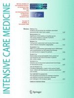
A 65-year-old lady was admitted to the intensive care unit (ICU) following emergency surgery for a perforated duodenal ulcer. The post-operative course was complicated by abdominal sepsis, multiple laparotomies, ventilator associated pneumonia and she required multi-organ support. Percutaneous tracheostomy was performed on day 11. At 6 weeks, she remained ventilator-dependent. High dose steroids were started. High resolution CT scan showed extensive consolidation, subpleural reticulation and interlobular septal thickening suggestive of early fibrosis. Incidentally, a metallic cylindrical object (15 mm × 5 mm) was apparent in the right lower lobe previously unseen on plain chest radiographs (Fig. 1).
Fig. 1
Detail of patient's CT scan and (a–c) the temperature probe
× ![]()
…