


Anzeige
Erschienen in:
27.06.2017 | Imaging in Intensive Care Medicine
Fat embolism syndrome
Erschienen in: Intensive Care Medicine | Ausgabe 9/2017
Einloggen, um Zugang zu erhaltenExcerpt
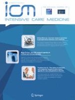
A 19-year-old man was admitted to our emergency department after a road accident. He presented with bilateral femoral fractures that were promptly treated with external fixators. After surgery, he was monitored in ICU. Twenty-four hours after admission, fever, tachycardia, dyspnea, and hypoxia appeared. He also became drowsy but arousable, confused, and agitated. Tracheal intubation was performed. A few hours later, reddish-brown nonpalpable axillary (Fig. 1a) and subconjunctival petechiae (Fig. 1b) appeared. Brain CT and chest X-ray were normal. Suspecting a fat embolism syndrome, we performed an MRI that showed multiple hyperintense puntiform lesions disseminated in deep white substance, basal ganglia, and thalamus on FLAIR imaging (Fig. 1c) and multiple microembolic infarcts (“starfield pattern”) on DWI (Fig. 1d). Transesophageal echocardiography excluded septal defects, and an electroencephalogram showed a generalized polymorphic delta activity. Urine analysis was normal, but lipiduria was observed. After 5 days the patient was extubated and 2 days later he was discharged by the ICU with good recovery. Skin alterations persisted for 4 days after ICU discharge.
Fig. 1
Classic cutaneous signs of fat embolism syndrome are shown: axillary (a) and subconjunctival petechiae (b). Multiple disseminated lesions are observed on FLAIR (c) and DWI (d)
× ![]()
…