Utilization of autologous stem cells has been proposed for the treatment of anal incontinence despite a lack of understanding of their mechanism of action and of the physiological healing process of anal sphincters after injury.
Aims
We aim to develop a technique allowing isolation and further study of local mesenchymal stem cells, directly from anal canal transition zone in pig.
Methods
Anal canal was resected “en bloc” from two young pigs and further microdissected. The anal canal transition zone was washed and digested with 0.1% type I collagenase for 45 min at 37 °C. The isolated cells were plated on dishes in mesenchymal stem cell medium and trypsinized when confluent. Cells were further used for flow cytometry analysis and differentiation assays.
Results
The anal canal transition zone localization was confirmed with H&E staining. Following culture, cells exhibited a typical “fibroblast-like” morphology typical of stem cells. Isolated cells were positive for CD90 and CD44 but negative for CD14, CD34, CD45, CD105, CD106, and SLA-DR. Following incubation with specific differentiation medium, isolated cells differentiated into adipocytes, osteoblasts, and chondrocytes, confirming in vitro multipotency.
Conclusions
Herein, we report for the first time the presence of mesenchymal stem cells in the anal canal transition zone in pigs and the feasibility of their isolation. This preliminary study opens the path to the isolation of human anal canal transition zone mesenchymal stem cells that might be used to study sphincters healing and to treat anal incontinence.
Springer Nature remains neutral with regard to jurisdictional claims in published maps and institutional affiliations.
Abkürzungen
ATZ
Anal canal transition zone
A-SCs
Adipose-tissue-derived stem cells
DMEM
Dulbecco’s modified Eagle medium
FBS
Fetal bovine serum
IGL-1
Institute Georges Lopez 1
IMDM
Iscove's modified Dulbecco’s medium
MSCs
Mesenchymal stem cells
M-SCs
Muscular-tissue-derived stem cells
PBS
Phosphate-buffered saline
Introduction
Stem cell therapy is a promising approach to cure degenerative diseases and has been proposed for the treatment of various disease, including anal sphincter incontinence (ASI) [1‐3]. Despite more than 20 years of research in this field with encouraging preclinical studies and clinical trials, there still is a lack of consensus regarding the cells that should be used [1‐4]. In the literature, two candidates emerged for clinical application: muscular tissue-derived stem cells (M-SCs) (satellite cells or myoblasts) and adipose-tissue derived stem cells (A-SCs) [3]. The precise effects of these cells on the damaged anal sphincters of animal models or patients remain unknown, but they undeniably increase the continence as measured by anorectal manometry and evaluated by incontinence scores [2]. However, it is supposed that stem cells and, particularly, mesenchymal stem cells (MSCs) act by the secretion of regenerative trophic factors and anti-inflammatory cytokines [3]. Different strategies of cell therapy utilization have been proposed, including ultrasound-guided injection of cells directly into sphincter defects or their utilization during surgical procedure to increase the success of sphincter repair surgery [5‐7]. Recently, Bitar et al. successfully assessed the potential of a bioengineered autologous BioSphincter made from enteroneurons and smooth muscle cells extracted from the internal anal sphincter of nonhuman primate that was further reimplanted to the animals to restore their continence [8]. Moreover, Son et al. investigated the possibility to extract stem cells from the external anal sphincter and internal anal sphincter in human from proctectomy specimens and assessed the vitality of cells in patients in whom pelvic radiotherapy was conducted [9].
Anal sphincters are a complex structure that is not only constituted of muscle tissue but also includes nerves and connective tissue surrounding the sphincters. Hemorrhoids localized at the anal canal transition zone (ATZ) are in close proximity to the internal anal sphincter and nerve tracts, and this is nicely demonstrated by the visualization of IAS during Ferguson or Milligan-Morgan hemorrhoidectomy procedures [10] but also by the potential implication of nerve injury in chronic pain after stapled hemorrhoidopexy [11]. Specific ATZ stem cells participated in rectal mucosa healing at the level of ATZ and beyond [12]. Therefore, it is highly probable that surrounding tissues participate in the response to injury and the healing of sphincter lesions after either primary injury or secondary repair of a chronic sphincter injury. Hemorrhoid tissue and ATZ are easily accessible and could in theory constitute a source of stem cells after their in vitro expansion. We report herein a preliminary study on the technical feasibility of stem cell isolation from the ATZ in pig.
Anzeige
Materials and Methods
Animals
This report was based on the reutilization of sacrificed animals, which is encouraged by the 3R principles (Reduction). Two female Large White piglets (Appel, Switzerland) were sacrificed by exsanguination, as part of a protocol for pancreas isolation (GE/151/16) after organ perfusion with cold Institute Georges Lopez 1 (IGL-1) solution (Institute Georges Lopez, Lyon, France) and rapid cooling of the animal with ice. This protocol was approved by the animal ethics committee of the Geneva Veterinarian Office (Geneva, Switzerland). Pigs were transferred 1 week before surgery and housed under standard conditions at the animal facility core of the University of Geneva with food and water ad libitum. Shortly after the death of the animal and the removal of the pancreas, the anus and distal rectum were resected “en bloc” and immersed in an IGL-1 bath (Fig. 1A). The removed specimen was microdissected on ice on a back table to leave only the mucosa of the transition zone, which was further immerged into cold IGL-1 and transported to our cell isolation facility (Fig. 1B).
Fig. 1
Anal canal transition zone stem cell isolation. A Anal canal with distal rectum was removed “en bloc.” B The specimen was further dissected on a back table using surgical loop to remove anoderm, sphincters, and remaining rectum. C Standard hematoxylin–eosin staining of a slide demonstrating that only the upper anal canal mucosa was isolated (anoderm was absent). We observed the rectal columnar single-layer epithelium, the transition epithelium [anal transition zone (AZT)], and the multilayer epithelium of the anal canal with numerous glands
×
Materials
Falcon tubes, culture plates, flasks, and 100 μm cell strainers were obtained from Corning (Corning, NY, USA). Centrifuges included a Rotixa 50 RS (Hettich Zentrifugen, Tuttlingen, Germany). Leica DM750 and Leica DMIL microscope were used for standard light analysis (Wetzlar, Germany). Flow cytometry sample acquisition was performed with an Accuri C6 (Becton Dickinson, Franklin Lakes, NJ, USA) and an Attune NxT (Invitrogen, Carlsbad, CA, USA).
Reagents and Antibodies
Culture-grade phosphate-buffered saline (PBS), Dulbecco’s modified Eagle medium high glucose (DMEM), Iscove’s modified Dulbecco’s medium (IMDM), fetal bovine serum (FBS), trypsin/EDTA 0.25%, and penicillin and streptomycin were from Gibco (Life Technologies, Carlsbad, CA, USA). Recombinant TGF-β1 and platelet-derived growth factor BB were purchased from PreproTech (Rocky Hill, NJ, USA). Dexamethasone, hydrocortisone, ascorbic acid, insulin–transferrin–selenium premix, l-proline, β-glycerolphosphate, isobutyl-1-methylxanthin, indomethacin, 10% formalin, Alizarin Red S and Oil-red-O solution, and rabbit serum were all from Sigma-Aldrich (Buchs, Switzerland).
For flow cytometry, owing to difficulties finding antibodies specific for pig, we used anti-human antibodies known to cross-react against pig tissue. Fluorescein isothiocyanate-conjugated anti-human CD34 was from Bioss Inc. (Boston, MA, USA). Fluorescein isothiocyanate-conjugated anti-human CD44 and anti-human CD105 was from GenTex, whereas fluorescein isothiocyanate-conjugated anti-human CD45 and phycoerythrin-conjugated and unconjugated mouse anti-human CD90 were purchased from BD (Franklin Lakes, NJ, USA). Fluorescein isothiocyanate-conjugated anti-human CD106 was from Life Technologies (Carlsbad, CA, USA). Alexa Fluor 488-conjugated anti-mouse antibody and anti-human vimentin antibody were obtained from Invitrogen (Carlsbad, CA, USA). Fluorescein isothiocyanate-conjugated anti-swine leukocyte antigen DR class 2 and anti-human CD14 were from Bio-Rad (Hercules, CA, USA). Fluorescein isothiocyanate-conjugated anti-swine leukocyte antigen DR class 2 and anti-human CD14 were from Bio-Rad (Hercules, CA, USA).
Anzeige
Isolation of Multipotent Cells
One-third of the specimen was used for standard histology (hematoxylin–eosin) and three-thirds for cell isolation. The isolated tissue was then transferred to a new tube with 50 ml cold PBS and 2% penicillin and streptomycin, and washed two times for 3 min at 1152g and 4 °C. Then, the specimen was transferred into a solution of lukewarm PBS with 0.1% collagenase type I (Sigma-Aldrich, Buchs, Switzerland). The specimen was chopped with scissors, and this process was repeated every 15 min while the tube was incubated for 45 min at 37 °C with 5% CO2. After digestion was completed, the preparation was centrifuged during 2 min at a speed of 512g at 4 °C. The tube was then hand-shaken vigorously to disrupt the pellet. Then the suspension was further centrifuged for 2 min at a speed of 512g at 4 °C. The obtained pellet was rinsed with 2 ml of STOP solution (DMEM and 10% fetal bovine serum). The pellet was further pulled down during 5 min at a speed of 184g at 4 °C. We used osmotic lysis to get rid of erythrocytes: the pellet was incubated in a solution of NaCl 0.2% for 30 s, then in a solution of NaCl 1.6% for 30 s. After centrifugation at a speed of 610g for 5 min at 4 °C, the pellet was resuspended in 20 ml of PBS and 2% penicillin and streptomycin and further centrifuged at 184g for 5 min at 4 °C. The pellet was then suspended in 2 ml culture medium and filtered through a 100 µm cell strainer. The filter was rinsed with 4 ml of culture medium, and the whole flow-through with 4 ml supplementary medium was plated on a culture flask. Cell yield was assessed with a Neubauer chamber.
Cell Culture and Differentiation Assays
MSC culture medium was prepared by adding 10% FBS, 10 ng/ml platelet-derived growth factor BB, and 2% penicillin and streptomycin to IMDM. For chondrocyte differentiation medium, we mixed dexamethasone 0.1 µM, ascorbic acid 50 µg/ml, l-proline 20 µM, TGF-β1 10 ng/ml, insulin–transferrin–selenium premix 50 ng/ml, and 2% penicillin and streptomycin to DMEM. For adipogenic differentiation medium, we used IMDM enriched with isobutyl-1-methylxanthin 0,5 mM, hydrocortisone 1 µM, indomethacin 0,1 mM, and 2% penicillin and streptomycin. Osteoblast differentiation medium was prepared with 10% rabbit serum, dexamethasone 0.1 µM, β-glycerophosphate 10 mM, ascorbic acid 50 µg/ml, and 2% penicillin and streptomycin with IMDM. Control differentiation medium was DMEM or IMDM with 2 or 10% FBS and 2% penicillin and streptomycin. Cells were cultured in plastic flasks at 37 °C and 5% CO2 until confluence. For each passage, cells were trypsinized. For experiments, cells between passages 3 and 5 were used.
For chondrogenic differentiation assay, cells were first washed with PBS and 300,000 MSCs were placed in a 15 ml Falcon tube with 2 ml differentiation medium or DMEM with 10% FBS and 2% penicillin and streptomycin. To obtain a wad, cells were spun at 200g for 5 min. The tubes were cultured for 3 weeks and medium changed every 72 h. Then the pellets were fixed with 10% cold formalin for 24 h and further dehydrated and embedded in paraffin. Five-μm-thick histological sections were obtained and stained according to Masson’s trichrome and Goldner. For adipogenic and osteogenic differentiation assay, 50,000 MSCs were cultured in six-well plate with either adipogenic differentiation medium, osteogenic differentiation medium, or IMDM with 2% FBS and 2% penicillin and streptomycin. Cells were cultured for 3 weeks with medium changed every 3 days. At the end, cells were fixed with 10% formalin for 15 min. Cytoplasmic triglyceride droplets were stained with Oil-red-O solution for 30 s. Calcium deposits were studied after incubation of cells with Alizarin Red S for 20 min. Pits were then analyzed with inverted microscope. Vimentin cytoimmunofluorescence was performed as previously described [13].
Flow Cytometry
For flow cytometry, 250,000 cells were resuspended in flow cytometry buffer (PBS with 2% FCS). Cells were incubated with antibodies (2 µl for 100 µl) for 20 min at a temperature of 4 °C. After incubation, cells were rinsed and suspended in 300 µl flow cytometry buffer. For CD90 detection, cells were further incubated with Alexa Fluor 488 conjugated-goat anti-mouse antibody for 20 min at a temperature of 4 °C and finally rinsed and resuspended in 300 µl flow cytometry buffer. As controls, we incubated cells without antibody or with only the secondary antibody. Cell populations were first gated from SSC-A and FSC-A, and doublets were eliminated.
Data Analysis
Flow cytometry data were acquired with BD Accuri C6 software (Franklin lakes, NJ, USA) and processed by FlowJo v.10.7.1 (FlowJo LLC, Ashland, OR, USA).
Results
Isolation
Isolation procedure was successfully conducted in the two animals (Fig. 1). The yield of primary cells was very low (< 100,000 cells/ml) (passage 0). At 24 h, half of the medium was changed. Four days after isolation, fibroblast-like cells were observed. Confluence was obtained after 7 days. Cells were trypsinized, and we obtained 3,280,000 cells and 5,040,000 cells for the first and second pig, respectively. A total of 250,000 cells were used to seed flasks for passage 1. Cells were then cultured until passage 5, and the cell population became more homogeneous (Fig. S1).
Histology
Topographic localization of the anal transition zone was verified with several transmural section of the specimen with a standard hematoxylin–eosin coloration (Fig. 1C), which demonstrated the absence of anoderm and hair follicles but the presence of glands and the transition with a columnar epithelium and a multilayer epithelium. Some zones of smooth-muscle were identified but probably correspond to muscularis mucosae as the anal sphincters were not identified. Vascular structures underneath the transition zone that could correspond to hemorrhoidal cushion were visualized.
Characterization
Cells were first characterized with flow cytometry (Fig. S2) and demonstrated positivity to CD44 and CD90 but were negative for CD14, CD34 CD45, CD105, CD106, and SLA-DR (swine leukocyte antigen class 2 DR). Further, trilineage differentiation assay demonstrated the presence of adipocytes, chondrocytes, and osteoblasts (Fig. 2 and Fig. S3). Differentiation was not observed when control medium was used. Moreover, using cytoimmunofluorescence, we demonstrated that all isolated cells were positive for vimentin, a classical MSC cytoskeleton marker (Fig. S5).
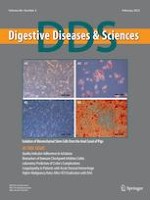
Fig. 2
Differentiation assay of pig anal canal transition zone stem cells. Representative pictures of differentiation assay. Isolated cells were incubated for 3 weeks with either adipogenic differentiation medium, chondrogenic differentiation medium, or osteogenic differentiation medium. A In contact with adipogenic differentiation medium, isolated cells transformed into adipocytes, as demonstrated by cell morphology and the presence of cytoplasm lipid inclusion after Oil-red-O staining (A2). This differentiation did not occur when control medium was used (A1). B Following incubation of isolated cells with osteogenic medium, we detected the presence of calcium deposits after Alizarin Red S staining, indicating the presence of osteoblasts (B2). Calcium deposits were not shown after control plate staining (B2). C Cell cultured in pellet for chondrocyte differentiation assay. In this Masson’s trichrome staining we observed in the control group (C1) a poor production of collagen type I, whereas cells incubated with chondrogenic differentiation medium (C2) exhibited type I collagen deposit (blue)
×
Anzeige
Discussion
We demonstrated the presence of stem cells in the region of the anal canal transition zone in pigs and the feasibility of their extraction. Isolated cells exhibited plastic adherence and homogeneous morphology with typical MSC fibroblast-like phenotype and clonal proliferation (Fig. S4), confirming their stemness (Fig. S1). Using flow cytometry, we demonstrated that the isolated cells were bearing the typical markers of MSC CD90 and CD44, whereas they were negative for CD14, CD34, CD45, CD105, CD106 and SLA-DR (Fig. S2). Interestingly, it has been suggested in human that CD106 is absent in A-MSCs but present in bone-marrow-derived MSCs [14‐16]. Recent evidence proposes CD106 to characterize a subset of MSCs with more immunomodulatory potential [17]. The isolated cells incubated with differentiation media differentiated nicely into adipocytes, osteoblasts, and chondrocytes, confirming their in vitro multipotency, a feature of MSCs.
The anal canal transition zone encompasses different tissues of endodermal and mesodermal origin [18]. Anal canal lining consists of anoderm, a squamous epithelium, a transition zone multilayer epithelium, and the single-layer columnar epithelium of rectal mucosa [19]. According to these characteristics, the cells we isolated could originate from the surrounding stromal tissue or from the glandular crypts of the mucosa while, our culture medium stimulates the proliferation of cells of mesenchymal origin. Indeed, in our culture condition, the absence of proliferation after passage of isolated ATZ mucosa fragments confirmed that the isolated cells originated from submucosa (data not shown). Mesenchymal stem cells have been isolated from uterine cervix transitional zone in human [20‐22], but to the best of our knowledge we report for the first time the possibility to isolate these cells from ATZ in pig. Our goal was to evaluate the feasibility of stem cell extraction in this localization, and further studies will need a more precise sampling to separate the different components of anal canal transition zone that should be considered separately. Considering the analogy existing between ATZ and uterine cervix transitional zone, ATZ might also be a source of stem cells in humans. These cells could be procured after hemorrhoid surgery, which could offer a high yield and success rate of cell culture establishment. After few passages, these cells could potentially be used in a strategy to manage anal incontinence.
Conclusion
In this work, we demonstrated the feasibility of ATZ region stem cell procurement in pigs. Further studies need to confirm the reproducibility of this procedure in human.
Acknowledgments
The authors are grateful to Ms. Nadja Perriraz-Mayer and Mr. Joël Pimenta and Cécile Gameiro for their advice and excellent technical assistance.
Anzeige
Declarations
Conflict of interest
None.
Open AccessThis article is licensed under a Creative Commons Attribution-NonCommercial 4.0 International License, which permits any non-commercial use, sharing, adaptation, distribution and reproduction in any medium or format, as long as you give appropriate credit to the original author(s) and the source, provide a link to the Creative Commons licence, and indicate if changes were made. The images or other third party material in this article are included in the article's Creative Commons licence, unless indicated otherwise in a credit line to the material. If material is not included in the article's Creative Commons licence and your intended use is not permitted by statutory regulation or exceeds the permitted use, you will need to obtain permission directly from the copyright holder. To view a copy of this licence, visit http://creativecommons.org/licenses/by-nc/4.0/.
Publisher’s Note
Springer Nature remains neutral with regard to jurisdictional claims in published maps and institutional affiliations.
Mit e.Med Innere Medizin erhalten Sie Zugang zu CME-Fortbildungen des Fachgebietes Innere Medizin, den Premium-Inhalten der internistischen Fachzeitschriften, inklusive einer gedruckten internistischen Zeitschrift Ihrer Wahl.
Mit e.Med Allgemeinmedizin erhalten Sie Zugang zu allen CME-Fortbildungen und Premium-Inhalten der allgemeinmedizinischen Zeitschriften, inklusive einer gedruckten Allgemeinmedizin-Zeitschrift Ihrer Wahl.