


Anzeige
Erschienen in:
19.01.2017 | Imaging in Intensive Care Medicine
Quick diagnosis of venous air embolism
Erschienen in: Intensive Care Medicine | Ausgabe 5/2017
Einloggen, um Zugang zu erhaltenExcerpt
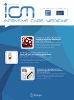
A 38-year-old man with idiopathic dilated cardiomyopathy and chronic atrial fibrillation presented with symptoms of heart failure. He underwent temporary override pacemaker insertion through right femoral access. A few hours later, he developed shock. Central venous (femoral) and arterial (radial) cannulations were performed for organ support and monitoring. In spite of cardiorespiratory support, his lactate level increased steadily. A CT scan was done to rule out mesenteric ischemia. Bedside echocardiography was performed to identify the type of shock. It showed gross air in the inferior vena cava and right ventricle as well as a dilated right ventricle with flattened interventricular septum and low ejection fraction (Video 1; Fig. 1). Management of air embolism was initiated with 100% oxygen, steep head-down with left lateral decubitus positioning, and aspiration of air through a newly inserted right atrial catheter. The patient developed acute kidney injury for which renal replacement therapy was instituted. CT scan confirmed presence of air in the inferior vena cava (IVC), femoral and hepatic veins (Fig. 1). Follow-up echocardiography showed resolution of air.
Fig. 1
Arrows highlight air in the venous system. Panel 1 echocardiography in subcostal view showing bubbles in inferior vena cava (IVC) and subhepatic veins (SHV). Panel 2 echocardiography in parasternal short-axis view showing bubbles in right ventricle (RV) and flattened interventricular septum (IVS). Panel 3 CT scan of abdomen in coronal plane showing air in IVC. Panel 4 CT scan of abdomen in coronal plane showing air in SHV
× ![]()
…