


Abstract
Orthopedic surgery for bone metastases is mainly a palliative treatment. Pathological central dislocation of the hip joint secondary to osteonecrosis of acetabular metastasis after radiation therapy brings severe suffering to cancer patients. We performed minimally invasive palliative surgery for an elderly woman, and excellent pain relief was achieved. An 80-year-old female suffering from right hip pain was referred to our hospital. She had undergone surgery for lung cancer 5 years previously and her right acetabulum was subsequently affected by metastasis. With the aim of controlling the metastasis, radiation therapy was performed. Two years later, pathological central dislocation of the hip joint occurred with sudden onset of severe pain, and she was unable to maintain a sitting position and became bedridden. After she was referred to our hospital, we created an intentional pseudarthrosis in the femoral neck for palliation. After the surgery, excellent pain relief and remarkably improved mobility were achieved during her limited remaining lifetime. In this report, we introduce a novel method of producing a pseudarthrosis in the femoral neck for pathological dislocation. This procedure is a minimally invasive treatment and an alternative option for palliative surgery for pathological dislocation of the hip joint due to osteonecrosis after radiation therapy.
INTRODUCTION
Acetabular metastases are associated with significant pain, disability and morbidity. Pathological fracture caused by metastasis with destruction of the acetabulum remains a difficult surgical challenge (1), considering the poor risk–benefit profile. Surgical resection of periacetabular metastasis and reconstruction are extremely invasive and complex (1–3). Given that the average life expectancy of patients with acetabular metastases is remarkably short, surgery is undesirable because a resection requires much of the remainder of life for postoperative recovery. Therefore, a non-surgical treatment, such as radiation therapy, is preferred in most cases. However, radiation therapy induces bone fragility and bone collapse, including bone and metastatic necrosis, and sometimes results in pathological dislocation of the hip joint (4). We describe a case of painful acetabular metastasis in an elderly woman that was successfully treated by a minimally invasive surgical procedure involving intentional creation of a pseudarthrosis in the femur, using a neck osteotomy for palliation during her limited remaining lifetime.
The patient and her family gave consent to the publication of her case.
CASE REPORT
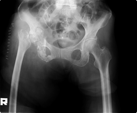
An 80-year-old female was referred to our institution for a surgical consultation for palliation. The patient had undergone surgery for lung cancer in the right lower lobe 5 years previously. The postoperative clinical stage had been pT2N0M0, and histopathological diagnosis was judged to be cystic mucinous adenocarcinoma with an uncommon histological subtype. She did not undergo postoperative chemotherapy because of her advanced age and lack of evidence of the efficiency of postoperative chemotherapy for her diagnosed histopathology at that time. One year later, she presented with gradually increasing pain in the right buttock. A plain film from the previous hospital revealed an osteolytic lesion with an ill-defined border in the right acetabulum, suggesting bone metastasis. Local radiation therapy of 39 Gy/13 fractions was performed. Subsequently, an intrapulmonary metastasis appeared in the middle lobe of the right lung. Therefore, 2 years and 5 months before the minimally invasive procedure, she underwent systemic chemotherapy consisting of carboplatin and gemcitabine hydrochloride, followed by molecular targeting drug therapy with an epidermal growth factor receptor tyrosine kinase inhibitor. However, neither of these anticancer drug therapies worked effectively, and she was judged to have a progressive disease. Consequently, the systemic treatment was suspended, and it was recommended that she receive supportive care. Two months later, she suddenly became unable to walk by herself, and had severe increasing pain in the right hip. A plain film of the right hip at the previous hospital revealed pathological central dislocation of the right hip with sclerotic changes between the acetabulum and ischium, suggesting osteonecrosis caused by the radiation therapy (Fig. 1a). A plain chest radiograph showed a residual intrapulmonary pneumonia shadow in the middle of the left lung (Fig. 1b).

(a) A plain radiograph shows pathological central dislocation of the hip joint after radiation therapy. The right acetabulum exhibits sclerotic changes, suggesting bone necrosis. (b) An anteroposterior plain chest radiograph shows a residual pneumonia shadow in the middle of the left lung.
At the first visit to our hospital, she was confined to bed all day because of an intolerance to a sitting posture and inadequate control of severe pain, scored 11 on an 11-point visual analog scale (VAS). Her symptoms were not controlled by a fentanyl patch. In addition, a urethral balloon catheter had been inserted for urination in the previous hospital, as she could not move to the toilet by herself.
We decided to intentionally produce a pseudarthrosis in the femoral neck using a neck osteotomy, instead of performing an invasive surgical procedure for tumor resection and megaprosthetic reconstruction, considering her advanced age of 80 years and primary lung cancer, which meant that her life expectancy was relatively short. Under general anesthesia, the surgery was performed in the right side up position. After a lateral approach to the hip joint with a small longitudinal skin incision, the femoral neck was visualized after an osteotomy of the greater trochanter. We produced a pseudarthrosis in the femoral neck as follows. We cut the femoral neck with a bone saw and excised a bone block with a width of ∼1.5 cm to avoid bony friction between the femoral head and neck (Fig. 2). The operation time and intraoperative blood loss were 36 min and 20 ml, respectively. There were no major or minor intraoperative complications.
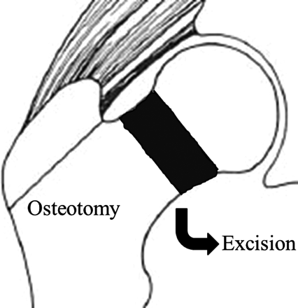
Schema of our surgical procedure. An osteotomy of the greater trochanter is performed using a lateral approach. Next, a femoral neck osteotomy is performed, and a bone block is excised.
A postoperative plain film demonstrated a pseudarthrosis in the right femoral neck that resembled an intracapsular femoral neck fracture, although the central dislocation of the femoral neck remained persistent (Fig. 3). The patient's recovery was uneventful. On the day after the surgery, the pain in her right hip was dramatically improved and she was able to sit on the bed. After 1 month, she was discharged from the hospital in a wheelchair. At that time, her pain was remarkably reduced and scored 1 on the 11-point VAS. Although she could not walk, she was able to move by herself to a portable toilet beside the bed (Fig. 4). She died 3 months after the surgery, because of worsening respiratory function caused by the lung cancer.
Postoperative plain film demonstrating a femoral neck pseudarthrosis resembling an intracapsular femoral neck fracture.
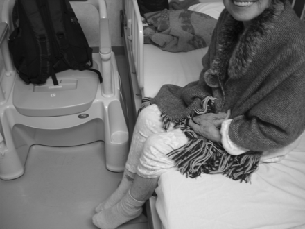
The patient could maintain a sitting position on a bed without aids and move to a portable toilet beside the bed by herself after discharge from the hospital.
DISCUSSION
The treatment of painful acetabular metastasis remains a major therapeutic challenge (1–3). With the aim of local curability, Harrington (1) classified 58 cases secondary to metastatic disease into four types on the basis of the extent of the metastatic destruction in the acetabulum, and proposed operative techniques accordingly. However, these treatments are considered to be highly invasive and to have high rates of complications, such as intraoperative massive bleeding, perioperative death, deep infection, dislocation and nerve palsy. Meanwhile, radiation therapy is only a mediocre option for durable pain relief. Furthermore, a previously irradiated acetabulum is always exposed to the risk of bone collapse, because it is a weight-bearing joint structure (4).
From the viewpoint of biomechanics, the forces acting on the hip joint change their position as the femoral head moves. They are affected by both the position and alterations of the muscle force contribution (5). Rydell et al. (6) previously measured the hip joint forces, and reported loads of 1.20–1.23 times at <10°, 0.98–1.05 times at 30° and 0.65–0.80 times at 90° of hip flexion in the supine position, compared with the body weight. These findings mean that dislocation of the hip joint of patients is continuously loaded toward the acetabular direction, even at rest in bed. This overloading may cause severe pain in patients with pathological central dislocation of the hip joint. We speculate that our procedure involving intentional creation of a pseudarthrosis in the femoral neck contributes to a change in the mechanical axis of the lower leg and a dispersal of the concentration of forces in the hip joint. This procedure may relieve pain dramatically.
The purpose of orthopedic surgery for bone metastases is mainly palliative. In the case presented here, we had to select a minimally invasive surgical treatment to alleviate the patient's suffering as much as possible, because of her advanced age of 80 years. Khan and Masterson (7) introduced defunctioning of the hip joint for acetabular and greater trochanter metastases, and carried out a femoral neck osteotomy through a posterior approach to the hip joint. However, we recommend a lateral approach and an osteotomy of the greater trochanter. This approach provides two advantages compared with the other procedure. First, the femoral neck is clearly visualized, which makes it easy to cut. After pathological central dislocation of the hip joint has occurred, the femoral neck may not be easily visible through a posterior approach, because of the loss of the normal anatomical position. If the metastasis is close, an intralesional procedure can trigger additional intraoperative bleeding. Second, an osteotomy of the greater trochanter is not technically demanding. A posterior approach requires the surgeon to cut the short rotator muscles, including the piriformis muscle, which have such abundant blood flow that the surgeon must be very careful to prevent bleeding. Our procedure is a less invasive, simple, safe, effective and reproducible technique that may recover patient function in the treatment of painful acetabular metastasis with central dislocation of the hip joint.
The concept of our surgical procedure appears to resemble that of the Girdlestone procedure, which is currently carried out as a salvage surgery for failed hip replacement in pyogenic arthritis and failed surgery for hip trauma (8). The defunctioning procedure after resection of the hip also contributes to the relief of patient suffering. Several surgeons have applied this procedure to primary or metastatic tumors around the hip joint (9,10). However, for the purpose of palliation alone, our procedure is less invasive than the Girdlestone method, which requires removal of the femoral head and possible handling of the affected acetabulum. Postoperative hematoma and deep infection in the dead space at the surgical site are possible representative complications for the Girdlestone method. Our procedure may have the demerit of residual persistent pain caused by bony irritation between the femoral head and neck after insufficient osteotectomy. Nevertheless, the shorter operation time and decreased blood loss are predominantly desirable for the general condition of cancer patients with a limited lifetime, provided that a postoperative functional outcome of at least achieving the use of a wheelchair can be expected.
In conclusion, the intentional creation of a pseudarthrosis in the femoral neck represents a minimally invasive treatment option for patients suffering from intolerable pain due to central migration of the hip joint associated with acetabular metastasis. This surgical procedure is not very technically demanding, meaning that any general orthopedic surgeon can easily attempt it. We confirm that this surgical procedure can be beneficial for cancer patients suffering from central dislocation of the hip joint.
Conflict of interest statement
None declared.
