


Abstract
Recent studies suggest a direct contribution of nicotine, the addictive component of tobacco and tobacco smoke, to human carcinogenesis. To assess the genotoxicity of nicotine, the DNA-damaging effect on human lymphocytes and target cells from lymphatic tissue of the palatine tonsils from 10 healthy patients was tested with the alkaline single-cell microgel electrophoresis (Comet) assay. The degree of DNA migration, a measure of possible DNA single strand breaks, alkali labile sites, and incomplete excision repair sites, was expressed as the Olive tail moment, the percentage of DNA in the tail, and the tail length. One hour exposure to nicotine at 0.125, 0.25, 0.5, 1, 2, and 4 mM induced a statistically significant dose-dependent increase of DNA migration up to 3.8-fold and 3.2-fold in tonsillar cells and lymphocytes, respectively. The lowest concentration eliciting significant DNA damage was 0.5 mM nicotine. The genotoxic effect was confirmed in a second series of experiments using nicotine of high purity from two different suppliers. There were no significant differences between the two series, excluding artifacts from the source of nicotine. Finally, DNA damage by nicotine was compared in cells incubated in medium strictly adjusted to neutral pH, with nonadjusted medium becoming alkaline with increasing nicotine concentrations. Again no differences in DNA migration were observed. The data indicate that nicotine expresses significant direct genotoxic effects in human target cells in vitro. However, no differences in DNA damage were observed in cells from smokers and nonsmokers incubated without nicotine. The lack of higher DNA damage in smokers compared to nonsmokers could be a question of nicotine dose, rapid DNA repair, or interactions with other smoke constituents. These results require further investigations on the contribution of nicotine to tobacco carcinogenesis.
Tobacco smoke is composed of a great variety of constituents. Some of these, including polycyclic hydrocarbons, nitrosamines, and aromatic amines, are known to contribute to the carcinogenic risk of tobacco smoke (Hoffmann and Hoffmann, 1997; Smith et al., 2003). Nicotine, the major tobacco alkaloid, however, is accused of the addictive potential of smoking, being mediated by neuronal nicotinic acetylcholine receptors in the central nervous system (Dajas-Bailador and Wonnacott, 2004). With regard to possible carcinogenic risk, nicotine and its demethylation product nornicotine have been discussed mainly for reasons of their nitrosation products 4-(methylnitrosamino)-1-(3-pyridyl)-1-butanone (NNK) or N′-nitrosonornicotine (NNN) (Hecht, 1998). Recently, direct genotoxic effects of nicotine have been shown in human gingival fibroblasts (Argentin and Cicchetti, 2004) and spermatozoa (Arabi, 2004). Furthermore, nicotine may also stimulate tumor development by non-genotoxic mechanisms such as angioneogenesis (Heeschen et al., 2001; Cooke and Bitterman, 2004), growth stimulation (Shin et al., 2004), antiapoptotic effects (Argentin and Cicchetti, 2004), and receptor-regulated cellular growth (Schuller, 1994).
This study focuses on a possible direct genotoxic effect of nicotine on cells of the lymphatic tissue of the palatine tonsils (tonsillar cells), a target of tobacco carcinogenesis in the human upper aerodigestive tract. In addition, DNA damage in lymphocytes of the peripheral blood, a well-established surrogate marker of systemic carcinogenic effects, was tested in the same donors (study A). To exclude artifactitious DNA damage, the genotoxic effect of nicotine was tested with two highly pure nicotine batches from two different commercial sources (study B) and under pH-controlled conditions, eliminating alkaline effects at high nicotine concentrations (study C).
The assessment of nicotine as a risk factor for carcinogenesis is of special interest. The health benefit of filter cigarettes and so-called light cigarettes with reduced fractions of tar and nicotine is controversially discussed (Harris et al., 2004; Hoffmann et al., 2001; Lee and Sanders, 2004; National Cancer Institute, 2001). Thus, the cancer risk in smokers of light cigarettes could in fact be much higher than expected because of the undetermined possible genotoxic effects of nicotine.
MATERIALS AND METHODS
Cell sources and study groups.
In surgery of the oropharynx, tonsillar tissue was harvested from 15 patients (22–47 years, 35.3 on average; 6 male, 9 female; 10 smokers, 5 nonsmokers; for further details see Table 1). This type of surgery is done after recurrent or chronic inflammation of the tonsils in an interval without signs of acute disease. Ten patients each were part of studies A and B, and cells from 5 patients were taken for study C. Study A represents the investigation of dose-dependent effects of nicotine on DNA migration. Study B delineates the comparison between nicotine of two different suppliers. Study C involves the evaluation of a possible impact of the pH of the incubation medium. For the benefit of the patient, only as much tissue as necessary was resected. Whole blood samples were obtained by venous puncture from each patient. The study was approved by the ethics commission of the University of Regensburg Medical Faculty, and all patients signed a written informed consent form.
Characterization of Patients and Study Groups
Patient/study | Gender | Age (years) | Profession | Alcohol (g/day) | Nicotine (py) |
|---|---|---|---|---|---|
| 1/A | m | 47 | Engineer | 10 | 30 |
| 2/A | f | 22 | Nurse | 5 | 3 |
| 3/AB | f | 31 | Communications | 0 | 0 |
| 4/AB | f | 45 | Sales clerk | 0–5 | 13 |
| 5/AB | m | 35 | Stove fitter | 0–5 | 16 |
| 6/AB | f | 23 | Housewife | 5 | 0 |
| 7/AB | m | 35 | Engineer | 15 | 5 |
| 8/AB | f | 41 | Clerk | 5 | 10 |
| 9/AB | m | 34 | Factory worker | 30 | 10 |
| 10/AB | m | 41 | Bricklayer | 20 | 15 |
| 11/BC | f | 29 | Factory worker | 5 | 1 |
| 12/BC | m | 39 | Bricklayer | 60 | 11 |
| 13/C | f | 33 | Governess | 3 | 0 |
| 14/C | f | 36 | Nurse | 3 | 0 |
| 15/C | f | 39 | Governess | 0 | 0 |
Note. Study A: investigation of dose dependency on DNA damage after incubation with nicotine; Study B: comparison of nicotine from two different suppliers; Study C: evaluation of pH adjustment of the incubation medium.
m: male, f: female; alcohol consumption is given in grams per day [g/day] and smoking habit in pack years [py] (1 pack year = 365 days × 20 cigarettes/day; e.g., 30 py = 1 pack/day for 30 years or = 2 packs/day for 15 years, etc.).
Cell separation.
Heparinized blood samples and biopsies stored in MEM-Joklik (without L-glutamine and NaHCO3; Linaris, Bettingen, Germany), were transferred to the laboratory immediately after collection. Lymphocytes from whole blood and tonsillar cells were isolated according to the procedure of Kleinsasser et al. (2003a). In brief, the cells were suspended in fetal calf serum (FCS; Gibco-BRL, Eggenstein, Germany) and 10% dimethyl sulfoxide (DMSO; Sigma-Aldrich, Taufkirchen, Germany), and stored at −80°C. After thawing, cells were washed in phosphate buffered saline (PBS) and resuspended in RPMI-1640 (Biochrom, Berlin, Germany) and 15% FCS. Tonsillar biopsies were digested with the aid of an enzyme mix of protease (5 mg/ml, Sigma), hyaluronidase from bovine testes (1 mg/ml, Roche, Mannheim, Germany), and collagenase P (1 mg/ml, Roche). Tonsillar cells and lymphocytes were counted and their viability was determined using trypan blue staining.
Exposure.
Cell suspensions with 5 × 10⁴ lymphocytes or tonsillar cells each were incubated for 60 min in an initial step with (-)-nicotine free base of >99% purity (N 3876, Sigma) and dissolved in PBS at concentrations of 0, 0.125, 0.25, 0.5, 1, 2, and 4 mM (study A). Concentrations of 0.5, 1, 2, and 4 mM of (-)-nicotine from either Sigma or TRC (N 412450, Toronto Research Chemicals, North York, ON, Canada) were used in study B. In study C, concentrations of 1, 2, and 4 mM nicotine (Sigma) were used. In half of the incubation medium the pH was adjusted to neutral (pH 7.2) with hydrochloric acid (Merck, Darmstadt, Germany), whereas in the other half the pH level was only monitored (study C). Nicotine doses were selected according to pilot studies and were held as low as possible to approach concentrations similar to those found in plasma and saliva of smokers (see Discussion). Additional experiments were performed with higher doses, allowing for viability results over 70%.
The directly alkylating mutagen N-methyl-N′-nitro-N-nitrosoguanidine (MNNG, 0.02 mM, Sigma) was used as a positive control, and the solvent PBS (1 or 10 %) served as a negative control.
Alkaline single-cell microgel electrophoresis (Comet) assay.
After incubation, cell viability was examined again using trypan blue staining. Once viabilities >80% were obtained, the cells were subjected to the Comet assay (Kleinsasser et al., 2003b). In brief, after resuspending the cells in 0.7% low melting agarose (Cambrex, Rockland, ME) and applying them to coated microscope slides, cell and core membranes were dissolved for at least 90 min in a lysis buffer (10% DMSO, 1% Triton-X in alkaline lysis solution: 2.5 M NaCl, 10 mM Tris, 100 mM Na2EDTA; pH 10). The slides were placed into a horizontal gel electrophoresis chamber (Renner, Dannstadt, Germany) and covered with alkaline buffer solution containing NaOH (10 mM) and Na2EDTA (200 mM), pH > 13. A 20-min “unwinding” period was followed by electrophoresis at 25 V and 300 mA for 20 min. Slides were neutralized (Trizma base, pH 7.5, Merck) and stained with ethidium bromide (Sigma). A DMLB microscope (Leica, Heerbrugg, Switzerland) equipped with an adapted CCD camera (Cohu Inc., San Diego, CA) was used to investigate the slides. The software Komet 4.0 (Kinetic Imaging, Liverpool, UK) was applied.
To quantify the induced DNA damage, 100 cells per probe were examined for the Olive tail moment (OTM) reflecting the percentage of DNA in the tail of the comet multiplied by the median migration distance (Olive et al., 1993), the percentage of DNA in the tail (DT), and the tail length (TL) (Lee et al., 2004).
Statistics.
Evaluation was based on mean OTM values of each individual incubation using Prism 4 (GraphPad Software, Inc., San Diego, CA). Concentration-dependent differences in DNA migration were analyzed by repeated measures analysis of variance (ANOVA) with post test for linear trend and Bonferroni's multiple comparison test for differences between untreated control cells and nicotine-treated cells. After testing for normal distribution, a paired Student's t-test was applied to compare DNA migration in tonsillar cells versus lymphocytes (study A), as well as for evaluation of possible effects with respect to the source of nicotine (study B) and different environmental pH values (study C).
RESULTS
Nicotine did not exert cytotoxic effects in any of the experiments based on the trypan blue test. Cell viability was well above 75% in both cell types before and after exposure to nicotine (data not shown).
In study A, 1 h of incubation with nicotine induced a significant concentration-dependent increase in DNA migration in the Comet assay in tonsillar cells (up to 3.8-fold, p < 0.0001), as well as in peripheral lymphocytes from the same donors (up to 3.2-fold, p < 0.0001) without affecting cell viability (Fig. 1, Table 2). Compared to the negative control, the effects in tonsillar cells and lymphocytes reached significance at 0.5 mM nicotine (p < 0.05). At all nicotine concentrations there were no significant differences in DNA damage between either cell type as assessed by OTM. The mean values of the positive control for tonsillar cells were 71.8 ± 20.7 (OTM), 67.3 ± 9.1 (DT), and 170.1 ± 33.2 (TL); and for lymphocytes, 76.0 ± 18.1 (OTM), 71.7 ± 11.9 (DT), and 179.5 ± 25.5 (TL).
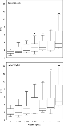
Concentration-dependent DNA damage in fresh specimens of lymphatic tissue cells of palatine tonsils (tonsillar cells) and peripheral lymphocytes from 10 patients after 1 h of incubation with nicotine as determined by the Comet assay. Lines in the boxes represent the median values of the Olive tail moments (OTM). Box plots show the lowest and highest values of OTM, as well as the 1st and 3rd quartiles. An increase in DNA migration with rising concentrations of nicotine was significant according to ANOVA with post test for linearity (p < 0.0001). Significant difference compared to control was observed in Bonferroni's multiple comparison test: p < 0.05 (*), p < 0.01 (**), p < 0.001 (***).
Study A: Individual Donor Data, Mean Values, and SD
Control | Nicotine (mM) | |||||||||||||||||||||||||||||||||||||||
|---|---|---|---|---|---|---|---|---|---|---|---|---|---|---|---|---|---|---|---|---|---|---|---|---|---|---|---|---|---|---|---|---|---|---|---|---|---|---|---|---|
| 0 | 0.125 | 0.25 | 0.5 | 1.0 | 2.0 | 4.0 | ||||||||||||||||||||||||||||||||||
| P | OTM | DT | TL | OTM | DT | TL | OTM | DT | TL | OTM | DT | TL | OTM | DT | TL | OTM | DT | TL | OTM | DT | TL | |||||||||||||||||||
| Tonsillar cells | ||||||||||||||||||||||||||||||||||||||||
| 1 | 1.81 | 3.18 | 50.5 | 3.95 | 5.42 | 60.4 | 3.93 | 6.40 | 62.3 | 5.72 | 6.68 | 60.4 | 5.85 | 7.18 | 71.5 | 7.15 | 9.06 | 60.5 | 11.2 | 13.3 | 94.9 | |||||||||||||||||||
| 2 | 0.56 | 1.25 | 36.9 | 2.36 | 3.14 | 39.8 | 3.13 | 3.62 | 40.0 | 3.85 | 4.32 | 46.1 | 4.75 | 5.12 | 47.0 | 3.15 | 3.39 | 42.4 | 7.62 | 6.91 | 56.3 | |||||||||||||||||||
| 3 | 1.73 | 2.85 | 48.0 | 1.46 | 2.43 | 37.7 | 1.98 | 2.74 | 37.2 | 3.00 | 3.91 | 39.7 | 1.90 | 2.63 | 34.0 | 2.75 | 3.66 | 38.7 | 3.50 | 4.28 | 41.3 | |||||||||||||||||||
| 4 | 1.05 | 1.82 | 39.4 | 1.23 | 2.15 | 34.3 | 1.62 | 2.47 | 30.0 | 0.92 | 1.76 | 27.1 | 2.59 | 3.34 | 33.0 | 1.52 | 2.29 | 29.3 | 3.52 | 4.56 | 36.9 | |||||||||||||||||||
| 5 | 1.56 | 2.40 | 31.9 | 1.72 | 2.53 | 29.2 | 1.77 | 2.90 | 30.6 | 2.11 | 3.31 | 28.5 | 2.22 | 3.53 | 31.9 | 2.15 | 3.19 | 27.9 | 2.50 | 3.84 | 32.2 | |||||||||||||||||||
| 6 | 1.20 | 2.41 | 29.0 | 1.48 | 2.23 | 28.9 | 1.50 | 2.82 | 24.4 | 1.46 | 2.77 | 30.6 | 1.71 | 2.69 | 36.4 | 1.76 | 2.85 | 25.1 | 2.31 | 3.59 | 36.4 | |||||||||||||||||||
| 7 | 2.24 | 3.09 | 30.8 | 1.62 | 2.67 | 31.1 | 2.00 | 3.04 | 29.7 | 2.18 | 3.10 | 32.0 | 2.72 | 3.81 | 42.8 | 2.23 | 3.26 | 37.9 | 3.91 | 5.51 | 45.3 | |||||||||||||||||||
| 8 | 2.13 | 3.34 | 36.1 | 1.18 | 2.07 | 35.5 | 1.38 | 2.26 | 35.5 | 3.39 | 5.13 | 48.0 | 2.88 | 5.01 | 37.0 | 2.85 | 6.01 | 43.5 | 4.09 | 5.12 | 41.7 | |||||||||||||||||||
| 9 | 1.52 | 2.49 | 39.7 | 3.26 | 4.43 | 47.9 | 4.44 | 5.61 | 48.3 | 3.01 | 3.96 | 42.8 | 4.04 | 5.28 | 49.7 | 5.64 | 6.89 | 46.2 | 6.34 | 7.63 | 51.7 | |||||||||||||||||||
| 10 | 1.92 | 3.32 | 42.3 | 1.61 | 2.81 | 37.1 | 1.20 | 2.03 | 34.4 | 1.90 | 2.94 | 41.2 | 2.22 | 3.29 | 40.0 | 2.85 | 4.26 | 43.8 | 2.85 | 5.03 | 44.3 | |||||||||||||||||||
| Mean | 1.57 | 2.62 | 38.5 | 1.99 | 2.99 | 38.2 | 2.29 | 3.39 | 37.2 | 2.75 | 3.79 | 39.6 | 3.09 | 4.19 | 42.3 | 3.20 | 4.49 | 39.5 | 4.78 | 5.98 | 48.1 | |||||||||||||||||||
| SD | 0.52 | 0.69 | 7.11 | 0.92 | 1.10 | 9.61 | 1.13 | 1.46 | 11.0 | 1.38 | 1.38 | 10.4 | 1.36 | 1.43 | 11.8 | 1.80 | 2.15 | 10.4 | 2.81 | 2.86 | 18.0 | |||||||||||||||||||
| Lymphocytes | ||||||||||||||||||||||||||||||||||||||||
| 1 | 3.42 | 4.60 | 46.7 | 5.48 | 6.37 | 58.2 | 3.75 | 4.78 | 57.0 | 9.26 | 9.88 | 77.8 | 6.33 | 7.57 | 64.6 | 6.71 | 7.89 | 69.5 | 11.0 | 11.5 | 89.6 | |||||||||||||||||||
| 2 | 1.61 | 2.42 | 36.9 | 1.58 | 2.55 | 39.8 | 2.76 | 3.36 | 40.0 | 3.17 | 4.28 | 46.1 | 3.89 | 4.77 | 47.0 | 2.81 | 3.62 | 42.4 | 4.11 | 5.07 | 56.3 | |||||||||||||||||||
| 3 | 3.87 | 5.38 | 57.1 | 4.80 | 5.75 | 55.3 | 7.42 | 7.88 | 78.2 | 7.82 | 7.55 | 68.5 | 9.82 | 10.7 | 73.7 | 12.8 | 12.6 | 91.8 | 13.8 | 14.2 | 101.7 | |||||||||||||||||||
| 4 | 1.65 | 2.55 | 37.1 | 2.70 | 3.90 | 37.1 | 3.14 | 3.85 | 38.6 | 3.88 | 5.05 | 46.2 | 4.10 | 4.61 | 49.3 | 4.78 | 5.55 | 54.7 | 7.26 | 8.29 | 66.9 | |||||||||||||||||||
| 5 | 2.49 | 3.93 | 31.5 | 2.67 | 3.70 | 32.5 | 3.06 | 3.90 | 40.0 | 1.71 | 2.92 | 29.1 | 2.57 | 3.70 | 34.4 | 3.28 | 4.19 | 37.3 | 3.60 | 4.85 | 40.7 | |||||||||||||||||||
| 6 | 1.85 | 2.76 | 29.3 | 1.60 | 2.72 | 35.1 | 2.25 | 2.92 | 33.7 | 2.35 | 3.05 | 33.6 | 1.99 | 2.69 | 32.0 | 1.61 | 2.28 | 35.0 | 4.48 | 5.76 | 61.4 | |||||||||||||||||||
| 7 | 1.64 | 2.34 | 41.5 | 1.85 | 3.92 | 37.1 | 3.46 | 4.20 | 42.4 | 3.21 | 4.52 | 47.3 | 3.37 | 4.00 | 37.4 | 3.21 | 3.95 | 44.5 | 3.37 | 4.72 | 51.1 | |||||||||||||||||||
| 8 | 1.29 | 2.05 | 38.6 | 2.00 | 2.70 | 40.9 | 1.77 | 2.56 | 35.4 | 2.56 | 3.24 | 41.9 | 2.51 | 4.80 | 47.9 | 2.95 | 3.88 | 36.9 | 4.64 | 5.57 | 49.0 | |||||||||||||||||||
| 9 | 0.89 | 1.63 | 24.2 | 0.72 | 1.42 | 20.6 | 1.47 | 2.60 | 28.2 | 1.78 | 2.76 | 27.6 | 2.73 | 3.36 | 33.2 | 3.05 | 4.02 | 37.9 | 5.06 | 5.96 | 45.1 | |||||||||||||||||||
| 10 | 1.84 | 3.41 | 26.8 | 0.90 | 1.74 | 30.4 | 1.27 | 2.08 | 29.2 | 1.76 | 2.60 | 34.6 | 2.43 | 3.71 | 37.5 | 3.43 | 4.48 | 49.7 | 4.83 | 5.59 | 62.1 | |||||||||||||||||||
| Mean | 2.05 | 3.11 | 37.0 | 2.43 | 3.48 | 38.7 | 3.04 | 3.81 | 42.3 | 3.75 | 4.59 | 45.3 | 3.97 | 4.99 | 45.7 | 4.46 | 5.24 | 50.0 | 6.21 | 7.14 | 62.4 | |||||||||||||||||||
| SD | 0.94 | 1.20 | 9.88 | 1.57 | 1.61 | 11.1 | 1.76 | 1.66 | 15.0 | 2.64 | 2.39 | 16.5 | 2.40 | 2.40 | 14.0 | 3.22 | 2.96 | 18.1 | 3.49 | 3.21 | 19.5 | |||||||||||||||||||
P: patient; OTM: Olive tail moment; DT: % of DNA in tail; TL: tail length.
Study B: Individual Donor Data, Mean Values, and SD
Control | Nicotine Sigma (mM) | Nicotine TRC (mM) | |||||||||||||||||||||||||||||||||||||||||||||||||
|---|---|---|---|---|---|---|---|---|---|---|---|---|---|---|---|---|---|---|---|---|---|---|---|---|---|---|---|---|---|---|---|---|---|---|---|---|---|---|---|---|---|---|---|---|---|---|---|---|---|---|---|
| 0 | 0.5 | 1.0 | 2.0 | 4.0 | 0.5 | 1.0 | 2.0 | 4.0 | |||||||||||||||||||||||||||||||||||||||||||
| P | OTM | DT | TL | OTM | DT | TL | OTM | DT | TL | OTM | DT | TL | OTM | DT | TL | OTM | DT | TL | OTM | DT | TL | OTM | DT | TL | OTM | DT | TL | ||||||||||||||||||||||||
| Tonsillar cells | |||||||||||||||||||||||||||||||||||||||||||||||||||
| 3 | 2.42 | 3.10 | 54.3 | 2.95 | 2.46 | 46.5 | 2.87 | 1.95 | 51.0 | 4.33 | 2.98 | 54.9 | 3.86 | 2.76 | 67.8 | 2.55 | 2.70 | 53.2 | 2.59 | 3.46 | 43.7 | 3.89 | 4.27 | 42.5 | 4.06 | 4.84 | 49.2 | ||||||||||||||||||||||||
| 4 | 1.98 | 1.82 | 43.6 | 3.35 | 2.64 | 55.8 | 2.52 | 2.64 | 42.0 | 3.60 | 2.77 | 51.0 | 3.46 | 3.39 | 43.7 | 2.49 | 2.80 | 40.3 | 3.01 | 3.98 | 49.0 | 3.98 | 3.76 | 45.8 | 5.37 | 2.91 | 58.4 | ||||||||||||||||||||||||
| 5 | 2.13 | 2.50 | 43.1 | 1.62 | 2.23 | 37.7 | 2.06 | 2.03 | 34.3 | 1.56 | 2.51 | 27.4 | 4.01 | 2.56 | 40.7 | 1.22 | 2.54 | 29.2 | 1.93 | 3.53 | 37.9 | 2.35 | 5.16 | 37.9 | 3.67 | 3.87 | 40.0 | ||||||||||||||||||||||||
| 6 | 1.25 | 2.69 | 29.3 | 1.85 | 2.51 | 29.7 | 1.82 | 2.02 | 29.3 | 2.11 | 2.60 | 32.8 | 2.54 | 4.75 | 40.0 | 1.70 | 4.15 | 25.0 | 2.44 | 3.61 | 34.9 | 2.81 | 3.79 | 45.6 | 2.13 | 3.37 | 39.2 | ||||||||||||||||||||||||
| 7 | 1.62 | 4.37 | 35.5 | 1.48 | 5.03 | 30.4 | 1.61 | 3.81 | 31.5 | 1.72 | 4.03 | 36.4 | 3.78 | 4.49 | 44.4 | 1.05 | 4.62 | 37.2 | 1.56 | 4.99 | 33.6 | 2.27 | 4.89 | 35.6 | 2.44 | 5.06 | 34.0 | ||||||||||||||||||||||||
| 8 | 1.78 | 3.96 | 33.4 | 1.39 | 3.82 | 34.2 | 1.46 | 3.56 | 34.5 | 2.84 | 5.68 | 42.3 | 2.25 | 3.97 | 42.5 | 1.07 | 3.94 | 34.8 | 2.30 | 5.52 | 40.9 | 1.36 | 6.06 | 40.1 | 2.21 | 4.03 | 44.0 | ||||||||||||||||||||||||
| 9 | 2.70 | 4.28 | 52.7 | 3.23 | 5.76 | 51.7 | 2.62 | 6.86 | 47.9 | 3.27 | 5.01 | 44.3 | 3.69 | 8.45 | 46.4 | 2.62 | 6.33 | 40.0 | 2.96 | 7.40 | 45.9 | 3.13 | 6.40 | 35.7 | 3.68 | 6.70 | 42.4 | ||||||||||||||||||||||||
| 10 | 2.26 | 3.70 | 45.5 | 1.74 | 3.36 | 38.7 | 4.07 | 4.43 | 51.5 | 2.58 | 4.23 | 41.7 | 4.44 | 4.26 | 52.9 | 2.29 | 4.20 | 39.6 | 2.59 | 4.89 | 43.0 | 3.56 | 6.09 | 50.0 | 2.51 | 5.66 | 40.2 | ||||||||||||||||||||||||
| 11 | 3.37 | 3.85 | 51.9 | 3.70 | 3.97 | 54.6 | 4.08 | 5.68 | 55.6 | 5.54 | 3.79 | 57.7 | 5.74 | 3.69 | 56.0 | 4.01 | 3.72 | 57.0 | 6.02 | 5.02 | 62.6 | 4.86 | 5.21 | 56.0 | 5.11 | 5.33 | 52.4 | ||||||||||||||||||||||||
| 12 | 2.11 | 3.27 | 47.5 | 2.08 | 4.45 | 41.0 | 2.81 | 4.79 | 38.4 | 2.93 | 3.41 | 48.1 | 4.66 | 4.56 | 47.8 | 1.96 | 3.55 | 36.4 | 2.83 | 5.56 | 38.5 | 3.31 | 4.58 | 43.8 | 3.94 | 6.34 | 42.5 | ||||||||||||||||||||||||
| Mean | 2.16 | 3.35 | 43.7 | 2.34 | 3.62 | 42.0 | 2.59 | 3.78 | 41.6 | 3.05 | 3.70 | 43.7 | 3.84 | 4.29 | 48.2 | 2.09 | 3.85 | 39.2 | 2.82 | 4.80 | 43.0 | 3.15 | 5.02 | 43.3 | 3.51 | 4.81 | 44.3 | ||||||||||||||||||||||||
| SD | 0.59 | 0.83 | 8.54 | 0.87 | 1.20 | 9.66 | 0.92 | 1.68 | 9.39 | 1.22 | 1.06 | 9.66 | 1.00 | 1.64 | 8.60 | 0.91 | 1.12 | 9.70 | 1.21 | 1.22 | 8.39 | 1.01 | 0.95 | 6.44 | 1.17 | 1.26 | 7.11 | ||||||||||||||||||||||||
| Lymphocytes | |||||||||||||||||||||||||||||||||||||||||||||||||||
| 3 | 3.15 | 4.15 | 34.0 | 4.68 | 5.24 | 52.2 | 7.62 | 5.54 | 53.8 | 8.33 | 8.08 | 69.5 | 10.8 | 8.66 | 50.6 | 4.81 | 8.97 | 40.8 | 7.48 | 8.94 | 40.0 | 7.67 | 10.71 | 50.5 | 9.34 | 9.85 | 62.9 | ||||||||||||||||||||||||
| 4 | 2.94 | 3.95 | 31.4 | 4.46 | 5.87 | 56.4 | 4.76 | 4.63 | 64.1 | 6.65 | 5.84 | 80.0 | 9.73 | 6.87 | 69.8 | 3.74 | 7.96 | 54.1 | 5.13 | 7.85 | 71.3 | 7.08 | 11.02 | 55.3 | 5.19 | 7.24 | 78.2 | ||||||||||||||||||||||||
| 5 | 2.58 | 4.03 | 47.7 | 3.94 | 5.12 | 56.4 | 4.37 | 4.34 | 78.8 | 4.68 | 5.68 | 77.4 | 6.22 | 5.70 | 84.8 | 3.45 | 5.74 | 52.6 | 4.58 | 7.69 | 61.0 | 6.36 | 7.12 | 65.9 | 5.33 | 6.33 | 85.5 | ||||||||||||||||||||||||
| 6 | 1.76 | 3.07 | 41.8 | 3.26 | 4.53 | 56.6 | 3.39 | 4.28 | 61.5 | 6.33 | 4.83 | 68.8 | 5.14 | 3.30 | 75.6 | 2.78 | 8.24 | 51.3 | 2.14 | 5.87 | 50.9 | 3.51 | 7.05 | 59.2 | 4.27 | 6.09 | 58.3 | ||||||||||||||||||||||||
| 7 | 1.69 | 2.61 | 43.2 | 1.78 | 2.60 | 43.8 | 2.68 | 4.36 | 56.1 | 4.12 | 3.88 | 50.6 | 4.70 | 4.42 | 60.1 | 3.41 | 5.12 | 49.1 | 2.95 | 5.04 | 51.4 | 3.85 | 6.16 | 54.8 | 3.71 | 4.63 | 53.8 | ||||||||||||||||||||||||
| 8 | 1.18 | 2.14 | 35.9 | 2.63 | 3.84 | 37.2 | 3.27 | 4.38 | 39.2 | 3.16 | 4.69 | 58.1 | 5.90 | 4.20 | 52.0 | 2.66 | 4.61 | 39.3 | 3.33 | 5.56 | 36.2 | 4.19 | 7.03 | 40.3 | 4.61 | 5.78 | 42.0 | ||||||||||||||||||||||||
| 9 | 1.74 | 2.85 | 35.0 | 3.41 | 4.74 | 32.3 | 3.15 | 3.86 | 51.5 | 4.70 | 4.48 | 53.2 | 5.67 | 6.08 | 58.8 | 2.58 | 5.96 | 50.4 | 3.82 | 4.74 | 37.9 | 3.98 | 6.65 | 40.6 | 5.76 | 7.60 | 45.4 | ||||||||||||||||||||||||
| 10 | 1.37 | 2.32 | 36.3 | 2.26 | 3.42 | 44.8 | 3.44 | 3.16 | 48.6 | 3.93 | 4.76 | 44.8 | 4.12 | 4.12 | 61.1 | 2.26 | 5.20 | 50.1 | 3.11 | 4.97 | 50.0 | 3.82 | 5.40 | 58.6 | 5.78 | 8.38 | 48.7 | ||||||||||||||||||||||||
| 11 | 1.69 | 3.09 | 30.0 | 3.26 | 3.89 | 40.7 | 3.72 | 3.83 | 46.1 | 6.78 | 4.38 | 45.1 | 6.89 | 4.60 | 59.5 | 3.13 | 7.60 | 38.4 | 2.94 | 4.70 | 44.9 | 3.91 | 7.76 | 46.1 | 6.68 | 6.77 | 55.5 | ||||||||||||||||||||||||
| 12 | 1.55 | 2.33 | 31.8 | 3.79 | 5.01 | 38.7 | 5.35 | 4.57 | 46.4 | 9.01 | 6.32 | 57.6 | 8.69 | 7.81 | 62.5 | 4.15 | 9.64 | 32.8 | 6.75 | 5.72 | 38.5 | 4.42 | 9.33 | 42.4 | 9.63 | 10.2 | 46.2 | ||||||||||||||||||||||||
| Mean | 1.97 | 3.06 | 36.7 | 3.35 | 4.43 | 45.9 | 4.17 | 4.30 | 54.6 | 5.77 | 5.29 | 60.5 | 6.79 | 5.58 | 63.5 | 3.30 | 6.90 | 45.9 | 4.22 | 6.11 | 48.2 | 4.88 | 7.82 | 51.4 | 6.03 | 7.3 | 57.7 | ||||||||||||||||||||||||
| SD | 0.68 | 0.75 | 5.75 | 0.93 | 0.98 | 8.93 | 1.46 | 0.62 | 11.3 | 1.96 | 1.23 | 12.8 | 2.24 | 1.76 | 10.5 | 0.78 | 1.79 | 7.34 | 1.76 | 1.51 | 11.3 | 1.54 | 1.90 | 8.80 | 2.00 | 1.77 | 14.3 | ||||||||||||||||||||||||
P: patient; OTM: Olive tail moment; DT: % of DNA in tail; TL: tail length.
Study C: Individual Donor Data, Mean Values, and SD
Control | Nicotine, adjusted pH (mM) | Nicotine, alkaline pH (mM) | |||||||||||||||||||||||||||||||||||||
|---|---|---|---|---|---|---|---|---|---|---|---|---|---|---|---|---|---|---|---|---|---|---|---|---|---|---|---|---|---|---|---|---|---|---|---|---|---|---|---|
| 0 | 1.0 | 2.0 | 4.0 | 1.0 | 2.0 | 4.0 | |||||||||||||||||||||||||||||||||
| P | OTM | DT | TL | OTM | DT | TL | OTM | DT | TL | OTM | DT | TL | OTM | DT | TL | OTM | DT | TL | OTM | DT | TL | ||||||||||||||||||
| Tonsillar cells | |||||||||||||||||||||||||||||||||||||||
| 11 | 1.82 | 3.12 | 43.3 | 3.07 | 3.59 | 53.5 | 4.14 | 5.33 | 51.3 | 5.10 | 6.22 | 54.2 | 2.63 | 4.45 | 46.4 | 4.35 | 5.20 | 48.0 | 5.24 | 6.74 | 54.7 | ||||||||||||||||||
| 12 | 2.69 | 4.75 | 52.1 | 1.55 | 2.54 | 30.9 | 3.17 | 4.31 | 43.9 | 3.32 | 4.72 | 45.5 | 2.49 | 3.68 | 44.7 | 3.36 | 4.74 | 54.2 | 5.09 | 6.38 | 60.5 | ||||||||||||||||||
| 13 | 2.18 | 3.27 | 32.3 | 3.08 | 4.21 | 49.9 | 3.69 | 5.04 | 41.1 | 3.62 | 4.91 | 41.4 | 3.54 | 4.36 | 52.2 | 4.77 | 6.97 | 49.2 | 6.24 | 6.97 | 49.9 | ||||||||||||||||||
| 14 | 2.02 | 3.21 | 37.9 | 3.97 | 5.13 | 54.4 | 5.38 | 6.58 | 62.2 | 4.53 | 5.53 | 55.5 | 2.36 | 3.53 | 40.3 | 4.32 | 5.58 | 52.8 | 5.07 | 6.37 | 52.4 | ||||||||||||||||||
| 15 | 1.82 | 2.92 | 43.3 | 4.16 | 5.06 | 62.5 | 3.23 | 3.91 | 47.3 | 5.80 | 6.56 | 61.0 | 3.26 | 4.13 | 49.0 | 4.15 | 4.97 | 49.0 | 4.93 | 5.83 | 48.5 | ||||||||||||||||||
| Mean | 2.11 | 3.45 | 41.8 | 3.16 | 4.11 | 50.3 | 3.92 | 5.04 | 49.2 | 4.47 | 5.59 | 51.5 | 2.86 | 4.03 | 46.5 | 4.19 | 5.49 | 50.6 | 5.31 | 6.46 | 53.2 | ||||||||||||||||||
| SD | 0.36 | 0.74 | 7.35 | 1.03 | 1.08 | 11.8 | 0.91 | 1.03 | 8.23 | 1.03 | 0.80 | 7.94 | 0.51 | 0.41 | 4.49 | 0.51 | 0.88 | 2.69 | 0.53 | 0.43 | 4.71 | ||||||||||||||||||
| Lymphocytes | |||||||||||||||||||||||||||||||||||||||
| 11 | 1.29 | 2.27 | 29.0 | 2.34 | 3.07 | 34.1 | 4.67 | 5.78 | 50.0 | 8.30 | 9.21 | 68.2 | 2.63 | 3.15 | 41.8 | 6.11 | 6.40 | 55.5 | 8.42 | 8.54 | 77.8 | ||||||||||||||||||
| 12 | 1.85 | 3.14 | 38.8 | 3.58 | 4.88 | 60.0 | 5.30 | 6.61 | 58.3 | 6.96 | 8.29 | 69.5 | 4.81 | 6.24 | 61.4 | 5.32 | 6.39 | 49.8 | 6.49 | 7.60 | 62.5 | ||||||||||||||||||
| 13 | 1.82 | 2.83 | 36.5 | 4.16 | 5.60 | 54.7 | 5.13 | 7.18 | 64.0 | 8.61 | 10.3 | 79.9 | 2.75 | 4.36 | 43.0 | 5.92 | 7.74 | 71.1 | 6.73 | 8.03 | 77.2 | ||||||||||||||||||
| 14 | 1.34 | 2.05 | 31.9 | 4.22 | 5.52 | 58.0 | 6.57 | 7.27 | 80.7 | 9.47 | 11.2 | 78.3 | 2.92 | 3.72 | 54.0 | 5.33 | 5.86 | 68.4 | 7.41 | 8.09 | 69.5 | ||||||||||||||||||
| 15 | 1.21 | 2.04 | 27.7 | 2.50 | 3.40 | 51.1 | 3.71 | 4.60 | 53.2 | 6.14 | 7.01 | 62.1 | 2.31 | 3.21 | 40.9 | 2.90 | 3.99 | 45.6 | 5.31 | 6.60 | 50.6 | ||||||||||||||||||
| Mean | 1.50 | 2.47 | 32.8 | 3.36 | 4.49 | 51.6 | 5.08 | 6.29 | 61.3 | 7.89 | 9.21 | 71.6 | 3.08 | 4.13 | 48.2 | 5.12 | 6.08 | 58.1 | 6.87 | 7.77 | 67.5 | ||||||||||||||||||
| SD | 0.31 | 0.49 | 4.78 | 0.89 | 1.19 | 10.3 | 1.04 | 1.11 | 12.1 | 1.34 | 1.65 | 7.41 | 0.99 | 1.27 | 9.06 | 1.29 | 1.36 | 11.3 | 1.15 | 0.74 | 11.4 | ||||||||||||||||||
P: patient; OTM: Olive tail moment; DT: % of DNA in tail; TL: tail length.
In study B, in which highly pure nicotine from two different commercial sources was used, the significant concentration-dependent DNA damage by nicotine was confirmed for both tonsillar cells and peripheral lymphocytes (p < 0.0001; Fig. 2). There were no differences in DNA migration as far as the source of nicotine was concerned. The mean values of the positive control for tonsillar cells were 80.1 ± 22.7 (OTM), 76.6 ± 12.2 (DT)m and 175.2 ± 30.7 (TL); and for lymphocytes, 79.4 ± 11.6 (OTM), 73.4 ± 8.1 (DT), and 188.0 ± 14.3 (TL).
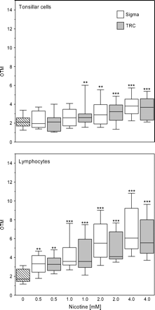
Comparison of DNA damage in fresh specimens of lymphatic tissue cells of palatine tonsils (tonsillar cells) and peripheral lymphocytes from 10 patients after 1 h of incubation with nicotine from two different suppliers (Sigma, TRC). Lines in the boxes represent the median values of the Olive tail moments (OTM). Box plots show the lowest and highest values of OTM, as well as the 1st and 3rd quartiles. An increase in DNA migration with rising concentrations of nicotine was significant according to ANOVA with post test for linearity (p < 0.0001). Significant difference compared to control were observed in Bonferroni's multiple comparison test: p < 0.05 (*), p < 0.001 (***).There were no significant differences in DNA migration at equal concentrations of nicotine from the two sources.
Without adjustment, the pH at higher nicotine concentrations increased up to pH 8.9. Therefore, the concentration-dependent effect of nicotine was studied under controlled pH conditions in study C. As can be seen from Figure 3, adjusting the pH to neutral conditions did not affect the extent of DNA damage from nicotine in lymphatic tissue of the palatine tonsils and lymphocytes. The linear trend of dose-dependent nicotine effects was again confirmed in this subset of patients for tonsillar cells (p = 0.0004), as well as for lymphocytes (p < 0.0001). The mean values of the positive control for tonsillar cells were 79.1 ± 4.9 (OTM), 76.7 ± 4.7 (DT), and 176.2 ± 13.5 (TL); and for lymphocytes, 78.2 ± 14.9 (OTM), 66.7 ± 11.5 (DT), and 183.4 ± 25.3 (TL).
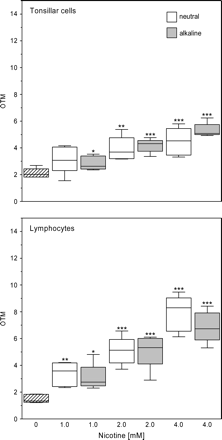
Comparison of DNA damage in fresh specimens of lymphatic tissue cells of palatine tonsils (tonsillar cells) and peripheral lymphocytes from five patients after 1 h of incubation with nicotine under unadjusted (alkaline) and pH-adjusted conditions (neutral; pH 7.2). Lines in the boxes represent the median values of the Olive tail moments (OTM). Box plots show the lowest and highest values of OTM, as well as the 1st and 3rd quartiles. An increase in DNA migration with rising concentrations of nicotine was significant according to ANOVA with post test for linearity in tonsillar cells (p = 0.0004) and lymphocytes (p < 0.0001). Significant difference compared to control was observed in Bonferroni's multiple comparison test: p < 0.05 (*), p < 0.01 (**), p < 0.001 (***). There were no significant differences in DNA migration at equal concentrations of nicotine with respect to pH level.
When comparing the negative controls of smokers with a history of 5–30 package years (py; N = 8) and nonsmokers and smokers with a history of 0–3 py (N = 7), the Mann-Whitney-U-test showed no differences (p = 0.613).
DISCUSSION
In research on the etiology of upper aerodigestive tract carcinogenesis, the mucosa in this region is of special interest because most tumors evolve from epithelial tissues. Oropharyngeal carcinomas are generally located in the palatine tonsils and show squamous cell epithelium with varying degrees of differentiation in histology. Consequently, in vitro monitoring for genotoxic effects by exposure to environmental, diet- or tobacco-related compounds may be performed in human epithelial cells, as well as in lymphocytes, to demonstrate both local cellular reactions and systemic effects. The alkaline single-cell microgel electrophoresis (Comet) assay is a sensitive tool for detection of DNA single strand breaks, as well as for alkali labile and incomplete excision repair sites (Lee et al., 2004; Tice et al., 2000). Lymphocytes and mucosal cells as human target cells have been analyzed by this method (Harréus et al., 1999; Kleinsasser et al., 2000a, 2000b, 2003b; Pool-Zobel et al., 1994; Schmezer et al., 2001).
Among the more than 4000 known components of cigarette mainstream smoke, up to 81 compounds are IARC classified carcinogens (Smith et al., 2003). Although both active and passive smoking, as well as use of smokeless tobacco products, have been classified by the IARC as carcinogenic for humans, nicotine as a major component of tobacco and tobacco smoke has never been classified (http://www-cie.iarc.fr/monoeval/grlist.html). Nicotine concentrations in smokers reach up to 100 ng/ml (=0.0006 mM) in plasma and up to 4000 ng/ml (= 0.025 mM) in saliva (Benowitz, 1990; Schneider et al., 2001; Teneggi et al., 2002). In the present study, the lowest concentration of nicotine eliciting significant DNA damage in tonsillar cells was observed after 1 h of incubation with 0.5 mM nicotine (Fig. 1). This concentration is only about 20-fold higher than saliva concentrations, to which a smoker is normally exposed for a much longer time. Therefore, nicotine could express significant direct genotoxic effects of carcinogenesis in human target cells. This possibility is consistent with recent data for human gingival fibroblasts (Argentin and Cicchetti, 2004) and spermatozoa (Arabi, 2004). A similar effect was shown previously for myosmine, a minor tobacco alkaloid that also occurs in a variety of foods, e.g., cereals, nuts, cocoa, and dairy products (Kleinsasser et al., 2003b; Tyroller et al., 2002; Zwickenpflug et al., 1998). This effect suggests a possible additional mechanism of induced carcinogenesis by these alkaloids in the upper aerodigestive tract. Other pathways of carcinogenesis for nicotine are non-neuronal acetylcholine receptor (nAchR)-mediated cell growth and suppression of apoptosis (Minna, 2003; Schuller et al., 2003), nAchR-mediated angioneogenesis in tumors (Natori et al., 2003; Cooke and Bitterman, 2004), epidermal growth factor–mediated tumor enhancement (Ye et al., 2004), and cyclooxygenase 2–dependent stimulation of gastric tumor growth (Wetscher et al., 1995). The mechanisms above all belong to the tumor promotion of carcinogenesis, whereas genotoxicity is a possible step in tumor initiation. Other steps of initiation include enhanced oxidative stress and free radical production (Crowley-Weber et al., 2003; Wetscher et al., 1995). Controversial results have been published in other classical in vitro tests for genotoxicity, showing either slight positive effects of nicotine— e.g., on sister chromatid exchange in mammalian cells (Trivedi et al., 1990)—no nicotine effects (Doolittle et al., 1995), or even protective effects of nicotine against tobacco smoke exposure and established tobacco carcinogens (Lee et al., 1996).
Nicotine was equally genotoxic to lymphocytes and tonsillar cells. This finding is in contrast to results obtained for other genotoxic compounds such as vanadium and, to a lesser extent, phthalates and myosmine, which were more genotoxic to lymphocytes than to mucosal cells (Kleinsasser et al., 2000a, 2003a, 2003b). One reason for this discrepancy could be the closer similarity of lymphocytes to tonsillar cells from lymphatic tissue as compared to cells from mucosal epithelium. Further studies with a higher number of probes will show more clearly whether a lack of difference in mutagen sensitivity between tonsillar cells and lymphocytes will prevail.
Nicotine impurities such as nornicotine could have a significant influence on nicotine effects (Carmella et al., 1997). However, DNA migration in the assay did not differ significantly with nicotine of high purity from two different suppliers (Fig. 2). Therefore the possibility that the nicotine effect could be due to impurities is minimal.
An analysis of a possible influence of smoking status on DNA migration in the negative controls showed no significant differences. This finding is in line with results of Hoffmann and Speit (2005), showing no differences in peripheral blood cells from heavy smokers and nonsmokers, either in the comet assay or in the micronucleus test. The apparent discrepancy between in vitro genotoxic effects of nicotine and no observable effects on DNA damage in vivo could have several sources. The comet assay as well as the micronucleus test might just be not sensitive enough to show a slight but still toxicologically relevant DNA damage by nicotine in vivo. Rapid in vivo repair of DNA damage by nicotine could also lower the chances of detecting genotoxic effects. In this context, the principle established by Druckrey and others (Druckrey et al., 1963; Peto et al., 1991), that no threshold values exist for genotoxic compounds, should be taken into account. Another possibility is that nicotine effects might be attenuated by other smoke constituents. This would not be the case in nicotine replacement therapy. To our knowledge, this aspect of nicotine safety has not been addressed in the literature so far.
The increasingly alkaline conditions resulting from higher nicotine concentrations did not have any significant effect on DNA damage by the alkaloid nicotine (Fig. 3). This was proven by adjusting the pH to about 7.2 at the start of the incubation. It will be interesting to look at the effect of pH levels well below neutral, which are commonly found in the gastric mucosa and with gastroesophageal reflux in the esophagus, pharynx, and larynx, taking into account the nitrosative stress from dietary nitrate (Iijima et al., 2003) and the excretion of high concentrations of nicotine with saliva (Boswell et al., 2000). These investigations will be performed with the aid of mini-organ cultures of epithelia of the upper aerodigestive tract combined with the Comet assay (Kleinsasser et al., 2001, 2004). This technique allows repetitive/chronic exposure under varying pH levels, simulating the in vivo situation in active and passive smokers. Further studies should be performed using saliva of smokers and nonsmokers (Reznick et al., 2004) and by co-incubation of nicotine with cigarette smoke condensate, established tobacco carcinogens as well as ethanol, a commonly found co-carcinogen in the pharynx. Finally, the possible role of metabolic activation for the ability of nicotine to induce DNA damage should be investigated.
In conclusion, nicotine expresses significant genotoxic effects in vitro in human target cells of upper aerodigestive tract carcinogenesis as well as in lymphocytes. The results suggest a direct tumor-initiating potency of this alkaloid.
The authors certify that all research involving human subjects was done under full compliance with all government policies and the Helsinki Declaration.
We thank Imperial Tobacco Ltd. for financial support of this study.
References
Arabi, M. (
Argentin, G., and Cicchetti, R. (
Benowitz, N. L. (
Boswell, C., Curvall, M., Elswick, R. K., and Leyden, D. (
Carmella, S. G., Borukhova, A., Desai, D., and Hecht, S. S. (
Cooke, J. P., and Bitterman, H. (
Crowley-Weber, C. L., Dvorakova, K., Crowley, C., Bernstein, H., Bernstein, C., Garewal, H., and Payne, C. M. (
Dajas-Bailador, F., and Wonnacott, S. (
Doolittle, D. J., Winegar, R., Lee, C. K., Caldwell, W. S., Hayes, A. W., and deBethizy, J. D. (
Druckrey, H., Schildbach, A., Schmähl, D., Preussmann, R., and Ivankovic, S. (
Harréus, U., Schmezer, P., Kuchenmeister, F., and Maier, H. (
Harris, J. E., Thun, M. J., Mondul, A. M., and Calle, E. E. (
Hecht, S. S. (
Heeschen, C., Jang, J. J., Weis, M., Pathak, A., Kaji, S., Hu, R. S., Tsao, P. S., Johnson, F. L., and Cooke, J. P. (
Hoffmann, D., and Hoffmann, I. (
Hoffmann, D., Hoffmann, I., and El-Bayoumy, K. (
Hoffmann, H., and Speit, G. (
Iijima, K., Grant, J., McElroy, K., Fyfe, V., Preston, T., and McColl, K. E. L. (
Kleinsasser, N., Dirschedl, P., Staudenmaier, R., Harreus, U., and Wallner, B. (
Kleinsasser, N. H., Gamarra, F., Bergner, A., Wallner, B. C., Harréus, U. A., Juchhoff, J., Kastenbauer, E. R., and Huber, R. M. (
Kleinsasser, N. H., Juchhoff, J., Wallner, B. C., Bergner, A., Harréus, U. A., Gamarra, F., Bührlen, M. B., Huber, R. M., and Rettenmeier, A. W. (
Kleinsasser, N. H., Wallner, B. C., Harréus, U. A., Zwickenpflug, W., and Richter, E. (
Kleinsasser, N. H., Wallner, B. C., Kastenbauer, E. R., Muenzenrieder, R. K., and Harréus, U. A. (
Kleinsasser, N. H., Weissacher, H., Kastenbauer, E. R., Dirschedl, P., Wallner, B. C., and Harréus, U. A. (
Lee, C. K., Fulp, C., Bombick, B. R., and Doolittle, D. J. (
Lee, E., Oh, E., Lee, J., Sul, D., and Lee, J. (
Lee, P. N., and Sanders, E. (
Minna, J. D. (
National Cancer Institute, Shopland, D. R., Burns, D. M., Benowitz, N. L., and Amacher, R. H., eds.
Natori, T., Sata, M., Washida, M., Hirata, Y., Nagai, R., and Makuuchi, M. (
Olive, P. L., Durand, R. E., Le Riche, J., Olivotto, I. A., and Jackson, S. M. (
Peto, R., Gray, R., Brantom, P., and Grasso, P. (
Pool-Zobel, B. L., Lotzmann, N., Knoll, M., Kuchenmeister, F., Lambertz, R., Leucht, U., Schröder, H.-G., and Schmezer, P. (
Reznick, A. Z., Hershkovich, O., and Nagler, R. M. (
Schmezer, P., Rajaee-Behbahani, N., Risch, A., Thiel, S., Rittgen, W., Drings, P., Dienemann, H., Kayser, K. W., Schulz, V., and Bartsch, H. (
Schneider, N. G., Olmstaed, R. E., Franzon, M. A., and Lunell, E. (
Schuller, H. M. (
Schuller, H. M., Plummer, H. K., and Jull, B. A. (
Shin, V. Y., Wu, W. K. K., Ye, Y.-N., So, W. H. L., Koo, M. W. L., Liu, E. S. L., Luo, J.-C., and Cho, C.-H. (
Smith, C. J., Perfetti, T. A., Garg, R., and Hansch, C. (
Teneggi, V., Squassante, L., Iavarone, L., Milleri, S., Bye, A., and Gomeni, R. (
Tice, R. R., Agurell, E., Anderson, D., Burlinson, B., Hartmann, A., Kobayashi, H., Miyamae, Y., Rojas, E., Ryu, J. C., and Sasaki, Y. F. (
Trivedi, A. H., Dave, B. J., and Adhvaryu, S. G. (
Tyroller, S., Zwickenpflug, W., and Richter, E. (
Wetscher, G. J., Bagchi, D., Perdikis, G., Bagchi, M., Redmond, E. J., Hinder, P. R., Glaser, K., and Hinder, R. A. (
Ye, Y. N., Liu, E. S. L., Shin, V. Y., Wu, W. K. K., Luo, J. C., and Cho, C. H. (
Author notes
*Department of Otolaryngology–Head and Neck Surgery, University of Regensburg, D-93053 Regensburg, Germany; †Department of Otolaryngology–Head and Neck Surgery, Ludwig-Maximilians University Munich, D-80337 Munich, Germany; ‡Walther Straub Institute of Pharmacology and Toxicology, Ludwig-Maximilians University Munich, D-80337 Munich, Germany

Comments