Open Access
01.02.2009 | Gastrointestinal Oncology
Cytoreductive Surgery Plus Hyperthermic Intraperitoneal Chemotherapy Improves Survival in Selected Patients with Peritoneal Carcinomatosis from Abdominal and Pelvic Malignancies: Results of 21 Cases
We evaluated the perioperative safety profile and efficacy of cytoreductive surgery (CRS) plus hyperthermic intraperitoneal chemotherapy (HIPEC) in 21 patients with peritoneal carcinomatosis (PC) from gastrointestinal and gynecological cancers. Twenty-one patients with PC (12 gastric cancer, 5 colorectal cancer, 2 ovarian cancer, 1 pseudomyxoma peritonei, 1 malignant mesothelioma) were treated with CRS + HIPEC with hydroxycamptothecin 20 mg and mitomycin C 30 mg in 12,000 mL of normal saline at 43 ± .5°C for 60 to 90 minutes. Vital signs were recorded for 5 days after surgery. We analyzed the following: local and systemic infections; gastrointestinal function recovery; hematological, hepatic, and renal parameters; wound healing time; adverse events; survival; and quality of life. The PC index was 2 to 33 (median, 11), the duration of operation 4 to 10 h (median, 8 h), and the highest temperature during 5 postoperative days 38.1°C. Two patients developed generalized edema and were successfully treated. Five patients developed hypoproteinemia on day 1 after surgery. All routine blood tests checked at 1 week after surgery were normal. Time of gastric tube removal was 2 to 7 days. Liquid food intake time was 3 to 8 days. Time of removal of stitches was 8 to 18 days. No local or systemic infections, wound disruption, or other clinically important adverse events occurred. The follow-up was 8 to 43 months (median, 22.5 months). Eleven patients died, three survived with tumor, and seven survived free of tumor. CRS + HIPEC was well tolerated in our selected patients with PC, some of whom had improved survival.
The locoregional progression of gastrointestinal and gynecological cancers usually results in peritoneal carcinomatosis (PC), which is characterized by the presence of tumor nodules of various size, number, and distribution on the peritoneal surface, with a very poor prognosis of <6 months’ median survival.1‐4 The most widely accepted therapies for such PC are systemic chemotherapy, best support care, and palliative treatment, without any hope of cure. Moreover, surgery alone can only remove the bulky visible tumor burden. For the micrometastases, invisible free cancer cells, and those tumors not suitable for resection, surgery cannot achieve any effect. Therefore, neither surgery nor chemotherapy alone can make an obvious difference in terms of quantity and quality of life in patients with PC.
To tackle this problem, a new treatment modality called cytoreductive surgery (CRS) plus hyperthermic intraperitoneal chemotherapy (HIPEC) has been developed as novel treatment for PC. Here, we summarize the results of a phase I clinical trail conducted in our center.
Anzeige
Patients and Methods
Patients
From January 2005 to January 2007, 21 patients with PC have been treated with CRS + HIPEC at the Cancer Center of Wuhan University, including 5 patients with colorectal cancer, 12 with gastric cancer, 2 with ovarian cancer, 1 with pseudomyxoma peritonei, and 1 with malignant mesothelioma. Major clinicopathological characteristics of the patients are listed in Table 1.
Table 1
Clinicopathologic characteristics of 21 patients with peritoneal carcinomatosis
The study protocol was approved by the ethical committee of Zhongnan Hospital of Wuhan University, and all the patients provided written informed consent to participate in the study. The abdominal exploration was performed under general anesthesia and hemodynamic monitoring through a midline xiphoid-pubic incision. Once the abdominal wall was open, a detailed evaluation of the PC was conducted, taking into consideration the size and distribution of disease, according to a principle previously described.5 When the PCI evaluation was finished, maximal CRS was performed, including the resection of the primary tumor with acceptable margins, any involved adjacent structures, lymphadenectomy, and peritoneotomies where peritoneal surfaces were involved by tumor, according to previously published surgical guidelines.5
After surgery, HIPEC was performed before the closure of abdominal cavity because this open technique is believed to provide optimal thermal homogeneity and spatial diffusion,5,6 with 12 L of heated saline containing 20 mg of hydroxycamptothecin and 30 mg of mitomycin C (MMC). An outflow tube for perfusion was placed in the Douglas pouch just before HIPEC. The perfusion solution was heated to 43.0 ± .5°C in a thermostatic water bath and infused into the peritoneal cavity at a rate of 200 mL/min through the inflow tube introduced from an automatic perfusion pump. The skin of the abdomen is attached to a retract ring, and a plastic sheet covered the open wound to keep the temperature stable. The perfusion in the peritoneal cavity was stirred manually with care not to infuse directly on the bowel surface. The first 1 L of perfusion solution was discarded through a drainage tube to wash out the residual debris and detached tumor cells, and the remaining solution was kept to circulate in the perfusion system. The temperature of the perfusion solution in the peritoneal space was monitored with a thermometer in real time. The total HIPEC procedure time was 60 to 90 min. After HIPEC, the perfusion solution in the abdominal cavity was removed through the suction tube, and drainage tubes were placed at appropriate sites depending on the type of primary operation. The wound was closed with a relaxing suture, and patient was sent to the recovery room.
The extent of CRS was determined by previously published criteria on the completeness of cytoreduction (CCR).7,8 A CCR score of 0 indicates no residual peritoneal disease after CRS; 1 represents <2.5 mm of residual disease; 2 indicates residual tumor between 2.5 mm and 2.5 cm; and 3 indicates >2.5 cm of residual tumor or the presence of a sheet of unresectable tumor nodules.
Anzeige
Postoperative Monitoring and Follow-Up
All patients were closely monitored for the following parameters: vital signs, bowel sound, flatus passage, drainage, and any discharges. The complete peripheral blood tests and blood chemistry were examined on the second postoperative day. Pulmonary cardiovascular functions were monitored.
Once the bowel function fully recovered, the nasogastric tube was removed, and the patient was placed on a trial liquid diet, which was gradually changed to semiliquid and soft food. The wound was examined daily, with the clinician paying particular attention to any signs of possible infection. All the information, including the time to drainage tube removal, time to suture removal, and the time on liquid food, was recorded on a special form. When patients could eat soft food and the wound suture was completely removed, they were discharged. The chemotherapy that followed was based on routine procedure.
All patients were routinely followed up by outpatient clinic or by telephone, and the information was recorded. The last time of follow-up was on August 1, 2008.
Statistical Analysis
Data were obtained from a database of clinical records, surgical reports, medical imaging reports, laboratory and pathology reports, and follow-up records. A serious adverse event was defined as a recurrence at any site or as a disease-related death. The survival time was calculated from the date of first CRS + HIPEC to the date of patient death due to any cause. The numerical data were directly recorded, and the category data were recorded into different categories. Data were analyzed by SPSS software, version 13.0 (SPSS, Chicago, IL), with P < .05 considered to be statistically significant.
Results
All patients underwent successful resection. The time of surgery ranged from approximately 4–10 h (median, 6 h, mean 6 ± 1.5 h). The volume of blood loss during surgery was 500 to 3000 mL, blood transfusion was 400 to 2200 mL, and fluid infusion was 2000 to 6500 mL. Eight patients received resection involving one organ part, 13 patients had resections involving more than one organ part, and 10 patients underwent partial peritonectomy greater than one quadrant of the abdomen. All patients received relaxation sutures. Ten patients transferred to the common ward after surgery, while 11 patients entered intensive care unit after surgery.
Successful wound healing was realized in all patients, and no wound infection or disruption occurred. The time to suture removal ranged from 8 to 18 days (median, 13 days), time of flatus passage from 2 to 7 days (median, 4 days), time of fluid intake from 3 to 8 days (median, 6 days), and time to drainage tubes removal from 3 to 9 days (median, 6 days). No major postoperative complications occurred, although two patients developed grade I generalized edema with puffiness of the face, arms, and legs and were treated with plasma and albumin transfusion and diuretics. The edema was relieved after 2 days.
Vital Signs
The numbers of patients with body temperature over 37°C were 13, 10, 3, and 1 on days 1, 2, 3, and 4 after surgery (Table 2). No patient experienced absorptive fever after the major operation. The hemodynamics parameters were stable, and no arrhythmia occurred.
Table 2
Important monitoring data during the 5 days after surgery
Index
Range (median)
Day 1
Day 2
Day 3
Day 4
Day 5
Temperature (°C)
36.7–38.1 (37.3)
36.7–38.0 (37.3)
36.2–37.2 (36.8)
36.3–37.2 (36.8)
36.1–37.1 (36.7)
Heart rate (bpm)
75–117 (92)
75–107 (88)
73–94 (83)
67–94 (80)
65–92 (82)
Laboratory Results
Among the 21 patients, 3 had aspartate aminotransferase of >46 U/L after surgery. Seven days postoperatively, only 1 patient had aspartate aminotransferase of >46 U/L. Five patients developed hypoproteinemia, but other laboratory results were normal (Table 3).
Table 3
Blood profile and biochemical test results
Parameter
Range (median)
Normal value
Day 1
Day 7
Peripheral blood test
Hemoglobin (g/L)
80.6–148 (118)
103–136 (113)
120–160
Red blood cell (×109/L)
3–4.91 (3.92)
3.47–4.94 (3.67)
4–5.5
White blood cell (×109/L)
6.71–30.1 (10.1)
8.77–15.6 (9.89)
4–10
Neutrophil count (×109/L)
4.99–27.4 (9.63)
6.61–14.8 (8.15)
2–7
Platelet count (×109/L)
95.4–356 (199)
105–336 (230)
100–300
Liver function tests
Aspartate aminotransferase (U/L)
14–99 (25)
14–43 (42)
0–46
Alanine aminotransferase (U/L)
10–116 (27)
13–65 (30)
0–46
TB (μmol/L)
.19–1.76 (1.25)
8.4–18.4 (13.2)
0–25
DBILI (μmol/L)
.2–12.5 (3.8)
1.8–6.2 (5.8)
0–7
IBILI (μmol/L)
2.9–17.9 (8.8)
5.5–12.56 (10.3)
1.5–18
TP (g/L)
41.3–71 (63.2)
54.1–72.3 (69.2)
60–80
ALB (g/L)
26.6–45.1 (24.2)
30.5–42.6 (40.2)
35–55
GLB (g/L)
14.6–30.4 (24.2)
18.5–29.7 (24.1)
20–30
Gamma glutamyl transferase (g/L)
8–84 (20)
19–135 (37)
5–55
Alkaline phosphatase (U/L)
41–99 (70)
65–146 (98)
35–134
Renal function tests
Blood urea nitrogen (mmol/L)
1.69–6.68 (3.27)
2.9–8.92 (4.08)
1.7–7.2
Creatine (μmol/L)
47.2–103.8 (59)
49.3–91.2 (70.2)
45–117
Electrolytes
K+ (mmol/L)
3.68–4.9 (4.2)
3.08–6.52 (4.22)
3.5–5.5
Na+ (mmol/L)
130–141.7 (136.1)
133.5–141 (137.4)
135–145
Cl− (mmol/L)
99–114.6 (103.7)
94.5–111.9 (102.9)
96–106
Ca2+ (mmol/L)
1.81–2.9 (2.25)
2.18–2.4 (2.26)
2–2.7
TB total bilirubin, DBILI direct bilirubin, IBILI indirect bilirubin, TP total protein, ALB albumin, GLB globulin
Anzeige
Adverse Events
No clinically important adverse events occurred in the perioperative period except for some minor abnormal laboratory results, and two patients (9.5%) experienced edema of the face and upper extremities.
Survival
The last follow-up was on August 1, 2008, by either outpatient clinic or by telephone. The follow-up time ranged from 8 to 43 months (median, 22.5 months) for all PC patients, 29 months (range, 8–43 months) for PC patients of gastric origin, and 18 months (range, 8.5–26.5 months) for PC of colorectal origin. Eleven patients died of disease progression, one due to rectal carcinoma recurrence to ovary with 8.5-month survival, one due to rectal cancer widespread metastases to abdominal cavity with 18-month survival, one due to colon cancer widespread metastases to abdominal cavity with 15.5-month survival, one due to ovarian cancer metastases to the whole abdominal cavity with 9-month survival, one due to malignant mesothelioma metastases to the whole abdominal cavity with 30.5-month survival, and the other six deaths due to gastric cancer progression with survivals of 8, 9.5, 10.5, 12, 15, and 29.5 months, respectively. Three patients survived with disease, while another seven patients experienced disease-free survival with satisfactory performance status.
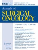
The overall survival curve of 21 patients and those PC patients from gastric, colorectal, and ovarian origins are presented in Fig. 1, and detailed information for each patient is summarized in Table 4.
Fig. 1
Overall survival curve of 21 patients with disease of gastric, colorectal, and ovarian origins
Table 4
Clinical and pathological features of 21 patients undergoing cytoreductive surgery (CRS) plus hyperthermic intraperitoneal chemotherapy (HIPEC)a
Subtotal gastrectomy, resection of part left upper quadrant of the abdomen peritoneum
0
SWOT
5
30
M
Gastric cancer
13
Total gastrectomy, splenectomy, partial pancreatectomy, resection of left upper quadrant of the abdomen peritoneum
3
8 mo
6
70
M
Gastric cancer
4
Subtotal gastrectomy, peritonectomy on the left upper quadrant
0
10.5 mo
7
37
F
Colon cancer
26
Right semicolectomy, jejunum and mesenterium tumor resection, lower abdominal and pelvic peritoneum resection
2
SWT
8
36
F
Rectal cancer recurrence in ovary
14
Rectum cancer resection, adnexa uteri resection, pelvic peritoneum resection, part bladder resection
3
8.5 mo
9
69
M
Gastric cancer
2
Subtotal gastrectomy, peritonectomy on upper left quadrant
0
SWOT
10
54
F
Gastric cancer
2
Gastric cancer resection, part upper quadrant of the abdomen peritoneum resection
0
SWOT
11
36
M
Colon cancer
6
Descending colon cancer resection, right lower quadrant of peritoneum resection
0
SWOT
12
67
F
Gastric cancer
6
Gastric cancer resection, left upper quadrant of the abdomen peritoneum resection
0
15 mo
13
43
M
Remnant gastric cancer
18
Remnant gastric cancer resection, spleen and cauda pancreatic resection, left upper quadrant of the abdomen peritoneum resection
3
9.5 mo
14
40
M
Gastric cancer
9
Total gastrectomy, left upper quadrant of the abdomen peritoneum resection
2
12 mo
15
38
F
Colon cancer
13
Ascending colon resection, right quadrant of peritoneum, and pelvic peritoneum resection
3
15.5 mo
16
69
M
Gastric cancer
2
Subtotal gastrectomy, left upper quadrant peritonectomy
0
SWOT
17
41
F
Gastric cancer
2
Subtotal gastrectomy, left upper quadrant peritonectomy
0
SWOT
18
59
F
Ovarian cancer
8
Adnexectomy, colic omentum resection, iliac fossa and pelvic peritoneum resection
1
SWT
19
67
F
Ovary cancer
29
Ovariectomy, resection of pelvic peritoneum, colic omentum, and diaphragmatic membrane
3
9 mo
20
64
F
Malignant peritoneum mesothelioma
30
Transverse colon resection, partial small intestine resection, colic omentum and epigastric peritoneum resection
3
30.5 mo
21
60
F
Pseudomyxoma peritonei
33
Hepatic region peritoneum, splenic region peritoneum, and pelvic peritoneum resection
3
SWT
CCR completeness of cryoreduction, SWT survival with tumor, SWOT survival without tumor, PCI peritoneal carcinomatosis index
aFollow-up continued until August 1, 2008, either by outpatient clinic or by telephone
bA CCR score of 0 indicates no residual peritoneal disease after CRS; 1, <2.5 mm of residual disease; 2, residual tumor between 2.5 mm and 2.5 cm; and 3, >2.5 cm of residual tumor or the presence of a sheet of unresectable tumor nodules
×
Discussion
Neither surgery nor chemotherapy or radiotherapy alone can make any important differences in terms of quantity and quality of life in patients with PC. To tackle this difficult problem, in the 1990s, a new treatment modality called CRS + HIPEC was developed, which has the advantages of surgery to reduce the visible tumor burden and regional hyperthermic chemotherapy to eradicate micrometastases and free cancer cells.9 Since then, over 20 phase I and II studies have been conducted with promising results, and this new treatment modality has gained increasingly wide acceptance in the treatment of PC from colorectal origin at cancer centers in North America, France, Italy, German, Holland, Spain, Australia, Japan, and South Korea.10‐13 After the randomized clinical trial by Netherland Cancer Center, a suggestion has been made to consider CRS and HIPEC the standard of care for patients with isolated colorectal PC.11,14‐17
Anzeige
This phase I clinical study was performed after an animal study, which showed important benefit of intraperitoneal chemotherapy for PC.18 For the 21 patients enrolled onto this study, the perioperative safety profile was satisfactory. There were no postoperative infections and no wound disruption because we used relaxation sutures during surgery and we delayed the time of removal of stitches. The heart rates of all patients were within normal limits during the 5 days after surgery, and all patients were hemodynamically stable. The highest temperature recorded was 38.1°C, and there was no absorption fever, which usually occurs after major abdominal surgery; the reason may be irrigation and clearance of abdominal cavity from blood residue during the HIPEC process. On the other hand, the heated chemotherapeutic agents can restrain the activity of inflammatory cells in the abdominal cavity and consequently reduce the release of cytokines, which leads to postoperative fever. Although there is no current evidence to support these hypotheses, they can be further tested in future trials. The gastrointestinal function recovery was optimal in these patients. Only two patients developed generalized edema, which might be due to the long duration of the operations, leading to more bleeding and hypoproteinemia, but the patients were successfully treated with plasma and albumin transfusion and diuretics. There was no intestinal perforation, no anastomosis leakage, no bowel obstruction, and no intra-abdominal hemorrhage or infections. These results indicate that our combined approach is a relatively safe modality of treatment with few postoperative complications.
To achieve effective cytoreduction, the field of surgery should be wide, and multiple organ parts maybe resected.19 This leads to high risks of major postoperative complications with 14% to 55% morbidity rate and 0% to 19% mortality rate, such as digestive fistulas and generalized sepsis in particular.20 In a prospective study by Yan et al. on 80 patients undergoing CRS + HIPEC for nonappendiceal PC, 1 patient died (1.3%) on the postoperative day 22 from multiorgan failure, 11 patients (14%) developed 12 grade I adverse events, 40 patients (50%) developed 71 grade II adverse events, 36 patients (45%) experienced 49 grade III adverse events, and 6 patients (8%) experienced 8 grade IV adverse events.21 In our study, however, no serious adverse events were observed, except for grade I generalized edema in two patients (9.5%) who were successfully treated. Such good results could be attributed to the following: (1) a fixed team of well-experienced surgeons, nursing, and operating room staff; (2) relatively mild HIPEC approach (hydroxycamptothecin, HCPT 20 mg plus MMC 30 mg for 60 min) compared with other approach (MMC 120 mg/m2 + cisplatin 200 mg/m2 for 90 to 120 min)20; (3) relaxation suture in closing abdominal wound; (4) postoperative use of effective broad-spectrum antibiotics to cover gram-positive and gram-negative aerobes and anaerobes; (5) transfusion of large-volume plasma and coagulation factors to quickly restore hypoproteinemia and stop bleeding; and (6) good nutritional support and quick restoration of electrolyte balance.
The median follow-up for the 21 patients was 22.5 months. The 1-year survival rate among all the patients was 66.7%. Among the 11 patients who died, the CCR scores were 3 in seven patients, 2 in two patients, 1 in one patient, and 0 in one patient, suggesting a close correlation between CCR and survival. The CCR is the most important prognostic indicator for survival.10,14,22,23 The patients with optimal CRS followed by HIPEC showed the best 5-year survival rate of 30%, whereas those underwent incomplete CRS gained little benefit, with a median survival comparable to that reported in historical controls.24 In a study by Cavaliere et al., patients with a CCR score of 0 had far higher 3-year survival rates than those with CCR scores of 1 and 2 (the P < .001).25 In 2004, Glehen et al. published a large-scale multicentric prospective study involving 506 patients who underwent CRS + HIPEC from 28 centers. The average follow-up was 53 months; the mean survival time was 19.2 months; and the 1, 3, and 5-year survival rates were 72%, 39%, and 19%, respectively. Thirty-eight patients survived for >5 years.26 In 2005, Netherland Cancer Research center summarized phase I, II, and III clinical studies and analyzed the long-term cure effects of 117 patients receiving CRS + HIPEC. The median survival time was 21.8 months, and the 1-, 3-, and 5-year survival rates were 75%, 28%, and 9%, respectively. Fifty-nine patients received complete cytoreduction; median survival time was 42.9 months, and 1-, 3-, and 5-year survival rates were 94%, 56%, and 43%, respectively.22
Despite CRS, the disease recurrence rate is still high, leading to treatment failure. In a multi-institutional study by Glehen et al., the overall incidence of recurrence was 73.3%.23 In a prospective study by Bijelic et al., among 70 patients with colorectal cancer undergoing combined treatment, 49 developed documented recurrence at median time for progression of 9 months, and most recurrent disease occurred inside the abdomen.24 One possible cause for such a high failure rate could be the marked differences in drug sensitivity between different PC types and between individuals with the same tumor types, as was found in a recent study of patient PC samples.27
Anzeige
In conclusion, CRS + HIPEC is a relatively safe treatment option in selected patients with PC that originates from gastrointestinal tract and gynecological malignancies, resulting in improved outcomes. There are still many problems about CRS + HIPEC, and a higher-level clinical trial is needed to provide more supportive evidence and to standardize the procedure.28‐30 On the basis of these results, we have registered and started a phase II randomized clinical trial (NCT00454519; http://www.clinicaltrials.gov). We hope that this trial will help gain better evidence to support this procedure.
Acknowledgments
This work was supported by New-Century Excellent Talents Supporting Program of the Ministry of Education of China (NCET-04-0669) and the Foundation for the Author of National Excellent Doctoral Dissertation of China (FANEDD-200464).
Open Access
This article is distributed under the terms of the Creative Commons Attribution Noncommercial License which permits any noncommercial use, distribution, and reproduction in any medium, provided the original author(s) and source are credited.
Open AccessThis is an open access article distributed under the terms of the Creative Commons Attribution Noncommercial License (https://creativecommons.org/licenses/by-nc/2.0), which permits any noncommercial use, distribution, and reproduction in any medium, provided the original author(s) and source are credited.
Mit der Zeitschrift Die Chirurgie erhalten Sie zusätzlich Online-Zugriff auf weitere 43 chirurgische Fachzeitschriften, CME-Fortbildungen, Webinare, Vorbereitungskursen zur Facharztprüfung und die digitale Enzyklopädie e.Medpedia.
Bis 30. April 2024 bestellen und im ersten Jahr nur 199 € zahlen!
Cytoreductive Surgery Plus Hyperthermic Intraperitoneal Chemotherapy Improves Survival in Selected Patients with Peritoneal Carcinomatosis from Abdominal and Pelvic Malignancies: Results of 21 Cases
Prof. Dr. med. Gregor Antoniadis Das Karpaltunnelsyndrom ist die häufigste Kompressionsneuropathie peripherer Nerven. Obwohl die Anamnese mit dem nächtlichen Einschlafen der Hand (Brachialgia parästhetica nocturna) sehr typisch ist, ist eine klinisch-neurologische Untersuchung und Elektroneurografie in manchen Fällen auch eine Neurosonografie erforderlich. Im Anfangsstadium sind konservative Maßnahmen (Handgelenksschiene, Ergotherapie) empfehlenswert. Bei nicht Ansprechen der konservativen Therapie oder Auftreten von neurologischen Ausfällen ist eine Dekompression des N. medianus am Karpaltunnel indiziert.
Dr. med. Benjamin Meyknecht, PD Dr. med. Oliver Pieske Das Webinar S2e-Leitlinie „Distale Radiusfraktur“ beschäftigt sich mit Fragen und Antworten zu Diagnostik und Klassifikation sowie Möglichkeiten des Ausschlusses von Zusatzverletzungen. Die Referenten erläutern, welche Frakturen konservativ behandelt werden können und wie. Das Webinar beantwortet die Frage nach aktuellen operativen Therapiekonzepten: Welcher Zugang, welches Osteosynthesematerial? Auf was muss bei der Nachbehandlung der distalen Radiusfraktur geachtet werden?
Dr. med. Mihailo Andric Inhalte des Webinars zur S1-Leitlinie „Empfehlungen zur Therapie der akuten Appendizitis bei Erwachsenen“ sind die Darstellung des Projektes und des Erstellungswegs zur S1-Leitlinie, die Erläuterung der klinischen Relevanz der Klassifikation EAES 2015, die wissenschaftliche Begründung der wichtigsten Empfehlungen und die Darstellung stadiengerechter Therapieoptionen.