Abstracts
This study evaluated the efficacy of low-level laser therapy (LLLT) and aluminum hydroxide (AH) in the prevention of oral mucositis (OM). A prospective, comparative and non-randomized study was conducted with 25 patients with head and neck cancer subjected to radiotherapy (RT) or radiochemotherapy (RCT). Twelve patients received LLLT (830 nm, 15 mW, 12 J/cm²) daily from the 1st day until the end of RT before each sessions during 5 consecutive days, and the other 13 patients received AH 310 mg/5 mL, 4 times/day, also throughout the duration of RT, including weekends. OM was measured using an oral toxicity scale (OTS) and pain was measured using the visual analogue scale (VAS). EORTC questionnaires were administered to the evaluate impact of OM on quality of life. The LLLT group showed lower mean OTS and VAS scores during the course of RT. A significant difference was observed in pain evaluation in the 13th RT session (p=0.036). In both groups, no interruption of RT was needed. The prophylactic use of both treatments proposed in this study seems to reduce the incidence of severe OM lesions. However, the LLLT was more effective in delaying the appearance of severe OM.
lasers; aluminum hydroxide; stomatitis; radiotherapy; head and neck cancer
Este estudo avaliou a eficácia da terapia do laser de baixa potência (LBP) e hidróxido de alumínio (HA) na prevenção da mucosite oral (MO). Um estudo prospectivo, comparativo e não-aleatorizado foi conduzido com 25 pacientes com câncer de cabeça e pescoço submetidos a radioterapia (RT) ou radioquimioterapia (RT/QT). Doze pacientes receberam LBP (830 nm, 15 mW, 12 J/cm²) diariamente desde o primeiro dia até o final da RT antes de cada sessão durante 5 dias consecutivos, e os outros 13 pacientes receberam HA 310 mg/5 mL, 4 vezes ao dia, também por toda a duração da RT, incluindo finais de semana. MO foi mensurada usando uma escala de toxicidade oral (ETO) e dor foi mensurada usando a escala visual analógica (EVA). Questionários da EORTC foram administrados para a avaliação do impacto da MO na qualidade de vida. O grupo LBP mostrou menores médias dos escores da ETO e EVA durante o curso da RT. Uma diferença significante foi observada na avaliação da dor na 13ª sessão de RT (p=0,036). Em ambos os grupos, nenhuma interrupção da RT foi necessária. O uso profilático de ambos os tratamentos propostos neste estudo parece reduzir a incidência de lesões severas de MO. No entanto, o LBP foi mais efetivo no atraso do aparecimento da MO severa.
Efficacy of low-level laser therapy and aluminum hydroxide in patients with chemotherapy and radiotherapy-induced oral mucositis
Aline Gouvêa de LimaI; Reynaldo AntequeraI; Maria Paula Siqueira de Melo PeresI; Igor Moysés Longo SnitocoskyII; Miriam Hatsue Honda FedericoII; Rosângela Correa VillarIII
IDentistry Division, Clinics Hospital, Medical School, University of São Paulo, São Paulo, SP, Brazil
IIOncology Divison, Medical School, University of São Paulo, São Paulo, SP, Brazil
IIIRadiotherapy Division, Clinics Hospital, Medical School, University of São Paulo, São Paulo, SP, Brazil
Correspondence Correspondence: Dra. Aline Gouvêa de Lima Rua Americo Nicoline, 75, Jardim Mosteiro 11740-000 Itanhaem, SP, Brasil Tel: +55 -13-9605-8322 e-mail: alinegov@yahoo.com
ABSTRACT
This study evaluated the efficacy of low-level laser therapy (LLLT) and aluminum hydroxide (AH) in the prevention of oral mucositis (OM). A prospective, comparative and non-randomized study was conducted with 25 patients with head and neck cancer subjected to radiotherapy (RT) or radiochemotherapy (RCT). Twelve patients received LLLT (830 nm, 15 mW, 12 J/cm2) daily from the 1st day until the end of RT before each sessions during 5 consecutive days, and the other 13 patients received AH 310 mg/5 mL, 4 times/day, also throughout the duration of RT, including weekends. OM was measured using an oral toxicity scale (OTS) and pain was measured using the visual analogue scale (VAS). EORTC questionnaires were administered to the evaluate impact of OM on quality of life. The LLLT group showed lower mean OTS and VAS scores during the course of RT. A significant difference was observed in pain evaluation in the 13th RT session (p=0.036). In both groups, no interruption of RT was needed. The prophylactic use of both treatments proposed in this study seems to reduce the incidence of severe OM lesions. However, the LLLT was more effective in delaying the appearance of severe OM.
KeyWords: lasers, aluminum hydroxide, stomatitis, radiotherapy, head and neck cancer.
RESUMO
Este estudo avaliou a eficácia da terapia do laser de baixa potência (LBP) e hidróxido de alumínio (HA) na prevenção da mucosite oral (MO). Um estudo prospectivo, comparativo e não-aleatorizado foi conduzido com 25 pacientes com câncer de cabeça e pescoço submetidos a radioterapia (RT) ou radioquimioterapia (RT/QT). Doze pacientes receberam LBP (830 nm, 15 mW, 12 J/cm2) diariamente desde o primeiro dia até o final da RT antes de cada sessão durante 5 dias consecutivos, e os outros 13 pacientes receberam HA 310 mg/5 mL, 4 vezes ao dia, também por toda a duração da RT, incluindo finais de semana. MO foi mensurada usando uma escala de toxicidade oral (ETO) e dor foi mensurada usando a escala visual analógica (EVA). Questionários da EORTC foram administrados para a avaliação do impacto da MO na qualidade de vida. O grupo LBP mostrou menores médias dos escores da ETO e EVA durante o curso da RT. Uma diferença significante foi observada na avaliação da dor na 13ª sessão de RT (p=0,036). Em ambos os grupos, nenhuma interrupção da RT foi necessária. O uso profilático de ambos os tratamentos propostos neste estudo parece reduzir a incidência de lesões severas de MO. No entanto, o LBP foi mais efetivo no atraso do aparecimento da MO severa.
INTRODUCTION
Oral mucositis (OM) is a common complication in patients subjected to chemotherapy (CT) and/or radiotherapy (RT), affecting approximately 80% of patients undergoing RT for treatment of tumors of the head and neck (1). Its occurrence is associated with severe pain, dysphasia, and increased risk of oral infection by opportunistic pathogens. OM has a great impact on patients' quality of life, increasing the morbidity and mortality, and adding a significant economic cost (2-4).
Prevention and treatment of OM have been mainly empirical and widely affected by basic oral care, analgesics, antibiotics and local anesthetics, yonder cryotherapy, growth factors, cytokines, biological mucosal protectants, antiinflammatory agents and complementary and alternative medicines (3).
Mucosal protective drugs, such as sucralfate, a complex salt of sucrose sulfate and aluminum hydroxide (AH), have been used for the treatment of OM (5). They provide a protective coating due to an ionic adhesion affinity with proteins of the damaged mucosa and provide local production of prostaglandin, increasing the blood flow and mitotic activity, as well as the surface migration of cells (6).
The use of sucralfate is considered a viable option due to the low cost, easy administration and absence of adverse effects. However, reports in the literature are conflicting (6-8).
Double-blind, randomized, prospective studies (6-8) have evaluated patients subjected to RT in the region of the head and neck in which the study group was treated with sucralfate and the control group received placebo. Carter et al. (6) observed OM grade III in 50% of the control patients and 43% of the patients in the study group (p=0.31), while significant reduction of OM after sucralfate use was found by Cengiz et al. (7) (p<0.05) and Etiz et al. (8) (p=0.0002).
Low-level laser therapy (LLLT) or prophylactic laser irradiation provides mainly the acceleration of wound healing. However, the major clinical indication of LLLT has historically been analgesia (9). LLLT produces wound healing by the transformation of fibroblasts into myofibroblasts, which are responsible for wound contraction (9,10).
The use of LLLT has been associated with the treatment of CT-induced OM (10-14), but few studies have been carried out with patients with OM lesions induced by RT (2,15,16).
The prophylactic effects of LLLT were first presented in a non-randomized retrospective study published in the early 1990's (10). Sixty-seven patients with cancer of different origins and locations were subjected to CT. The sample was divided into 3 groups: a control group and two other groups that received curative and prophylactic laser. The group treated with LLLT presented lower incidence of OM and was the only group in which the protocol did not have to be modified.
Barasch et al. (11) and Cowen et al. (12) conducted double-blind and randomized studies in patients undergoing bone marrow transplant. In both studies, the patients were divided into 2 groups: the study group received LLLT to evaluate its effect on the prevention of OM and the control group received placebo. Barasch et al. (11) found significant better results with the use of LLLT for pain reduction (p=0.027) and for evaluation of OM index (p<0.005). Although no statistically significant difference was found in the evaluation of the oral toxicity scale (OTS), Cowen et al. (12) observed a reduction in OM severity (p=0.01) and pain (p=0.05) in the laser-treated group.
Bensadoun et al. (2) were the first to carry out a double-blind and randomized study to evaluate effects of prophylactic laser only in patients with RT-induced OM. Thirty patients with head and neck cancer were treated with either LLLT or placebo. The frequency of OM grade III was 7.6% in the LLLT group and 35.2% in the placebo group. Severe pain occurred in 1.9% of the patients in LLLT group and 23.8% of those in the placebo group (p<0.05). The authors concluded that the laser is a safe and efficient method for the prevention of RT-induced OM.
A prospective and controlled study has been developed to evaluate the efficacy of LLLT on the prevention and treatment of RT-induced OM (16). Twenty-four patients with oral cancer were allocated to a group treated with laser or a group that served as a control. The LLLT group presented significantly better results in both pain and OM evaluations at the 2nd week (p=0.004), 3rd week (p=0.000), 4th week (p=0.029), 5th week (p=0.031), 6th week (p=0.019) and 7th week (p=0.045).
Although the literature presents effective results of LLLT in the reduction of OM severity, the scarcity of studies with patients subjected to RT in the region of the head and neck, and the morbidity of the treatment have stimulated the development of the present investigation. There are still many aspects to be clarified about laser therapy: the mechanism of action, the best application mode, the ideal wavelength, and the amount of released energy, among other questions. Regarding AH, the therapeutic role of this drug in OM treatment and the practicality of its use have motivated its inclusion in the study methodology.
The present study evaluated the capacity of LLLT and prophylactic AH in preventing or delaying the appearance of CT- and RT-induced OM, and the pain deriving from the lesions. The impact of OM on the ability to swallow and the patients' life quality were evaluated.
MATERIAL AND METHODS
The study was conducted between July and December 2005 in patients with diagnosis of head and neck tumors treated with RT associated or not with CT at the Radiotherapy and Oncology Division of the Institute of Radiology and Dentistry Division of the Clinics Hospital's Central Institute at the Medical School, University of São Paulo, Brazil. All patients were subjected to dental treatment before the oncology therapy to remove focus of infections, and received oral hygiene instructions.
Study Design
A prospective, comparative and non-randomized study was conducted with 25 patients after approval of the research protocol by the local Research Ethics Committee. The sample was divided into 2 groups: Group I was subjected to LLLT and Group II received AH suspension.
Inclusion Criteria
To be considered as eligible for this study, the patients had to present all the following characteristics: tumor in the head and neck region; ongoing treatment by external RT with a total dose ranging from 4,000 to 7,000 cGy at a rate of 1 fraction of 1, 8 to 2 Gy/day, 5 days a week (Monday to Friday), from linear accelerator (photons and electrons); age ranging from 18 to 80 years in an attempt to evaluate all adult patients with KPS>70 treated in the Radiotherapy Division; written informed consent for participation in the study.
Treatments
Group I: Twelve patients were subjected to LLLT applications by a diode (Laser Unit KM 3000; DMC, São Carlos, SP, Brazil) with wavelength 830 nm, nominal power 60 mW, effective power 15 mW and 0.2 cm2 aperture. The laser was delivered in a punctual form and the energy released by point was 2.4 J, giving an energy density of 12 J/cm2. Patients had laser applications daily since the first day of RT up to the end of the therapy during 5 consecutive days (Monday to Friday), before RT sessions. The laser was applied in 12 areas of the oral cavity, including the region of the upper and the lower labial mucosa, soft palate, buccal mucosa, lateral region of the tongue and floor of the mouth bilaterally. Laser was applied in the areas considered to the most susceptible for the occurrence of OM, that is, non-keratinized mucosas. The tumor areas were avoided and the patients wore specific protective glasses during the laser sessions to prevent eye injuries.
Group II: Thirteen patients received the AH suspension (310 mg/5 mL) since the first day of RT up to the end of the therapy, including weekends. Patients were instructed to use 10 mL of the mouthwash 4 times daily, and then to swallow the suspension. They were told to avoid eating during the first hour after treatment.
The association of RT and CT was allowed since the CT was to be performed with cisplatin, a drug that presents low potential for stomatotoxicity. In such case, the risk of occurrence of OM would be similar in both treatments. The use of concomitant antiinflammatory and/or analgesic was also allowed in both groups. Nistatin was prescribed in case there was any sign of fungal infection.
The National Cancer Institute's (NCI) oral toxicity scale (OTS) was used for OM evaluation and the visual analogue scale (VAS) was used for pain evaluation. The VAS was modified according to the scale proposed by Bensadoun et al. (2): scores 1 and 2 (mild pain) were considered grade I, scores 3 and 4 (moderate pain) were grade II, scores of 5 to 7 (severe pain) were grade III and scores of 8 to 10 (very severe pain) were grade IV. The evaluations were performed twice a week during the entire RT period.
Quality of life was evaluated by two questionnaires developed by the European Organization for Research and Treatment of Cancer (EORTC), an international non-profit organization that develops, coordinates and stimulates cancer laboratory and clinical research in Europe (17). The questionnaires QLQ-C30 and QLQ-H&N35 were applied at the beginning and end of RT.
Outcomes
Data referring to OM severe grades as defined by the NCI, time of appearance of severe OM, severe pain according to the VAS, swallowing capacity according to the NCI and quality of life according to the EORTC's questionnaires, were analyzed statistically by the Mann-Whitney and Wilcoxon tests (α=0.05).
RESULTS
Three patients were excluded from the sample: two patients missed the clinical appointments for oral examination and one patient died. All excluded patients belonged to the AH group. The sample of this study was composed by 22 patients, 20 men (90.91%) and 2 women (9.08%), with ages ranging from 33 to 80 years (mean age = 55.82 years). Squamous cell carcinoma was the most common type of tumor, corresponding to 77.27% of the sample. The most frequent location was the region of the oropharynx, corresponding to 40.91% of the patients. Nineteen patients could be classified according to the TNM clinical staging system; 89.47% of these patients were classified as stage IV and 10.53% as stage III.
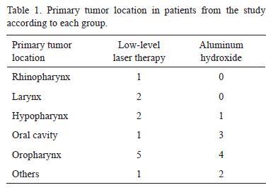
The RCT was the choice of treatment for 81.82% of the sample (18 patients) and the other patients were subjected to RT only. The daily radiation dose was 200 cGy in 86.36% of the sample (19 patients). The total dose was over 6000 cGy in 90.91% (20 patients). The areas more frequently involved in the irradiation field between the areas evaluated in the study were: the soft palate, the buccal mucosa, the lateral surface of tongue and the floor of the mouth (Tables 1 and 2).
Clinical Evaluation
According to the OTS, lower mean OM scores were observed in the LLLT group throughout the duration of the RT period, with values near to the significance level (p=0.061) between the 18th and 20th RT sessions (Fig. 1). During the RT treatment, grade IV OM was not observed. OM grade III occurred in 33.33% of the patients in the LLLT group compared to 50% in the AH group. The LLLT group (Fig. 2A) presented severe OM only after the 5th week of RT, while in the AH group severe OM occurred in the 2nd treatment week (Fig. 2B).
Functional Evaluation
The LLLT group also presented lower scores during the RT treatment (Fig. 3), though without statistical significance. Severe grades (III and IV) of dysphagia were found in approximately 33% of the LLLT group versus 50% of the AH group.
Pain Evaluation
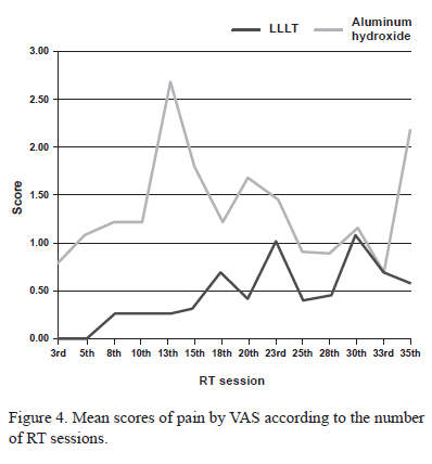
Lower mean pain scores were observed in the laser group during the whole RT treatment, except in the 33rd RT session. A statistically significant difference was observed only in the 13th RT session, in which the LLLT group presented important mean pain reduction compared to the AH group (p=0.036) (Fig. 4). During the whole RT treatment, very severe pain (grade IV) was reported by only 1 patient of the AH group, while 3 other patients of the same group and 1 patient of the LLLT group presented severe pain (grade III).
Quality of Life Evaluation
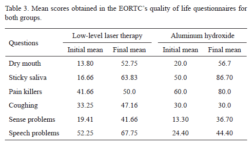
The analysis of the EORTC's quality of life questionnaires showed a marked worsening in the final evaluation compared to the initial evaluation in almost all questions asked during RT in both groups. The main mean scores of questions evaluated in this study can be seen in Table 3, keeping in mind that a high score for a symptom indicates a high level of symptomatology (17).
The questions about dry mouth, sticky saliva and pain killers presented worse index in the final evaluation compared to the initial index for both groups. However, the LLLT group presented better scores compared to the AH group in all these questions cited above, though without statistical significance. In the same way, the questions involving coughing, sense and speech problems presented a worse index in the final evaluation for both groups, except for the AH group that presented the same score in the question about coughing in both evaluations. In addition, the AH group presented better scores for these questions compared to the LLLT group, with statistically significant difference in the speech problems item (p=0.05).
At the end of the treatment, none of the patients of either of the groups needed to suspend the RT or RCT, due to the severity of the OM.
DISCUSSION
The best approach to treat advanced head and neck tumors is the association of treatments: surgery plus RCT or RCT. However, the combination of treatments in general is also associated with morbidity. A worsening in the quality of life questions scores was observed in the questionnaires for RCT therapy in the present study, which shows its high toxicity and the importance of using treatments to decrease side-effects.
Previous studies (2,15,16) performed in patients treated only with RT found good results with the use of LLLT in the prevention of OM. In the present study, which was performed with most patients receiving RCT, the prophylactic use of both treatments, compared to historical controls, apparently reduced the incidence of severe OM. This meant no interruption of RT, which clearly has a positive impact on treatment outcome. According to Buffa et al. (18), for each day of treatment suspended, a reduction of nearly 1% in survival for the patient is expected.
The mechanisms of action of laser are not completely known. Attenuating pain, stimulating endorphin release and modulating the immune system are some of the effects caused by LLLT. LLLT can also influence the wound healing process by the transformation of fibroblasts into myofibroblasts, which are responsible for wound contraction (19,20). In addition, the use of prophylactic laser seems to be more efficient than the AH prophylactic use (12).
To date, a great variety of doses ranging from 0.75 J/cm2 (10) to 35 J/cm2 (13) have been evaluated for the prophylactic and curative treatment of OM. Before the beginning of the present study, the laser was calibrated at the University of São Paulo's Technological Research Institute, obtaining an effective power of 15 mW. As we used to apply the laser during 40 s, a 160-s-irradition time was used to try to recover the effect lost with power reduction, resulting in an energy density of 12 J/cm2. The use of a high energy density did not cause adverse effects.
Although the visible red laser has been more frequently used for the healing of OM lesions, an infrared laser was used in this study because we have experience with the use of this laser in the treatment of OM with the above-mentioned dose. Satisfactory results were obtained in the reduction of OM and pain in non-prophylactic treatments. The decision to use daily LLLT was based on the study by Bensadoun et al. (2), who used LLLT in patients with OM lesions induced by RT and presented beneficial results with the daily application of LLLT.
In the present study, signs of OM were seen only between the 2nd and 3rd weeks in the LLLT group (peak in the 6th week). The present results showed that the use of LLLT was also effective to delay the appearance of the severe OM lesions, in the same way as reported by previous studies comparing LLLT and control groups of patients (2,15,16). In the present investigation, the lower incidence of severe OM in the LLLT group compared to the AH group was not highly evident probably because a placebo group was not included.
The powerful analgesic effect of LLLT has been reported. It has been demonstrated that low-power lasers can promote a decrease in the frequency of nociceptors and an increase in endorphin synthesis (9).
The AH group also showed a decrease in OM and no patient of this group had the RT or RCT treatment suspended. According to the EORTC questionnaire, AH presented higher efficacy than laser in coughing control, speech problems, sense problems and trouble with social contact (Table 2). This is probably because it is an oral suspension that can be swallowed, entering in contact not only with the oropharynx and the oral mucosa, but also with the esophagus.
Although the literature is controversial about the use of sucralfate, there is evidence that this drug can promote mucus production and local production of prostaglandin, thus increasing blood flow. Additionally, sucralfate can increase the mitotic activity and surface migration of cells, and provide binding of epithelial growth factors and basic fibroblastic growth factors to tissues (5). In our estimation, this fact justifies the prophylactic use of the drug in the absence of other resources. Moreover, some authors have recommended the use of sucralfate (7,8), a drug that contains AH, mainly because it is a low-cost drug, with easy administration and no adverse effects. The association of two prophylactic measures can probably produce more satisfactory results.
However, several questions should be clarified about the use of laser, regarding not only the ideal frequency and wavelength, but also the best description of its mechanism of action, which will certainly contribute to a more widespread the indication of this therapeutic resource.
In conclusion, the prophylactic use of both treatments proposed in this study seems to reduce the incidence of severe OM lesions, but LLLT was more effective in delaying their appearance. The results obtained with laser radiation in the present study have motivated us to keep on studying its application in our outpatient clinic. Randomized studies to assess mainly RCT-induced OM lesions must be developed because reports in the literature for this therapeutic modality are scarce.
ACKNOWLEDGEMENTS
The authors wish to thank the physicist Antonio Francisco Gentil Ferreira Júnior from the Electricity and Mechanics Division of the Optics Laboratory of the University of São Paulo's Technological Research Institute for his assistance with the LLLT equipment.
Accepted September 16, 2009
- 1. Sonis ST, Eilers JP, Epstein JB, LeVeque FG, Liggett WH, Mulagha MT, et al.. Validation of a new scoring system for the assessment of clinical trial research of oral mucositis induced by radiation or chemotherapy. Am Cancer Soc 1999;85:2103-2113.
- 2. Bensadoun RJ, Ciais G, Darcourt V, Schubert MM, Viot M, Dejou J, et al.. Low-energy He/Ne laser in the prevention of radiation-induced mucositis. A multicenter phase III randomized study in patients with head and neck cancer. Support Care Cancer 1999;7:244-252.
- 3. Genot MT, Klastersky J. Low-level laser for prevention and therapy of oral mucositis induced by chemotherapy or radiotherapy. Curr Opin Oncol 2005;17:236-240.
- 4. Mañas A, Palacios A, Contreras J, Sánchez-Magro I, Blanco P, et al.. Incidence of oral mucositis, its treatment and pain management in patients receiving cancer treatment at Radiation Oncology Departments in Spanish hospitals (MUCODOL Study). Clin Transl Oncol 2009;11:669-676.
- 5. McCarthy DM. Sucralfate. N Engl J Med 1991;325:1017-1025.
- 6. Carter DL, Hebert ME, Smink K, Leopold KA, Clough RL, Brizel DM. Double blind randomized trial of sucralfate vs. placebo during radical radiotherapy for head and neck cancers. Head Neck 1999;21:760-766.
- 7. Cengiz M, Özyar E, Öztürk D, Akyol F, Atahan IL, Hayran M. Sucralfate in the prevention of radiation-induced oral mucositis. J Clin Gastroenterol 1999;28:40-43.
- 8. Etiz D, Erkal HS, Serin M, Küçük B, Hepari A, Elhan AH, et al.. Clinical and histopathological evaluation of sucralfate in prevention of oral mucositis induced by radiation therapy in patients with head and neck malignancies. Oral Oncol 2000;36:116-120.
- 9. Walsh LJ. The current status of low level laser therapy in dentistry. Part 1. Soft tissues applications. Austr Dent J 1997;42:247-254.
- 10. Ciais G, Namer M, Schneider M, Demard F, Pourreau-Schneider N, Martin PM, et al.. La laserthérapie dans la prévention et lê traitement des mucites liées à la chimiothérapie anticancéreuse. Bull Cancer 1992;79:183-191.
- 11. Barasch A, Tanzer JM, Nuki K, Franquin JC. Helium-neon laser effects on conditioning-induced oral mucositis in bone marrow transplantation patients. Cancer 1995;76:2550-2556.
- 12. Cowen D, Tardieu C, Schubert M, Peterson D, Resbeut M, Faucher C, et al.. Low energy helium-neon laser in the prevention of oral mucositis in patients undergoing bone marrow transplant: results of a double blind randomized trial. Int J Radiat Oncol Biol Phys 1997;38:697-703.
- 13. Nes AG, Posso MB. Patients with moderate chemotherapy-induced mucositis: pain therapy using low intensity lasers. Int Nurs Rev 2005;52:68-72.
- 14. Genot-Klastersky MT, Klastersky J, Awada F, Awada A, Crombez P, Martinez MD, et al.. The use of low-energy laser (LEL) for the prevention of chemotherapy- and/or radiotherapy-induced oral mucositis in cancer patients: results from two prospective studies. Support Care Cancer 2008;16:1381-1387.
- 15. Maiya GA, Fernandes D. Effect of low level energy helium-neon (He-Ne) laser therapy in the prevention & treatment of radiation induced mucositis in head & neck cancer patients. Indian J Med Res 2006;124:399-402.
- 16. Arora H, Pai KM, Maiya A, Vidyasagar MS, Rajeev A. Efficacy of He-Ne laser in the prevention and treatment of radiotherapy-induced oral mucositis in oral cancer patients. Oral Surg Oral Med Oral Pathol Oral Radiol Oral Endod 2008;105:180-186.
- 17. Fayers PM, Aaronson NK, Bjordal K, Groenvold M, Curran D, Bottomley A. On behalf of the EORTC Quality of Life Group. The EORTC Scoring Manual (3rd Edition). Brussels: European Organization for Research and Treatment of Cancer; 2001.
- 18. Buffa FM, Bentzen SM, Daley FM, Dische S, Saunders MI, Richman PI, et al.. Molecular marker profiles predict locoregional control of head and neck squamous cell carcinoma in a randomized trial of continuous hyperfractionated accelerated radiotherapy. Clin Cancer Res 2004;10:3745-3754.
- 19. Safavi SM, Kazemi B, Esmaeili M, Fallah A, Modarresi A, Mir M. Effects of low-level He-Ne laser irradiation on the gene expression of IL-1beta, TNF-alpha, IFN-gamma, TGF-beta, bFGF, and PDGF in rat's gingiva. Lasers Med Sci 2008;23:331-335.
- 20. Silveira LB, Prates RA, Novelli MD, Marigo HA, Garrocho AA, Amorim JC, et al.. Investigation of mast cells in human gingiva following low-intensity laser irradiation. Photomed Laser Surg 2008;26:315-321.
Publication Dates
-
Publication in this collection
08 Sept 2010 -
Date of issue
2010
History
-
Accepted
16 Sept 2009 -
Received
16 Sept 2009


