Abstract
Cranial osteomyelitis is a potentially fatal lesion. White blood cell scanning (WBC) with 99mTc-hexamethylpropylene amine oxime (HMPAO) has proven highly sensitive and specific in the diagnosis and follow-up of patients with suspected osteomyelitis. In this report we show the usefulness of SPECT and transmission CT performed simultaneously using a hybrid imaging device for the functional anatomic mapping of soft tissue and cranial bone infections. 99mTc-HMPAO-labeled leukocytes scintigraphy was performed on an elderly diabetic man with an intracranial mass lesion and with suspected temporal bone infection. Planar scans were acquired 30 min, 4 h, and 24 h after injection. SPECT/CT was obtained 6 h after tracer injection, using a dual-head camera coupled with a low-power X-ray tube. The scintigraphic results were matched with the results of surgery and of clinical follow-up. The planar images alone were true-positives for abscess in this patient. SPECT/CT improves the accuracy of99mTc-HMPAO scintigraphy especially in discriminating between soft-tissue and bone involvement. In fact, SPECT/CT also showed temporal bone osteomyelitis. This result indicates that SPECT/CT performed using a hybrid device can improve imaging with 99mTc-HMPAO-labeled leukocytes in patients with suspected osteomyelitis by providing accurate anatomic localization and precise definition of the extent of infection.
SPECT/CT; brain abscess; 99mTc-HMPAO-labeled leukocyte scintigraphy
CASE REPORTS
Usefulness of hybrid SPECT/CT for the 99mTc-HMPAO-labeled leukocyte scintigraphy in a case of cranial osteomyelitis
Chiara Bruni; Federico Padovano; Laura Travascio; Orazio Schillaci; Giovanni Simonetti
Department of Diagnostic Imaging, Molecular Imaging, Interventional Radiology and Radiotherapy University of Rome "Tor Vergata"; Rome, Italy
Address for correspondence Address for correspondence: Dr.Chiara Bruni Viale Oxford 81, 00133, Rome, Italy Phone= +390620902418; fax= +390620902469 Email: chiarabruni79@hotmail.com
ABSTRACT
Cranial osteomyelitis is a potentially fatal lesion. White blood cell scanning (WBC) with 99mTc-hexamethylpropylene amine oxime (HMPAO) has proven highly sensitive and specific in the diagnosis and follow-up of patients with suspected osteomyelitis. In this report we show the usefulness of SPECT and transmission CT performed simultaneously using a hybrid imaging device for the functional anatomic mapping of soft tissue and cranial bone infections. 99mTc-HMPAO-labeled leukocytes scintigraphy was performed on an elderly diabetic man with an intracranial mass lesion and with suspected temporal bone infection. Planar scans were acquired 30 min, 4 h, and 24 h after injection. SPECT/CT was obtained 6 h after tracer injection, using a dual-head camera coupled with a low-power X-ray tube. The scintigraphic results were matched with the results of surgery and of clinical follow-up. The planar images alone were true-positives for abscess in this patient. SPECT/CT improves the accuracy of 99mTc-HMPAO scintigraphy especially in discriminating between soft-tissue and bone involvement. In fact, SPECT/CT also showed temporal bone osteomyelitis. This result indicates that SPECT/CT performed using a hybrid device can improve imaging with 99mTc-HMPAO-labeled leukocytes in patients with suspected osteomyelitis by providing accurate anatomic localization and precise definition of the extent of infection.
Key-Words: SPECT/CT, brain abscess, 99mTc-HMPAO-labeled leukocyte scintigraphy.
Cranial osteomyelitis arises from complications of paranasal sinus infection, trauma, dental extractions, chronic mastoiditis, necrotizing otitis externa and various surgical procedures [1]. The diagnosis and localization of infection still representa challenge for physicians. Because laboratory tests are relativelynonspecific, imaging is required to reach a correct diagnosis. Locating the site of infection is important for planning adequate treatment and evaluation the response to it [2].Various approaches have been developed to visualize inflammation and infection by nuclear medicine techniques. 99mTc-hexamethylpropylene amine oxime (HMPAO)-labeled leukocytes are often preferred for imaging inflammation, despite the relatively complex and time-consuming labeling procedure.These scintigraphic modalities are, however, limited by theirlow image resolution and lack of anatomic landmarks. The fusionof functional (i.e., SPECT) and morphologic (i.e., CT) images has significantly improved diagnostic accuracy as compared to that of using SPECT alone [3].
We describe a case of brain abscess presenting as a high uptake lesion on labeled leukocyte.
SPECT/CT had, in this case, a significantly higher incremental contributory value for WBC, contributing to the accurate identificationof infection in a patient with suspected temporal osteomyelitis.
Case Report
A 65 year old man with a one month history of pain in the right temporal region and fever (38ºC), presented with weakness, disphagia and high blood pressure (200/95). His vital signs and his motor function were normal. His pupils were isocoric and reacted normally to light. Laboratory studies revealed elevation of the inflammatory indices (ESR, CRP) and clinical examination showed a middle ear infection.
CT with contrast revealed a heterogeneous, dense lesion widespread from the oropharynx to the right mastoid. Intravenous antibiotic therapy was initiated. With the purpose to rule-out the presence of an initial osteomyelitis, follow-up three days later with 99mTc-HMPAO-labeled white blood cell scan (WBC) was performed at the three-day follow-up.
After blood sampling, leukocytes were isolated and labeled asdescribed by Biancone et al. [4]. The average labeling yield was 70%-85%. The labeled cells were reinjected into each patient, and their activity ranged from 400 to 555MBq.
A hybrid SPECT/CT system (Millennium VG and Hawkeye; GE Healthcare) consisted of a dual-head, variable-angle camera equipped with high-resolution low-energy collimators and an x-ray tube with detectors mounted on the opposite site of the camera gantry. Multiple planar images of the suspected area were acquired 2 hours after injection. The images were acquired in a 128 x 128 matrix using an imaging time of 15 min. SPECT/CT was performed 3 h after tracer injection. CT data were acquired over 360º during 14 s for each transaxial slice. Multiple slices were obtained by moving the table by 1 slice step before acquisition of each subsequent slice. The full field of view consisted of 40 slices.
SPECT was acquired in a 128 x 128 matrix, obtaining multiple views over 360º at a 30-s acquisition time per projection with an angular step of 3º. Images were reconstructed using Butterworth filtered backprojection (cutoff, 0.5; order, 10). Transverse, sagittal, and coronal slices were generated.
Transmission data were reconstructed at a nuclear medicine workstation (eNTEGRA; GE Healthcare) to obtain cross-sectional attenuation images (256 x 256 matrix) in which each pixel represents the attenuation value of the corresponding tissue. The reconstructed CT data and nuclear medicine data were transmitted to a nuclear medicine database. The matching SPECT and CT data were subsequently fused.
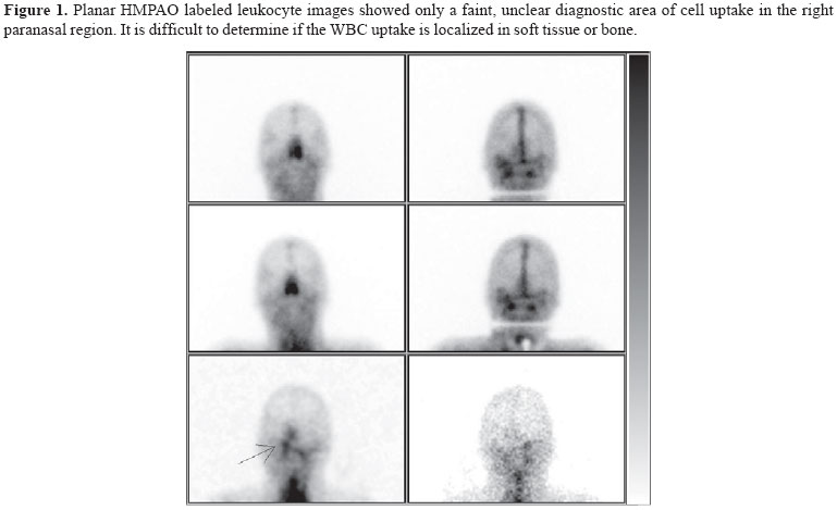
Planar WBC images showed only a faint, unclear diagnostic area of cell uptake in the right paranasal region. It was difficult to determine if the WBC uptake was localized only in soft tissue or also in bone. The hybrid SPECT/CT fusion imaging confirms an inflammatory focus involving the right temporal bone region. It also characterized the extension of infection (soft tissue only versus bone) more accurately. Right temporal bone biopsy subsequently confirmed the presence of osteomyelitis.
Discussion
Osteomyelitis of the central skull base is an uncommon condition that is potentially life threatening if not promptly recognized and properly treated. Several potentially serious complications can arise as a resultof skull base osteomyelitis, including cranial neuropathy, soft tissue involvement of the cavernous sinus with or without cavernoussinus thrombosis, and meningeal and brain parenchymal extension. This is usually a result of inadequately treated necrotizing otitis externa in diabetic or immunosuppressed patients and sometimes occurs after surgical debridement or drainage of a mastoid infection [5].
Periostal reaction and bone formation may progress slowly in diabetic patients, therefore the radiographic changes are even less sensitive in diabetics compared to non-diabetics [6]. Therefore conventional imaging is often unable to provide early detection of osteomyelitis. By contrast, 99mTc-HMPAO-labeled leukocytes are positive at the time of initial presentation of this pathology [7].
Moreover, conventional planar and SPECT images are often unable to precisely define the site of infection. It is a well known fact that nuclear imaging is characterized by a relatively limited spatial resolution,compared with that of other imaging methods (CT and MRI).
The hybrid SPECT/CT system delivers the high sensitivity of scinitigraphic technology with the high specificity of CT. This reduces the disadvantage of the SPECT's low spatial resolution [8]. The synergic use of images acquired in different manners such as SPECT/CT is particularly efficient, because it can help to overcome partial limitations of each technology.
In a recent study Filippi et al. affirm that the sensitivity in identifying the infected focus is identical in SPECT and SPECT/CT(100%). However, the specificity increases from 78% to 89% using SPECT/CT rather than SPECT alone. SPECT/CT, infact, is useful in patients with planar images positive for active infectionbut equivocal for localization [7].
In particular, in this case of bone infection with adjacent soft-tissue involvement, conventional CT of the head fails to demonstrate any abnormality of the right petrous bone. Planar images alone of scinitigraphy did not allow differentiation of soft tissue from bone. However, hybrid SPECT/CT allowed better characterization of the extension of infection and provided an accurateanatomic localization of the positive focus not only in the soft tissue but also in the temporal bone. This ability is of particular importance since the therapeutic approaches to soft-tissue and bone infections are different [9]. There are many studies that underline the usefulness of HMPAO labeled leukocyte SPECT imaging to assess for active cranial osteomyelitis after treatment and follow-up of this patients.
Received on 22 June 2008; revised 18 November 2008.
- 1. Chandler J.R., Grobman L., Quencer R., Serafini A. Osteomyelitis of the base of the skull. Laryngoscope 1986;96:245-51.
- 2. Rote N.S.V. Inflammation. In: McCance KL, Huerther SE, eds. Pathophysiology. St. Louis, MO: Mosby; 1998:205-36.
- 3. Schillaci O., Simonetti G. Fusion imaging in nuclear medicine: applications of dual-modality systems in oncology. Cancer Biother Radiopharm 2004;19:1-10.
- 4. Biancone L., Schillaci O., Capoccetti F., et al. Technetium-99mHMPAO labeled leukocyte single photon emission computerized tomography (SPECT) for assessing Crohns disease extent and intestinal infiltration. Am J Gastroenterol. 2005;100:344-54.
- 5. Seabold J.E., Simonson T.M., Weber P.C., et al. Cranial osteomyelitis: diagnosis and follow-up with In-111 white blood cell and Tc-99m methylene diphosphonate bone SPECT, CT, and MR imaging. Radiology 1995;196(3):779-88.
- 6. Seldin D.W., Heiken J.P., Feldman F., Alderson P.O. Effect of oft tissue pathology on detection of pedal osteomyelitis in diabetics. J Nucl Med 1985;26:988-93.
- 7. Filippi L., Schillaci O. Usefulness of hybrid SPECT/CT in 99mTc-HMPAO-labeled leukocyte scintigraphy for bone and joint infections. J Nucl Med 2006;47(12):1908-13.
- 8. Bar-Shalom R., Yefremov N., Guralnik L., et al. SPECT/CT using 67Ga and 111In-labeled leukocyte scintigraphy for diagnosis of infection. J Nucl Med 2006;47(4):587-94.
- 9. Horger M., Eschmann S.M., Pfannenberg C., et al. The value of SPET/CT in chronic osteomyelitis. Eur J Nucl Med Mol Imaging 2003;30:1665-73.
Publication Dates
-
Publication in this collection
10 Mar 2009 -
Date of issue
Dec 2008
History
-
Accepted
18 Nov 2008 -
Received
22 June 2008