Abstract:
Tinea nigra is a superficial mycosis whose diagnosis is confirmed by isolating the infectious agent Hortae werneckii through mycological examinations. In vivo reflectance confocal microscopy, initially used in melanocytic dermatosis, has been used with skin infectious diseases to identify the parasite at the cellular level. We report, for the first time in the scientific literature, the use of reflectance confocal microscopy in a case of tinea nigra and compare its findings to dermoscopy and mycological examination results.
Keywords:
Bacterial infections and mycoses; Culture; Culture media; Dermatology; Dermoscopy; Microscopy, confocal; Mycoses; Phaeohyphomycosis; Skin diseases; Tinea
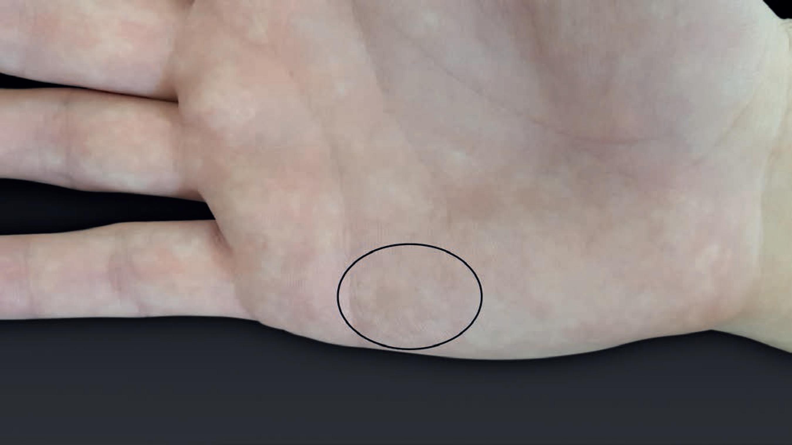
We report a patient with an asymptomatic macula in the palmar region (Figure 1). Due to the hypothesis of tinea nigra, we performed a dermoscopy examination. The results showed a non-melanocytic pattern with speckled and superficial pigmentation that did not follow the dermatoglyphs (Figure 2).11 Zaitz C, Campbell I, Marques SA, Ruiz LRB, Framil VMS. Compêndio de Micologia Médica. 2.ed. Rio de Janeiro: Guanabara Koogan; 2010.
2 Darrigade AS, Saint-Marie D, Dufour J, Edouard S, Graille J, Cheuret M, et al. The value of dermoscopy in the diagnosis of tinea nigra. Ann Dermatol Venereol. 2014;141:167-9.-33 Nazzaro G, Ponziani A, Cavicchini S. Tinea nigra: A diagnostic pitfall. J Am Acad Dermatol. 2016;75:e219-e220. Direct mycological examination (DME) revealed septate and dematiaceous tortuous hyphae and the fungal culture isolated the agent Hortae werneckii (Figure 3), confirming the diagnosis of tinea nigra.11 Zaitz C, Campbell I, Marques SA, Ruiz LRB, Framil VMS. Compêndio de Micologia Médica. 2.ed. Rio de Janeiro: Guanabara Koogan; 2010.,44 Odds FC, Arai T, Disalvo AF, Evans EG, Hay RJ, Randhawa HS, et al. Nomenclature of fungal diseases: a report and recomendations from a Sub-Committee of the International Society for Human and Animal Mycology (ISHAM). J Med Vet Mycol. 1992;30:1-10.
Clinical appearance of tinea nigra (highlighted by the circle): hyperchromic macula with ill-defined borders in hypothenar eminence on the right palm
Dermoscopic aspect of the lesion: a non-melanocytic pattern, composed of linear spiked structures that do not follow the palmar dermatoglyphs
Mycological examination. A: direct mycological examination under optical microscopy (Hematoxylin & eosin X100) clarified with 10% KOH showing mycelium composed of short and dematiaceous tortuous hyphae. B: fungal culture in Sabouraud agar with growth of dematiaceous colonies. C: microculture under optical microscopy (Hematoxylin & eosin X200) cylindrical-shaped yeast-like cells with darkly pigmented septa, compatible with Hortae werneckii agent
Before collecting samples for mycological examination, we performed an in vivo reflective confocal microscopy (RCM) of the lesion, which showed to a lesser extent linear structures with high reflectivity of speckled appearance, similar to the ones revealed by dermoscopy.22 Darrigade AS, Saint-Marie D, Dufour J, Edouard S, Graille J, Cheuret M, et al. The value of dermoscopy in the diagnosis of tinea nigra. Ann Dermatol Venereol. 2014;141:167-9.,33 Nazzaro G, Ponziani A, Cavicchini S. Tinea nigra: A diagnostic pitfall. J Am Acad Dermatol. 2016;75:e219-e220.,55 Veasey JV, Framil VM, Nadal SR, Marta AC, Lellis RF. Genital warts: comparing clinical findings to dermatoscopic aspects, in vivo reflectance confocal features and histopathologic exam. An Bras Dermatol. 2014;89:137-40.
6 Guarenti IM, Almeida HL Jr, Leitão AH, Rocha NM, Silva RM. Scanning electron microscopy of Tinea nigra. An Bras Dermatol. 2014;89:334-6.-77 Veasey J, Framil V, Ribeiro A, Lellis R. Reflectance confocal microscopy use in one case of Pityriasis folliculorum: a Demodex folliculorum analysis and comparison to other diagnostic methods. Int J Dermatol. 2014;53:e254-7. The image showed that the speckled aspect, when isolated, was morphologically similar to the hyphae identified on DME (Figure 4).
Reflective confocal microscopy in vivo. A: examination being performed on the patient. B: hyperreflectivity of tortuous linear structures. C: detail of structures, identifying short, thick, and tortuous structures
In vivo reflective confocal microscopy (RCM) is a valuable method for evaluating pigmented and oncological lesions. The method fills the limitations of dermoscopy by displaying structural details similar to those seen on histopathological examinations. Recent studies have described the efficacy of RCM in identifying the infectious agent in vivo in infectious diseases.55 Veasey JV, Framil VM, Nadal SR, Marta AC, Lellis RF. Genital warts: comparing clinical findings to dermatoscopic aspects, in vivo reflectance confocal features and histopathologic exam. An Bras Dermatol. 2014;89:137-40.,77 Veasey J, Framil V, Ribeiro A, Lellis R. Reflectance confocal microscopy use in one case of Pityriasis folliculorum: a Demodex folliculorum analysis and comparison to other diagnostic methods. Int J Dermatol. 2014;53:e254-7. However, no scientific publications are found about its use for tinea nigra diagnosis. Therefore, this is the first report to do so.
In the present study, we identified that the fungus is hyperreflective, possibly due to the melanin present in its cell wall. The fibrillar aspect described with dermoscopy corresponds to the vegetative mycelium of the fungus. It is worth mentioning that the aspect of the hyphae identified in RCM is tortuous, irregular, and short, similar to that found in DME, and different from the morphology of the thin and elongated hyphae of the dermatophytes visualized in both DME and RCM.
-
*
Work performed at Dermatology Clinic at Hospital Santa Casa de São Paulo - São Paulo (SP), Brazil.
-
Financial support: None.
REFERENCES
-
1Zaitz C, Campbell I, Marques SA, Ruiz LRB, Framil VMS. Compêndio de Micologia Médica. 2.ed. Rio de Janeiro: Guanabara Koogan; 2010.
-
2Darrigade AS, Saint-Marie D, Dufour J, Edouard S, Graille J, Cheuret M, et al. The value of dermoscopy in the diagnosis of tinea nigra. Ann Dermatol Venereol. 2014;141:167-9.
-
3Nazzaro G, Ponziani A, Cavicchini S. Tinea nigra: A diagnostic pitfall. J Am Acad Dermatol. 2016;75:e219-e220.
-
4Odds FC, Arai T, Disalvo AF, Evans EG, Hay RJ, Randhawa HS, et al. Nomenclature of fungal diseases: a report and recomendations from a Sub-Committee of the International Society for Human and Animal Mycology (ISHAM). J Med Vet Mycol. 1992;30:1-10.
-
5Veasey JV, Framil VM, Nadal SR, Marta AC, Lellis RF. Genital warts: comparing clinical findings to dermatoscopic aspects, in vivo reflectance confocal features and histopathologic exam. An Bras Dermatol. 2014;89:137-40.
-
6Guarenti IM, Almeida HL Jr, Leitão AH, Rocha NM, Silva RM. Scanning electron microscopy of Tinea nigra. An Bras Dermatol. 2014;89:334-6.
-
7Veasey J, Framil V, Ribeiro A, Lellis R. Reflectance confocal microscopy use in one case of Pityriasis folliculorum: a Demodex folliculorum analysis and comparison to other diagnostic methods. Int J Dermatol. 2014;53:e254-7.
Publication Dates
-
Publication in this collection
Jul-Aug 2017
History
-
Received
13 Dec 2016 -
Accepted
10 Jan 2017