 Julien Andreani1
 Jacques Y. B. Khalil1,2
 Emeline Baptiste1Issam Hasni1
 Caroline Michelle1
 Didier Raoult1
 Anthony Levasseur1
 Bernard La Scola1*
- 1Aix Marseille Université, IRD, APHM, MEPHI, IHU-Méditerranée Infection, Marseille, France
- 2Centre National de la Recherche Scientifique, Marseille, France
Giant viruses continue to invade the world of virology, in gigantic genome sizes and various particles shapes. Strains discoveries and metagenomic studies make it possible to reveal the complexity of these microorganisms, their origins, ecosystems and putative roles. We isolated from a rat stool sample a new giant virus “Orpheovirus IHUMI-LCC2,” using Vermamoeba vermiformis as host cell. In this paper, we describe the main genomic features and replicative cycle of Orpheovirus IHUMI-LCC2. It possesses a circular genome exceeding 1.4 Megabases with 25% G+C content and ovoidal-shaped particles ranging from 900 to 1300 nm. Particles are closed by at least one thick membrane in a single ostiole-like shape in their apex. Phylogenetic analysis and the reciprocal best hit for Orpheovirus show a connection to the proposed Pithoviridae family. However, some genomic characteristics bear witness to a completely divergent evolution for Orpheovirus IHUMI-LCC2 when compared to Cedratviruses or Pithoviruses.
Introduction
‘Giant viruses’ is a name commonly given to all viruses which are characterized by a capsid or ovoid shape, a size larger than 0.2 μm and a genome containing more than approximately 200,000 base pairs. This term encompasses a monophyletic group of large double stranded DNA viruses known as the nucleo-cytoplasmic large DNA viruses (NCLDV) (Iyer et al., 2001). The discovery of many Mimiviruses (La Scola et al., 2003; Arslan et al., 2011), Marseilleviruses (Boyer et al., 2009; Dornas et al., 2016) and Pandoraviruses (Philippe et al., 2013; Antwerpen et al., 2015), broke the paradigm of the previously held definition of the frontier between prokaryote and viruses. The “Megavirales” order proposed by Colson et al. (2013) continues to expand to host new arrivals with the potential of replacing the current NCLDV families (Colson et al., 2013; Aherfi et al., 2016). All these viruses share fundamental genes, for example, the conserved five ancestral genes and some others established into clusters of orthologous genes named NCVOGs (Yutin et al., 2009). Their replicative strategies appear to have adapted through their own evolution, as is the case for Pandoraviruses or Mollivirus sibericum (Abergel et al., 2015; Colson et al., 2017). Major improvements in taxonomy would be needed to definitely classify viruses in their families and in the putative “Megavirales” order. Further investigations should be focused on their genome content, hosts, ecosystems, tropisms and infectivity in order to determine whether their evolution is expansive or reductive or if it happens in a more dynamic accordion-like pattern (Moreira and Brochier-Armanet, 2008; Filée, 2014, 2015; Yutin et al., 2014; Moreira and López-García, 2015).
For now, co-culture on amoeba remains the major tool for isolating giant viruses (Pagnier et al., 2013; Khalil et al., 2016a). We recently combined co-culture with flow cytometry to come up with a faster and more sensitive way of detecting, presumably identifying and purifying the causative agent of lysis (Khalil et al., 2016b, 2017). In 2013, Pithovirus sibericum was isolated from a 30,000-year-old sample in the Siberian permafrost, and was described as being the most elongated-ovoid shape currently known for a virus with a maximum length of 1.5 μm. Surprisingly, the circular genome size is “only” of 610,033 base pairs, which appears to be astonishing given their viral particle size. The genome of P. sibericum is delivered via a single cork. Two years after the description of Pithovirus, a modern one that we named “Pithovirus massiliensis LC8” (Levasseur et al., 2016a) was also isolated and displayed amazing and extreme genomic conservation regarding its ancestor P. sibericum, which enabled us to estimate a molecular clock about the evolution of Pithoviruses. Moreover, we recently described a new virus Cedratvirus A11 (Andreani et al., 2016) a possible new genus in the putative Pithoviridae family. This virus presented two corks, one at each extremity and a circular genome estimated at 589,068 base pairs. In addition, a new strain, close to Cedratvirus A11, known as Cedratvirus lausannensis, was recently isolated (Bertelli et al., 2017) with a genome size estimated at 575,161 base pairs. This latter appears to represent a fourth member of this new emerging family. Our isolated Faustovirus (Bou Khalil et al., 2016) and Pithovirus (Levasseur et al., 2016a) indeed came from the same sampling area. For this reasons and after successfully isolating these viruses, we decided to investigate the same location once again, 4 months later in order to search for the same isolates that could be circulating and to explore the relation to ecosystemic or environmental changes. The result of this work was a new isolate from rat stool sample, which we named Orpheovirus, the genome and replicative cycle features of which we describe in this paper.
Materials and Methods
Sample Collection
Twelve different rat stools and nine water samples contaminated by proximity sewage were collected. Rat stools were taken from a dry place one meter from the water sample area. Samples were harvested in November 2015 in La Ciotat, France, at the same GPS location where P. massiliensis LC8 (Levasseur et al., 2016a) and Faustovirus LC9 samples had been collected (Bou Khalil et al., 2016; Cherif Louazani et al., 2017) (N43.181834, E5.614423).
Virus Isolation
Vermamoeba vermiformis stain CDC19 was used as cell support. The amoebas were harvested after 48 h of culture in homemade peptone yeast extract glucose medium (PYG) when a concentration of 1.106 amoebas/mL was reached. Cells were then rinsed twice in homemade page’s amoeba saline (PAS) and pelleted at 700 × g for 10 min. The amoebas were then re-suspended in the starvation medium (Bou Khalil et al., 2016) at a concentration of 1.106 amoebas/mL. An antibiotic and antifungal mixture with vancomycin (10 μg/mL), ciprofloxacin (20 μg/mL), imipenem (10 μg/mL), and voriconazole (20 μg/mL) was added to the suspension in order to decrease or eliminate bacterial or fungal contamination. A cell suspension of 250 μL per well was then distributed onto a 48-well plate. The samples were then vortexed and 50 μL were added to each well. The rest of the wells served as negative controls by adding 50 μL of PAS. The plate was incubated at 30°C for 4 days in order to monitor any potential cytopathic effect. This co-culture was repeated twice in the same order. When confronted with a high degree of contamination detected in some wells, filtration using 1.2 μm syringe filter (Merck Millipore) was carried out and gentamycin (20 μg/mL) was added 24 h before the second plate of co-culture (sub-culture 1).
Viral Production and Purity Control
End-point dilution was performed in order to clone the virus before its production. To do so, we successively inoculated diluted viral supernatant on V. vermiformis at a dilution factor of 10. End point dilution was assessed for 5 days and the lysis was controlled by inverted microscopy.
For the production and purification processes, 14 infected flasks of 150 cm2 (Corning®, Corning, NY, United States) were pelleted using the Beckman coulter® centrifuge Avanti® J-26 XP (Beckman, France) at 14,000 × g for 30 min (Andreani et al., 2016; Levasseur et al., 2016a). A 25% sucrose gradient was used for the final purification step. After finalizing production, we proceeded with genome sequencing.
Genome Sequencing
Genomic DNA was sequenced on the MiSeq Technology (Illumina Inc., San Diego, CA, United States) using the paired end and mate pair applications. The DNA was barcoded in order to be mixed with 11 other projects for the Nextera Mate Pair sample prep kit (Illumina) and with 16 other projects for the Nextera XT DNA sample prep kit (Illumina).
gDNA was quantified using a Qubit assay with the high sensitivity kit (Life Technologies, Carlsbad, CA, United States) to 131.3 ng/μl.
For the paired end library, dilution was performed requiring 1ng of each genome as input. The “tagmentation” step fragmented and tagged the DNA. Limited cycle PCR amplification (12 cycles) then completed the tag adapters and introduced dual-index barcodes. The library profile was validated on an Agilent 2100 BioAnalyzer (Agilent Technologies Inc., Santa Clara, CA, United States) with a DNA High sensitivity labchip and the fragment size was estimated to 1.5 kb. After purification on AMPure XP beads (Beckman Coulter Inc., Fullerton, CA, United States), the libraries were then normalized on specific beads according to the Nextera XT protocol (Illumina). Normalized libraries were pooled for MiSeq sequencing. Automated cluster generation and paired end sequencing with dual index reads were performed in a single 39-h run in 2 × 250-bp.
A total of 6.6 Gb of information was obtained from a 697,000 per mm2 for the density cluster with a cluster passing quality control filters of 94.6% (12,733,000 passed filtered clusters). Within this run, the index representation for Orpheovirus IHUMI-LCC2 was determined to 12.93%. The 1,942,146 paired end reads were trimmed and filtered according to the read qualities.
The mate pair library was prepared with 1.5 μg of genomic DNA using the Nextera mate pair Illumina guide and two libraries were constructed. The genomic DNA sample was simultaneously fragmented and tagged with a mate pair junction adapter. The pattern of the fragmentation was validated on an Agilent 2100 BioAnalyzer (Agilent Technologies Inc., Santa Clara, CA, United States) with a DNA 7500 labchip. The DNA fragments ranged from 1.5 kb to 11 kb with an optimal size at 6.57 and 2.89 kb, respectively. No size selection was performed and 600 and 117 ng, respectively, of tagged fragments were circularized.
The circularized DNA was mechanically sheared to small fragments with an optimal size of 1029 and 1253 bp, respectively, on the Covaris device S2 in T6 tubes (Covaris, Woburn, MA, United States).
The library profile was visualized using a High Sensitivity Bioanalyzer LabChip (Agilent Technologies Inc., Santa Clara, CA, United States) and the final concentration libraries were measured at 5.13 and 5.4 nmol/l, respectively.
In each construction, the libraries were normalized at 2 nM and pooled. After a denaturation step and dilution at 15 pM, the pool of libraries was loaded onto the reagent cartridge and then onto the instrument along with the flow cell. Automated cluster generation and sequencing run were performed in a single 39-h run in a 2 × 151-bp.
Total information of the two flowcells at 6.2 and 7.9 Gb was obtained from a 648,000 and 863,000 cluster density per mm2 with a cluster passing quality control filters of 96.1 and 94% (12,144,000 and 15,627,000 passing filter paired reads). Within these runs, the index representation for Orpheovirus IHUMI-LCC2 was determined at 3.16 and 12.43%. The 725,401 and 1,942,196 paired reads were trimmed and assembled with the paired end reads.
Genome Assembly
Mate pair and paired-end reads were trimmed using CLC Genomics Workbench v7.51. De novo assembly of all reads was conducted using 64-word size and 50 bubble size parameters. We obtained 20 scaffolds representing a total size 1,461,620 bp with an average coverage reads ranged from 423 to 551. In parallel, we used an A5 pipeline assembler (Tritt et al., 2012) with standard parameters on 3,884,384 raw reads (paired end reads) representing 621,103,741 nucleotides. We obtained one scaffold of 1,473,699 with a median coverage reads of 295 with a 10th percentile at a coverage of 226. However, two regions of repeats were not completely resolved. Blast alignments of the two different assembling strategies confirmed these two regions and also underlined a high degree of identity between the two methods of assembly (>99%). For these two regions on the A5 assembly, we used GapCloser (Luo et al., 2012) and GapFiller (Nadalin et al., 2012) to fill two gaps and obtained a final single scaffold of 1,473,573 base pairs.
Genome Alignments and Genome Organization
The MAUVE program (Darling et al., 2004) was used to align and determine nucleotide divergence between genomes. BLAST nucleotide online was used to generate dot plots to explore large repeats in the whole genome and in all specific coding sequences. Emboss Explorer was used online using the following different software programs: palindrome of a 200 maximum length2, an e-inverted program, an equicktandem for a fast detection.
Genome Analysis
Gene prediction was computed using Genemarks software (Besemer et al., 2001). We deleted predicted proteins having a size less than 50 amino acids, and 85 predicted protein from 50 to 99 amino acids were detected by Phyre2 (Kelley et al., 2015) as having abnormal tri-dimensional folding and finally were discarded from our dataset. A Blast protein was performed against the non-redundant (nr) protein database (June 19, 2017). Annotation was performed using a combination of Interpro3 version 63.0, a CD-search tool online (Marchler-Bauer and Bryant, 2004) and delta-blastp (Boratyn et al., 2012). Interpro detected 100 transmembrane domain-containing proteins, and with CD-search and delta-blastp they congruently identified domains in 443 proteins.
tRNA prediction was computed online4 (Lowe and Chan, 2016) following different standard parameters successively with eukaryotes, archaea and bacteria. We identified orthologous and paralogous genes by using Proteinortho v5 (Lechner et al., 2011) with 60% coverage and 20% amino acid identity and an e-value of 10-2 as significance thresholds. Moreover, we generated pan-genomic tree on GET_HOMOLOGUES package (Contreras-Moreira and Vinuesa, 2013) using OrthoMCL algorithm with the standard parameters expected for the coverage and e-value. We choose 60% as minimum coverage in Blastp pairwise alignments and 1 × 10⁻² as maximum e-value.
Genome Submission
Orpheovirus IHUMI-LCC2 is available in the EMBL-EBI database under accession number LT906555.
Phylogenetic Analysis
All phylogenetic analyses were conducted using the following procedures. Blastp was used to find close homologous proteins. Then, the MUSCLE program (Edgar, 2004) was used to align amino acid sequences. The FastTree program (Price et al., 2009) was computed with standard parameters using the maximum likelihood method with 1,000 bootstrap replicates and the Jones–Taylor–Thornton (JTT) model for amino acid substitution. Phylogenetic trees were then visualized using iTOL v3 online (Letunic and Bork, 2016).
Results
Virus Isolation
Bacterial contamination is common in viral co-cultures when using stool and sewage samples, with the frequent presence of resistance to the antibiotics and anti-fungal mixtures used. For this, we used a classic mix of antibiotics notably, vancomycin, ciprofloxacin, and imipenem, as previously reported. However, we added gentamicin to our first sub-culture plate, 24 h before inoculating the new plate in order to eliminate resistant bacteria from the stool samples. After three passages on V. vermiformis, lysis referring to the cytopathic effect was detected in some wells. We performed negative staining on the supernatant of a rat stool in well LCC2 and observed particles with an elongated aspect (Figure 1), some of them appear to be irregular, with a concave shape compared to Pandoravirus and Pithoviruses. In contrast, the apex appears to be more similar to Pandoravirus. We named it Orpheovirus.
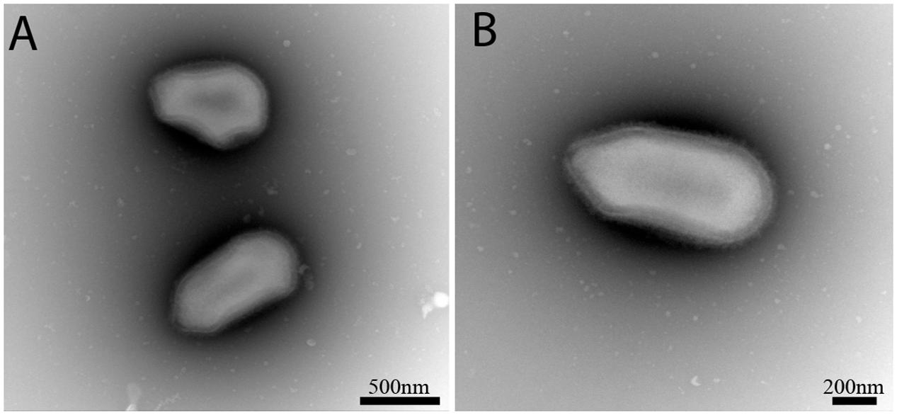
FIGURE 1. Negative staining obtained from the co-culture supernatant of well LCC2. Scale bars are indicated on each panel. (A) Two specimens of Orpheovirus, the top one has a curious irregular form with highly concave forms. (B) Classic ovoid shape of Orpheovirus IHUMI-LCC2.
Replicative Cycle
The length of the Orpheovirus virions range from 900 to 1,100 nm (N = 10) with a maximum diameter of about 500 nm (N = 13). Some virions could reach 1,300 nm in length; this process was sometimes observed in the host cytoplasm. The cork does not seem to seal by a grid as opposed to Pithoviruses and Cedratviruses. We noticed shapes which were similar to the ostiole-like apex observed in Pandoraviruses (Philippe et al., 2013) with a diameter ranging from 70 to 80 nm (N = 8) obstructed by a thick membrane (Figures 2F,I). Nevertheless, in Pandoraviruses the tegument is composed of three layers, each measuring about 20 ∼ 25 nm. The Orpheovirus’ particles presented a dark dense outside layer coated with short, sparse fibrils on their external surface (black arrow). This dark layer is followed by a medium dense space (white arrow) which is in direct contact with the thin inner hyperdense membrane surrounding the viral core cavity containing the nucleic acid (Figure 2I). Altogether, these layers measure ≈40 nm. The replicative cycle of Orpheovirus showed classical stages of infection and replication in amoeba. Briefly, the virus entry by phagocytosis is the start of the cycle, where particles escape the phagosomal process. DNA delivery occurs in the amoeba cytoplasm via the ostiole-like apex (Figures 2A,B). An eclipse phase takes place at 4 h post entry. Functional viral factories (Figure 2C) are well installed and detected around 14–16 h post-infection. Similar forms corresponding to early virion synthesis are also observed, as it is the case for Pithoviruses and Cedratviruses (Figure 2D). At 20 h post-infection, the host cells’ cytoplasm is fully occupied by newly synthesized virions (Figures 2E,G,H). We were also able to detect viruses outside the amoeba due to cell burst or viruses exiting by exocytosis. Complete cell burst occurred between 24 and 38 h post-infection. This slow viral cycle is often observed in the case of V. vermiformis used as cell support, which is not the case when using Acanthamoeba spp. (Reteno et al., 2015; Andreani et al., 2017).
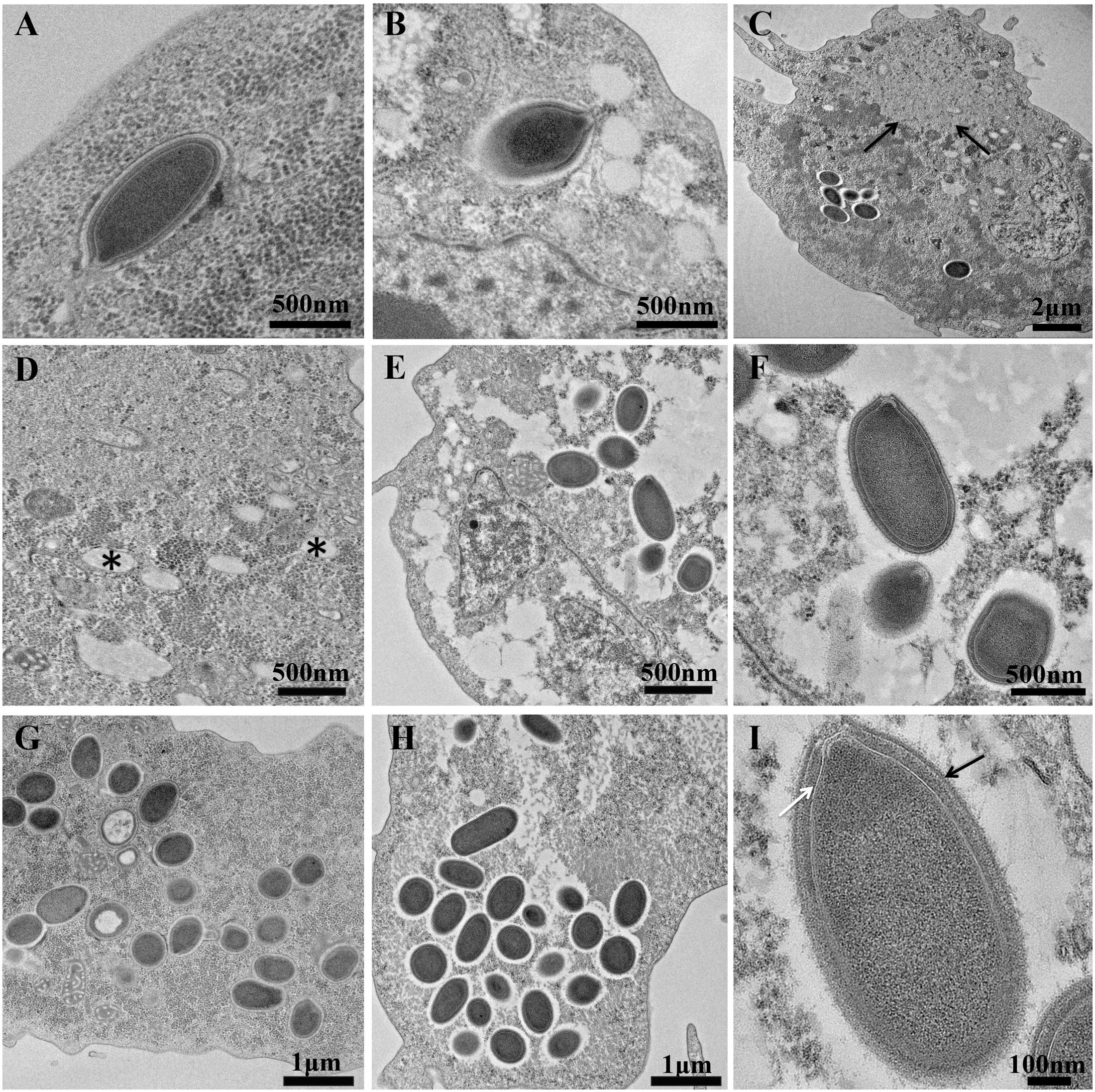
FIGURE 2. Ultrathin sections of Orpheovirus’s replicative cycle. Scale bars are indicated on each panel. (A,B) Represent viral entry at 2 h and 4 h post-infection. (C) Represents a section of Vermamoeba vermiformis 16 h post-infection, Black arrows delimitate the viral factory. (D) High magnification of (C) picture, (∗) represents some curious vacuoles in contact with the viral factory in the cytoplasm. (E–G) Show some cytoplasms and new virus synthetized 20 h post-infection. (H) Accumulation of assembled virions at 20 h post-infection. (I) Single virion into the cytoplasm of V. vermiformis at 24 post-infection, Black arrow points to the external membrane and white arrow indicates the medium dense space.
Orpheovirus: Main Genomic Characteristics
Orpheovirus has a circular genome estimated at 1,473,573 base pair (including 100 N due to an incomplete elucidate region in its genome) with a GC%-content established at around 25% (Table 1). A megablast or a simple blastn against the nr/nt nucleotide collection database revealed no match with other known giant viruses. Dot plots show various areas of repeats (Supplementary Figures S1–S3). We found 57 palindromic sequences, 1,527 tandem repeats and 832 inverted sequence candidates. The number of repeats explains the complexity observed during the genome assembly steps. A comparison with other giant viruses (Supplementary Table S1) showed an extremely high number of tandem repeats and inverted repeats for Orpheovirus.
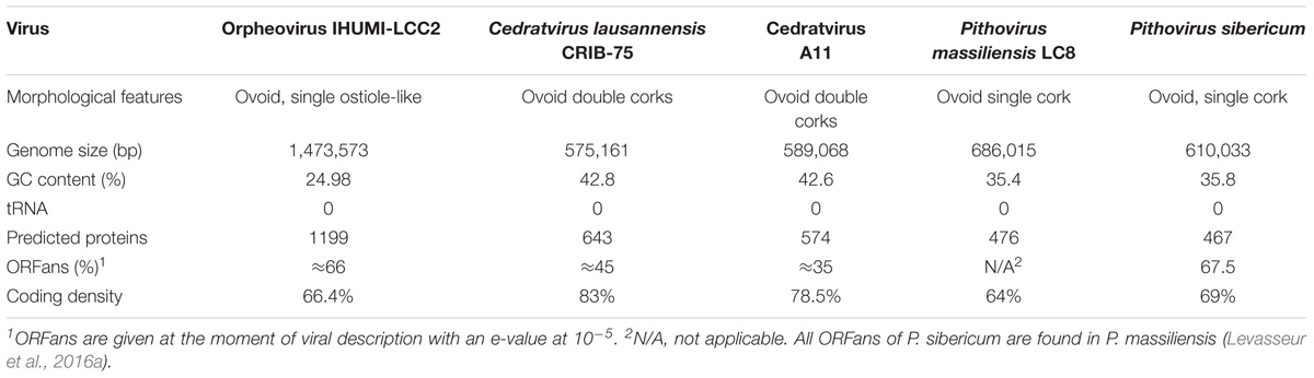
TABLE 1. Main genomic characteristics of Orpheovirus and other closely related viruses.
1,512 genes were predicted but, following our method, 313 genes with an abnormal conformation already cited in the material and methods section were discarded. We only retained 1,199 genes, resulting in a coding density of around 66.4% (979,005 base pairs). This value is close to that of Pithoviruses but lower than that of Cedratvirus A11. A Blast against the nr database retrieved 509 matched proteins with at least one known protein (≈42.5% of all predicted proteins), and 690 unmatched, which are classified like ORFans genes (≈57.5% of all predicted genes). Of the 509 proteins, two had a hit with unclassified sequences, 148 had a best hit with a virus (≈12.3% of all conserved proteins), 176 with eukaryotes (≈14.7%), and 183 with prokaryotes (≈15.3%) (Figure 3). Regarding the 148 best hits with viruses, we observed 27 best hits with P. sibericum, 11 with P. massiliensis, 15 with Cedratvirus A11, 24 with Mimivirus A, B, and C lineages, and 18 with Klosneuvirinae. Hence, the highest best hit viral was obtained with the putative family Pithoviridae with 53 best hits, although the value was also important with Mimiviridae and associated extend family.
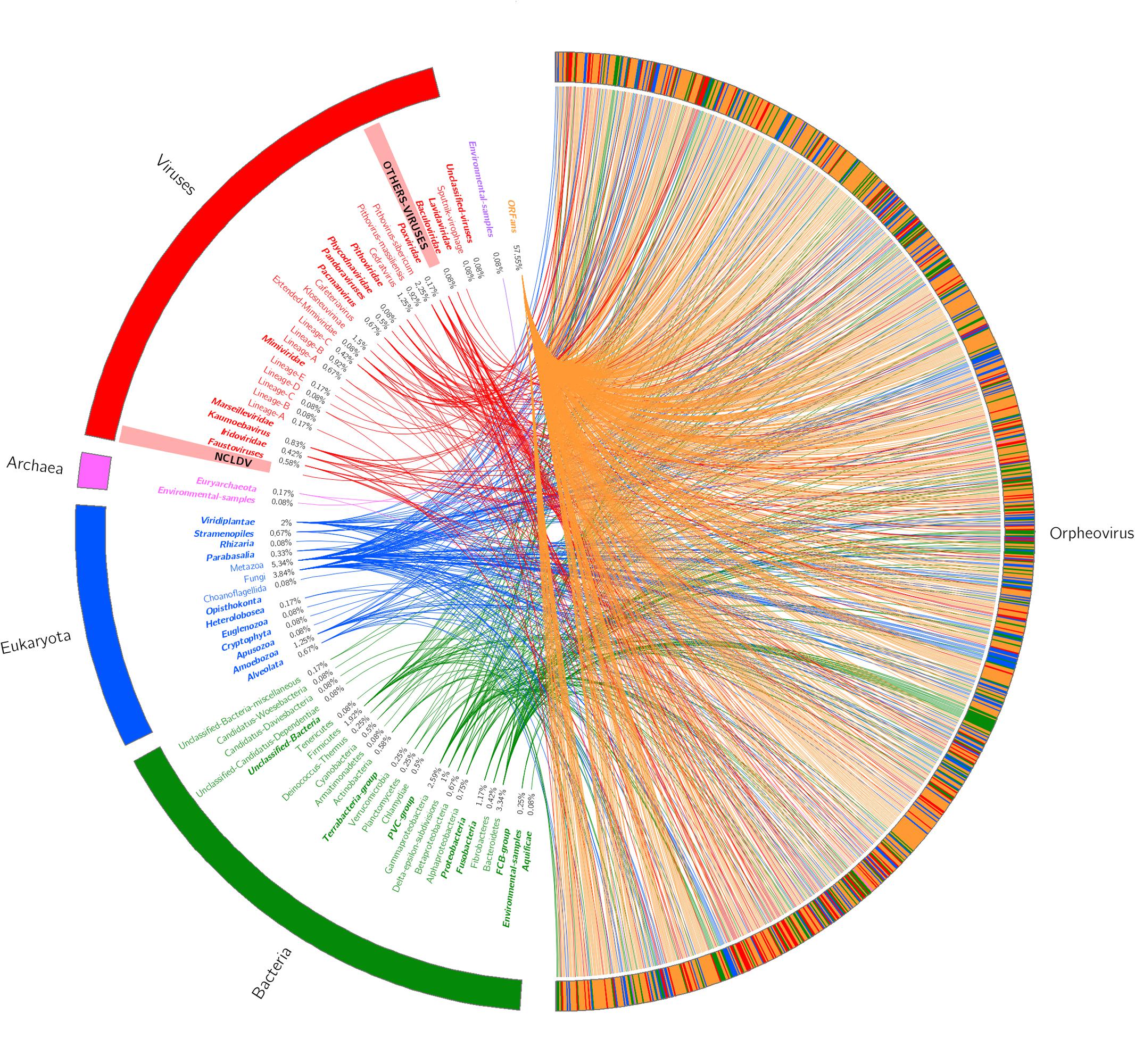
FIGURE 3. Best hit schema obtained for Orpheovirus IHUMI-LCC2 against the non-redundant database.
Despite this, the 57.5% of Orpheovirus’ genes are ORFans, with an e-value cut-off of 10-2. This could increase at ≈66% when we chose a more stringent cut-off value for the blastp at 10-5. We found 343 genes that formed 167 clusters of paralogous genes in Proteinortho, regrouping few numbers of genes ranging from two to a maximum of four by cluster. Annotation of these clusters revealed predominant predicted proteins mainly as MORN-repeat (55 sequences), Ankyrin-repeat (37 sequences), and F-box domain-containing (81 sequences).
The Orpheovirus annotation presented translation system components as follows: eight aminoacyl tRNA synthetases (aaRS), four translation factors: three initiation factors and one release factor (Supplementary Table S2). Surprisingly, Orpheovirus didn’t present any tRNA. We used the aminoacyl tRNA synthetase, which appeared to be a good way to distinguish and classify some lineages and to describe hypothetical common ancestor (Abrahão et al., 2017; Schulz et al., 2017). The Glycyl-tRNA synthetase of Orpheovirus was found to branch with the Asgard Glycyl-tRNA synthetase, and not with Catovirus CTV1 nor Klosneuvirus KNV1 homologs. This Asgard superphylum described by metagenomic studies seems to be a controversial bridge between prokaryotes and eukaryotes (Spang et al., 2015; Da Cunha et al., 2017; Zaremba-Niedzwiedzka et al., 2017). Regarding the phylogenetic analysis of each tRNA synthetase (Supplementary Figures S4–S11), we observed different patterns for amino acyl tRNA synthetase. While some are monophyletic with other described giant viruses, others appear to be polyphyletic resulting from potential lateral gene transfer.
Orpheovirus and Its Divergent Viral Neighborhood
First of all, we searched for five ancestral genes of NCLDV (Colson et al., 2013) encoding the major capsid protein, the helicase-primase (D5), the DNA polymerase elongation subunit family B, the DNA-packaging ATPase (A32), and the viral late transcription factor 3 VLTF3. Four of these genes were found with the exception of the A32-like packaging ATPase, which was absent in all four viruses (Legendre et al., 2014; Andreani et al., 2016). As reported for Cedratvirus A11, P. massiliensis, and P. sibericum, Orpheovirus presented two distinct RNA polymerase II subunit 1. Multiple ribonucleases such as Ribonuclease R, two Ribonuclease III and one ribonuclease HI were detected in Orpheovirus. Orpheovirus presented glycosyltransferase and numerous proteins involved in lipid pathways. We also identified two proteins presenting multiple fusion bacteria domains involved in Riboflavin (Vitamin B2) biosynthesis. Indeed, we observed in ORPV_596 Tri-functional domains of “Di-Hydro-Folate-Reductase/deoxycytidylate deaminase/Riboflavin biosynthesis protein RibD” presenting a homology with Indivirus ILV1. And the second protein is ORPV_666, annotated like Tri-functional domains “3,4 dihydroxy-2-butanone 4-Phosphate synthase/GTP cyclohydrolase II/Lumazine synthase (RibA+RibB+RibH)” presenting homologies with Indivirus ILV1, Bacillus subtilis, and Acanthamoeba castellanii strain Neff. Vitreschak (2002) demonstrated that Riboflavin operon gene fusion is frequently found in bacteria (Vitreschak, 2002).
After that, phylogenetic analysis based on the DNA polymerase B protein, VLTF3 and RNA polymerase II subunit 1 showed deep branching with Cedratvirus and Pithoviruses (Figure 4 and Supplementary Figures S12, S13). Moreover, 58 reciprocal best hit proteins were only shared between Orpheovirus IHUMI-LCC2, Cedratvirus A11, P. sibericum P1084-T and P. massiliensis LC8. In addition, 14 reciprocal best hit proteins were found to be shared between Orpheovirus and Pithoviruses (14+58) and 15 between Cedratvirus and Orpheovirus (15+58) (Figure 5), while Cedratvirus shared 151 proteins (58+93) with Pithoviruses. Meanwhile, 946 of 1,034 protein clusters (≈91.4%) are unique to Orpheovirus, 319 of 497 clusters (≈64.2%) to Cedratvirus A11, and 114 clusters of 543 (≈21%) to Pithoviruses. There were only two colinearity blocks and nine lines connecting Orpheovirus to other viruses (Supplementary Figure S14).
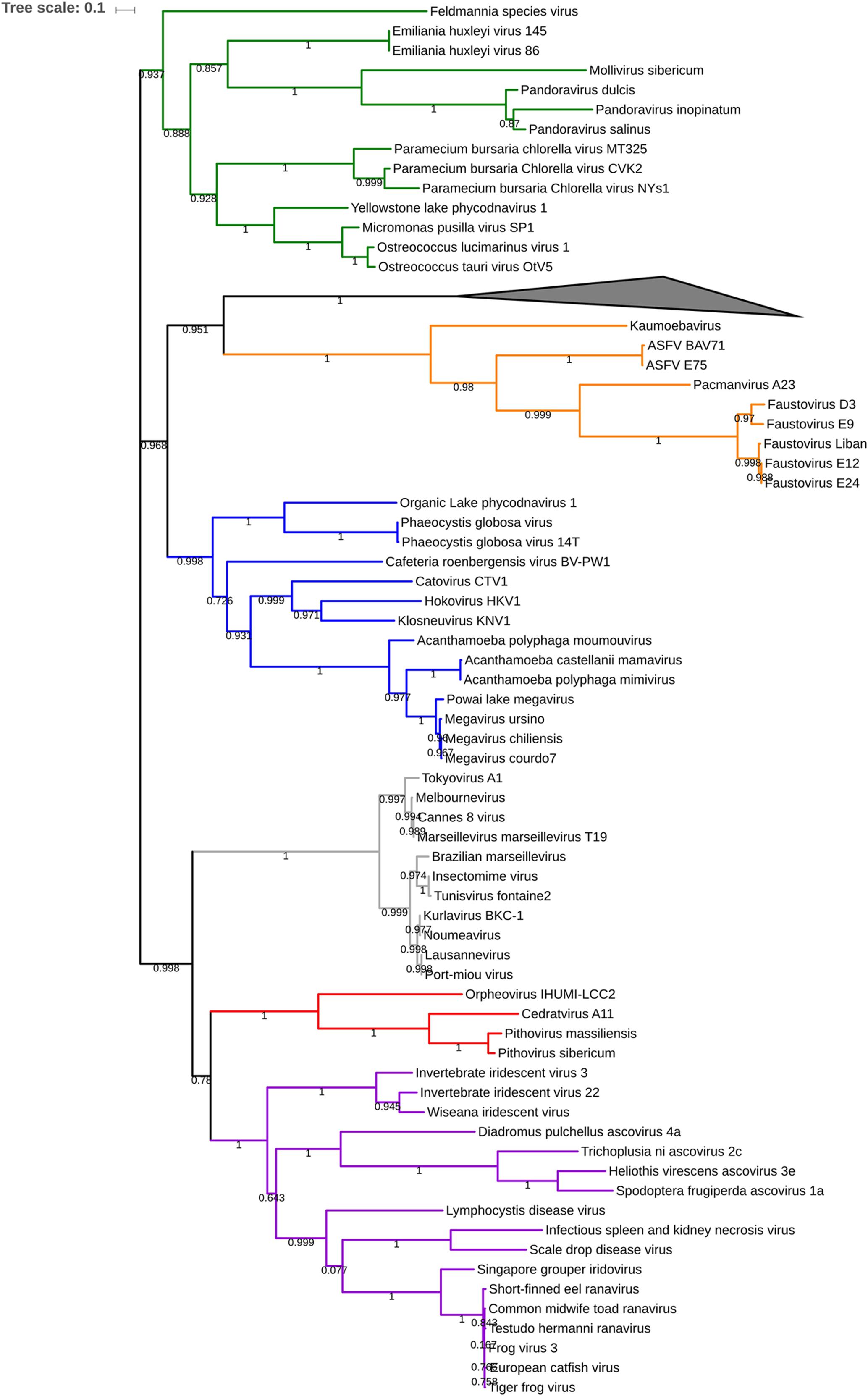
FIGURE 4. Phylogenetic tree based on 84 DNA polymerase b protein of nucleo-cytoplasmic large DNA viruses (NCLDV). Branch values lower than a bootstrap value of 0.5 were deleted. Colors were assigned for different group of viruses: blue for Mimivirus and extended Mimiviridae; green for Pandoraviruses, Mollivirus sibericum and Phycodnaviridae; orange for groups of Asfarviridae, Faustoviruses, Pacmanvirus and Kaumoabevirus; gray for Marseilleviridae; red for Orpheovirus, Cedratvirus, and Pithoviruses and purple for Asco-Iridoviridae. The collapsed branch represented by a black triangle was used for 15 Poxviridae members. The corresponding alignment is available on Supplementary Data Sheet 2 visualized by automatic MView software (https://www.ebi.ac.uk/Tools/msa/mview/). 3,450 positions were used to build the tree.
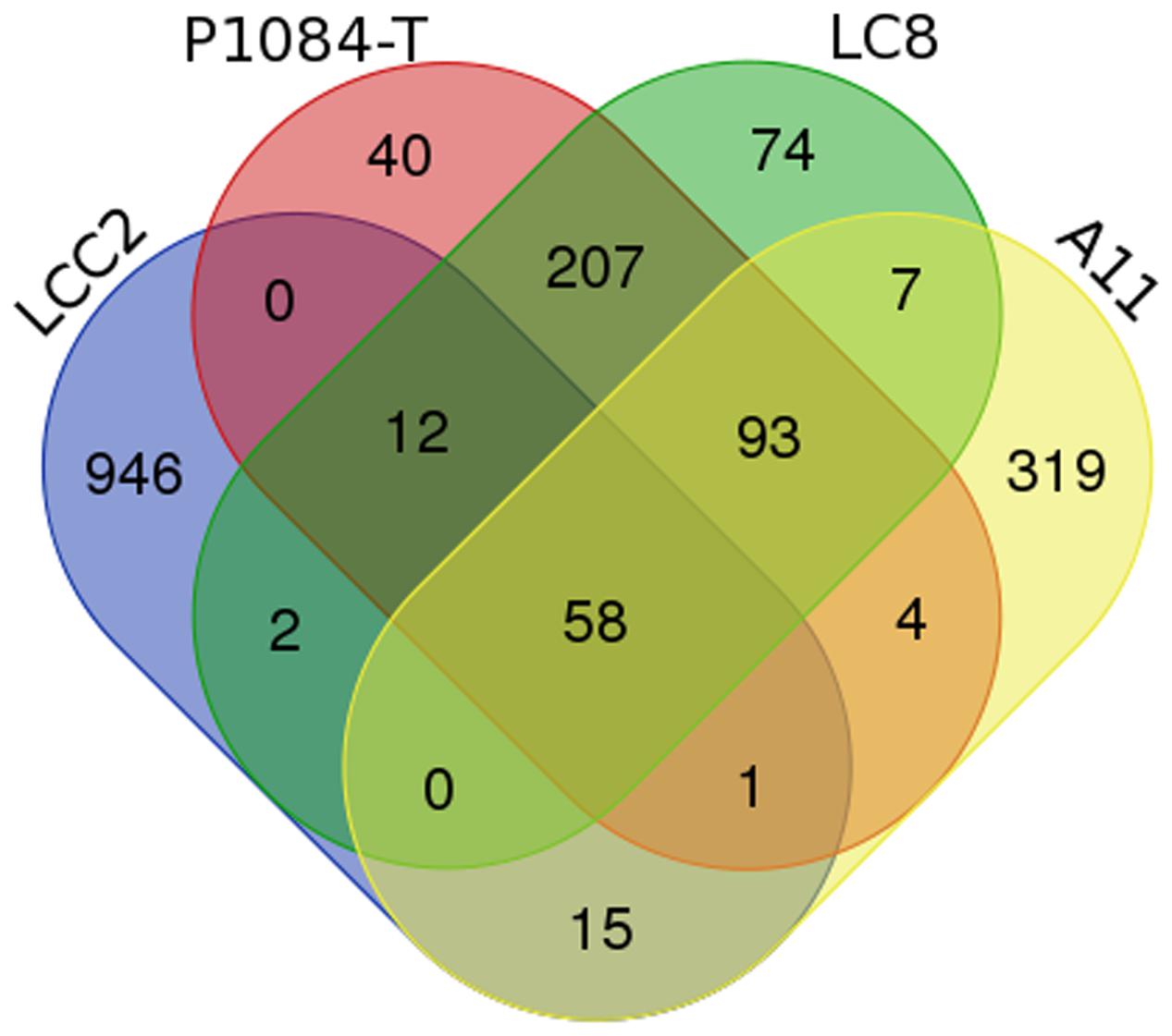
FIGURE 5. Venn diagram comparison of best reciprocal hit obtained belonging to the predicted proteins of Orpheovirus, Cedratvirus, Pithoviruses. LCC2 represents Orpheovirus IHUMI-LCC2, P1084-T represents Pithovirus sibericum, LC8 represents Pithovirus massiliensis and A11 represents Cedratvirus A11. The diagram was created online http://bioinformatics.psb.ugent.be/webtools/Venn/.
Following the discovery of this divergence between the four viruses, we decided to investigate Orpheovirus position in the “Megavirales” order further with the help of a parsimonious pan-genomic tree (Supplementary Figure S15). The long branch length observed for Klosneuvirus, Pandoravirus inopinatum and Orpheovirus is explained by the large genome size and by the number of predicted proteins compared to the close relative strains in the tree. These long branches could be a new common marker to explain the emergence of new viral family or lineages in the proposed “Megavirales” order. In the case of Orpheovirus, the pan-genomic analysis confirms this distant relation with the proposed Pithoviridae family.
Orpheovirus and Virophage: A Curious Homologous Sequence
Orpheovirus has a predicted gene a 434 amino acids protein that we called V21-like protein. This protein had homologs in Blastp, respectively, at 86% coverage, 21% identity with Sputnik virophage V21 protein (La Scola et al., 2008), and 85% coverage, 27% identity with Zamilon (Gaia et al., 2014). These two homologous proteins are annotated as hypothetical proteins, and showed no other homology using the blast strategy. However, HHpred online (Supplementary Data Sheet 3) and Phyre2 (Supplementary Figure S16) detected homology between the V21-like Orpheovirus sequence, Sputnik virophage V21 protein, the Zamilon protein, and a putative transferase present in the genomes of Mimivirus lineages A, B, and C. This V21-like protein also shared a common ancestor with all Sputnik virophages, and Zamilon virophage (Figure 6). No transposase or other mobile elements could be detected, no other special interest homology with other proteins was detected although a Ribonuclease III such as that in MIMIVIRE (Levasseur et al., 2016b) was present near this V21-like sequence in the genome of Orpheovirus.
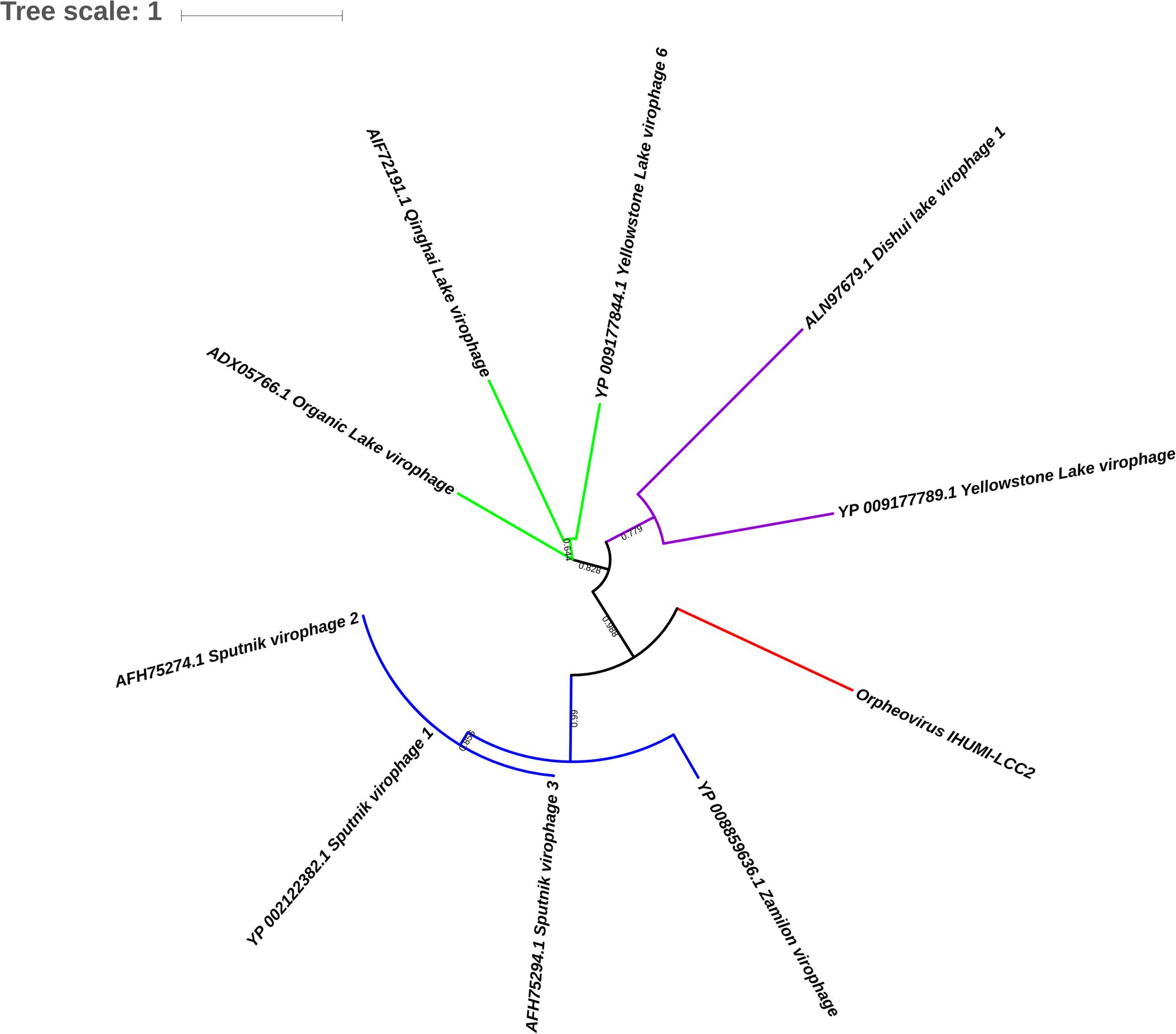
FIGURE 6. Phylogenetic tree based on V21-like protein of virophages and Orpheovirus IHUMI-LCC2 sequences. 484 positions were used to build the tree. Branch values inferior to a bootstrap value of 0.5 were deleted.
Discussion
Since the isolation of Faustovirus in 2015, all positive samples have been sewages samples or samples collected near to sewage areas (Reteno et al., 2015; Benamar et al., 2016; Bou Khalil et al., 2016; Cherif Louazani et al., 2017). We suspected that rats could also be a potential reservoir of Faustoviruses. In order to decipher the Faustovirus’ reservoirs, and in attempt to study the viral frequency and persistence in the environment, notably during seasonality (Martínez et al., 2007; Johannessen et al., 2017), we decided to explore the same area of sampling 4 months later. We succeeded in re-isolating, in the same area, more Faustoviruses in sewage samples (data not shown) but not in rat stools samples, and a new giant virus was revealed, that we called Orpheovirus IHUMI-LCC2. This virus represents a new virus, the first to come with an ovoid form at a size higher than 1 μm isolated from V. vermiformis as a new host cell, and a genome of 1,473,573 bp largely exceeding the genomes of Cedratvirus A11, P. massiliensis LC8, and P. sibericum. Orpheovirus conserved a replicative cycle which is typical but delayed in terms of cell burst or complete lysis, which could be due to its host V. vermiformis showing different features regarding the routinely used host Acanthamoeba spp. (Andreani et al., 2017).
Although Orpheovirus appears to share some replicative elements and genomic bases with Cedratvirus A11, P. massiliensis LC8, and P. sibericum, some other elements highlighted a complete divergent evolution. With its genomic size, high number of paralogs, its eight aminoacyl tRNA synthetase aaRS, its low GC content, and its high number of ORFans (≈66% at a 10-5 e-value), we propose Orpheovirus as a potential member of a new putative family; the Orpheoviridae closely related to the recently proposed Pithoviridae. To do so, more viral descriptions including new isolates are needed to understand genomic links in these novel expanding and complex families. Nevertheless, some new viral descriptions such as that of Klosneuvirus (Schulz et al., 2017), have reported complete translational components, and this could create a broader understanding of the viral lifestyle and tRNA synthetase usages. In contrast, aminoacyl tRNA synthetases (aaRS) are frequently (but not entirely) found and described in isolated viruses (Abrahão et al., 2017), and we are still unaware of the viral benefits of possessing aminoacyl tRNA synthetase or tRNA during the infectious cycle. Simultaneously, and following the discovery of the MIMIVIRE system, it has become more widespread to search for virophages sequences in giant viruses genomes. We found a high conserved size and similar V21-like sequence in Orpheovirus that made us investigate the probability of an integrated virophage sequence in the Orpheovirus genome, as is the case for Mavirus (Fischer and Hackl, 2016) in its protist host Cafeteria roenbergensis or the Sputnik 2 virophage in the Lentille virus (Desnues et al., 2012). However, no mobile elements could be detected and no relationship could be found even when this sequence was closely located to the Ribonuclease III as is the case in MIMIVIRE. In contrast, the fact that the V21-like sequence of Orpheovirus, together with the V21 of Sputnik and Zamilon, showed homology with a putative transferase present in the genomes of Mimivirus lineages A, B, and C, led us to postulate that these sequences could either have a similar function to transferase or a protein that simply interacts with the putative transferase.
Despite all these findings, the description of Orpheovirus, along with the previous findings in Pandoraviruses, Pithoviruses, and Cedratviruses, has revealed a large range of viruses with various extraordinary ovoid shapes, which have expanded the research characteristics for viral isolation. Some more sewers should be investigated at different time stages or seasonal dates. In addition, animal stool samples should be more commonly considered as potential new reservoirs for giant viruses. Finally, a large part of this vast world of giant viruses is still unknown, particularly its evolution and ancestors. For this reason, more strains should be isolated and described, and more data is needed. It is likely that further descriptions will increase knowledge and diversity across the NCLDV.
Author Contributions
JA and BL designed the study and experiments. JA, JK, EB, IH, CM, and AL performed the sample collection, virus isolation, experiments and/or analyses. JA, DR, and BL wrote the manuscript. All authors approved the final manuscript.
Funding
This work received a help of the ANR (National Agency for Research) through “future investments” program n°10-IAHU-03.
Conflict of Interest Statement
The authors declare that the research was conducted in the absence of any commercial or financial relationships that could be construed as a potential conflict of interest.
Acknowledgments
The authors would particularly like to thank Aurélia Magnien for her help with the sample collection and Claire Andréani for her help in the improvement of the English correction.
Supplementary Material
The Supplementary Material for this article can be found online at: https://www.frontiersin.org/articles/10.3389/fmicb.2017.02643/full#supplementary-material
Footnotes
- ^ http://www.clcbio.com/blog/clc-genomics-workbench-7-5/
- ^ http://emboss.bioinformatics.nl/cgi-bin/emboss/palindrome
- ^ https://www.ebi.ac.uk/interpro/
- ^ http://trna.ucsc.edu/tRNAscan-SE/
References
Abergel, C., Legendre, M., and Claverie, J.-M. (2015). The rapidly expanding universe of giant viruses: Mimivirus, Pandoravirus, Pithovirus and Mollivirus. FEMS Microbiol. Rev. 39, 779–796. doi: 10.1093/femsre/fuv037
Abrahão, J. S., Araújo, R., Colson, P., and La Scola, B. (2017). The analysis of translation-related gene set boosts debates around origin and evolution of mimiviruses. PLOS Genet. 13:e1006532. doi: 10.1371/journal.pgen.1006532
Aherfi, S., Colson, P., La Scola, B., and Raoult, D. (2016). Giant viruses of amoebas: an update. Front. Microbiol. 7:349. doi: 10.3389/fmicb.2016.00349
Andreani, J., Aherfi, S., Bou Khalil, J. Y., Di Pinto, F., Bitam, I., Raoult, D., et al. (2016). Cedratvirus, a double-cork structured giant virus, is a distant relative of pithoviruses. Viruses 8:300. doi: 10.3390/v8110300
Andreani, J., Bou Khalil, J. Y., Sevvana, M., Benamar, S., Di Pinto, F., Bitam, I., et al. (2017). Pacmanvirus, a new giant icosahedral virus at the crossroads between asfarviridae and faustoviruses. J. Virol. 91:e00212-17. doi: 10.1128/JVI.00212-17
Antwerpen, M. H., Georgi, E., Zoeller, L., Woelfel, R., Stoecker, K., and Scheid, P. (2015). Whole-genome sequencing of a pandoravirus isolated from keratitis-inducing acanthamoeba. Genome Announc. 3:e00136-15. doi: 10.1128/genomeA.00136-15
Arslan, D., Legendre, M., Seltzer, V., Abergel, C., and Claverie, J.-M. (2011). Distant mimivirus relative with a larger genome highlights the fundamental features of megaviridae. Proc. Natl. Acad. Sci. U.S.A. 108, 17486–17491. doi: 10.1073/pnas.1110889108
Benamar, S., Reteno, D. G. I., Bandaly, V., Labas, N., Raoult, D., and La Scola, B. (2016). Faustoviruses: comparative genomics of new megavirales family members. Front. Microbiol. 7:3. doi: 10.3389/fmicb.2016.00003
Bertelli, C., Mueller, L., Thomas, V., Pillonel, T., Jacquier, N., and Greub, G. (2017). Cedratvirus lausannensis – digging into pithoviridaediversity. Environ. Microbiol. 19, 4022–4034. doi: 10.1111/1462-2920.13813
Besemer, J., Lomsadze, A., and Borodovsky, M. (2001). GeneMarkS: a self-training method for prediction of gene starts in microbial genomes. Implications for finding sequence motifs in regulatory regions. Nucleic Acids Res. 29, 2607–2618. doi: 10.1093/nar/29.12.2607
Boratyn, G. M., Schäffer, A. A., Agarwala, R., and Altschul, S. F. (2012). Domain enhanced lookup time accelerated BLAST. Biol. Direct 7:12. doi: 10.1186/1745-6150-7-12
Bou Khalil, J. Y., Andreani, J., Raoult, D., and La Scola, B. (2016). A Rapid strategy for the isolation of new faustoviruses from environmental samples using Vermamoeba vermiformis. J. Vis. Exp. 112:e54104. doi: 10.3791/54104
Boyer, M., Yutin, N., Pagnier, I., Barrassi, L., Fournous, G., Espinosa, L., et al. (2009). Giant Marseillevirus highlights the role of amoebae as a melting pot in emergence of chimeric microorganisms. Proc. Natl. Acad. Sci. U.S.A. 106, 21848–21853. doi: 10.1073/pnas.0911354106
Cherif Louazani, A., Andreani, J., Ouarhache, M., Aherfi, S., Baptiste, E., Levasseur, A., et al. (2017). Genome sequences of new Faustovirus strains ST1 and LC9, isolated from the South of France. Genome Announc. 5:e00613-17. doi: 10.1128/genomeA.00613-17
Colson, P., De Lamballerie, X., Yutin, N., Asgari, S., Bigot, Y., Bideshi, D. K., et al. (2013). “Megavirales,” a proposed new order for eukaryotic nucleocytoplasmic large DNA viruses. Arch. Virol. 158, 2517–2521. doi: 10.1007/s00705-013-1768-6
Colson, P., La Scola, B., Levasseur, A., Caetano-Anollés, G., and Raoult, D. (2017). Mimivirus: leading the way in the discovery of giant viruses of amoebae. Nat. Rev. Microbiol. 15, 243–254. doi: 10.1038/nrmicro.2016.197
Contreras-Moreira, B., and Vinuesa, P. (2013). GET_HOMOLOGUES, a versatile software package for scalable and robust microbial pangenome analysis. Appl. Environ. Microbiol. 79, 7696–7701. doi: 10.1128/AEM.02411-13
Da Cunha, V., Gaia, M., Gadelle, D., Nasir, A., and Forterre, P. (2017). Lokiarchaea are close relatives of Euryarchaeota, not bridging the gap between prokaryotes and eukaryotes. PLOS Genet. 13:e1006810. doi: 10.1371/journal.pgen.1006810
Darling, A. C. E., Mau, B., Blattner, F. R., and Perna, N. T. (2004). Mauve: multiple alignment of conserved genomic sequence with rearrangements. Genome Res. 14, 1394–1403. doi: 10.1101/gr.2289704
Desnues, C., La Scola, B., Yutin, N., Fournous, G., Robert, C., Azza, S., et al. (2012). Provirophages and transpovirons as the diverse mobilome of giant viruses. Proc. Natl. Acad. Sci. U.S.A. 109, 18078–18083. doi: 10.1073/pnas.1208835109
Dornas, F. P., Assis, F. L., Aherfi, S., Arantes, T., Abrahão, J. S., Colson, P., et al. (2016). A Brazilian Marseillevirus is the founding member of a lineage in family Marseilleviridae. Viruses 8:76. doi: 10.3390/v8030076
Edgar, R. C. (2004). MUSCLE: a multiple sequence alignment method with reduced time and space complexity. BMC Bioinformatics 5:113. doi: 10.1186/1471-2105-5-113
Filée, J. (2014). Multiple occurrences of giant virus core genes acquired by eukaryotic genomes: the visible part of the iceberg? Virology 466–467, 53–59. doi: 10.1016/j.virol.2014.06.004
Filée, J. (2015). Genomic comparison of closely related giant viruses supports an accordion-like model of evolution. Front. Microbiol. 6:593. doi: 10.3389/fmicb.2015.00593
Fischer, M. G., and Hackl, T. (2016). Host genome integration and giant virus-induced reactivation of the virophage mavirus. Nature 540, 288–291. doi: 10.1038/nature20593
Gaia, M., Benamar, S., Boughalmi, M., Pagnier, I., Croce, O., Colson, P., et al. (2014). Zamilon, a novel virophage with Mimiviridae host specificity. PLOS ONE 9:e94923. doi: 10.1371/journal.pone.0094923
Iyer, L. M., Aravind, L., and Koonin, E. V. (2001). Common origin of four diverse families of large eukaryotic DNA viruses. J. Virol. 75, 11720–11734. doi: 10.1128/JVI.75.23.11720-11734.2001
Johannessen, T. V., Larsen, A., Bratbak, G., Pagarete, A., Edvardsen, B., Egge, E. D., et al. (2017). Seasonal dynamics of haptophytes and dsDNA algal viruses suggest complex virus-host relationship. Viruses 9:84. doi: 10.3390/v9040084
Kelley, L. A., Mezulis, S., Yates, C. M., Wass, M. N., and Sternberg, M. J. E. (2015). The phyre2 web portal for protein modeling, prediction and analysis. Nat. Protoc. 10, 845–858. doi: 10.1038/nprot.2015.053
Khalil, J. Y. B., Andreani, J., and La Scola, B. (2016a). Updating strategies for isolating and discovering giant viruses. Curr. Opin. Microbiol. 31, 80–87. doi: 10.1016/j.mib.2016.03.004
Khalil, J. Y. B., Langlois, T., Andreani, J., Sorraing, J.-M., Raoult, D., Camoin, L., et al. (2017). Flow cytometry sorting to separate viable giant viruses from amoeba co-culture supernatants. Front. Cell. Infect. Microbiol. 6:202. doi: 10.3389/fcimb.2016.00202
Khalil, J. Y. B., Robert, S., Reteno, D. G., Andreani, J., Raoult, D., and La Scola, B. (2016b). High-throughput isolation of giant viruses in liquid medium using automated flow cytometry and fluorescence staining. Front. Microbiol. 7:26. doi: 10.3389/fmicb.2016.00026
La Scola, B., Audic, S., Robert, C., Jungang, L., Lamballerie, X. D., Drancourt, M., et al. (2003). A giant virus in amoebae. Science 299:2033. doi: 10.1126/science.1081867
La Scola, B., Desnues, C., Pagnier, I., Robert, C., Barrassi, L., Fournous, G., et al. (2008). The virophage as a unique parasite of the giant mimivirus. Nature 455, 100–104. doi: 10.1038/nature07218
Lechner, M., Findeiß, S., Steiner, L., Marz, M., Stadler, P. F., and Prohaska, S. J. (2011). Proteinortho: detection of (Co-)orthologs in large-scale analysis. BMC Bioinformatics 12:124. doi: 10.1186/1471-2105-12-124
Legendre, M., Bartoli, J., Shmakova, L., Jeudy, S., Labadie, K., Adrait, A., et al. (2014). Thirty-thousand-year-old distant relative of giant icosahedral DNA viruses with a pandoravirus morphology. Proc. Natl. Acad. Sci. U.S.A. 111, 4274–4279. doi: 10.1073/pnas.1320670111
Letunic, I., and Bork, P. (2016). Interactive tree of life (iTOL) v3: an online tool for the display and annotation of phylogenetic and other trees. Nucleic Acids Res. 44, W242–W245. doi: 10.1093/nar/gkw290
Levasseur, A., Andreani, J., Delerce, J., Bou Khalil, J., Robert, C., La Scola, B., et al. (2016a). Comparison of a modern and fossil Pithovirus reveals its genetic conservation and evolution. Genome Biol. Evol. 8, 2333–2339. doi: 10.1093/gbe/evw153
Levasseur, A., Bekliz, M., Chabrière, E., Pontarotti, P., La Scola, B., and Raoult, D. (2016b). MIMIVIRE is a defence system in mimivirus that confers resistance to virophage. Nature 531, 249–252. doi: 10.1038/nature17146
Lowe, T. M., and Chan, P. P. (2016). tRNAscan-SE on-line: integrating search and context for analysis of transfer RNA genes. Nucleic Acids Res. 44, W54–W57. doi: 10.1093/nar/gkw413
Luo, R., Liu, B., Xie, Y., Li, Z., Huang, W., Yuan, J., et al. (2012). SOAPdenovo2: an empirically improved memory-efficient short-read de novo assembler. Gigascience 1:18. doi: 10.1186/2047-217X-1-18
Marchler-Bauer, A., and Bryant, S. H. (2004). CD-Search: protein domain annotations on the fly. Nucleic Acids Res. 32, W327–W331. doi: 10.1093/nar/gkh454
Martínez, J. M., Schroeder, D. C., Larsen, A., Bratbak, G., and Wilson, W. H. (2007). Molecular dynamics of Emiliania huxleyi and cooccurring viruses during two separate mesocosm studies. Appl. Environ. Microbiol. 73, 554–562. doi: 10.1128/AEM.00864-06
Moreira, D., and Brochier-Armanet, C. (2008). Giant viruses, giant chimeras: the multiple evolutionary histories of mimivirus genes. BMC Evol. Biol. 8:12. doi: 10.1186/1471-2148-8-12
Moreira, D., and López-García, P. (2015). Evolution of viruses and cells: do we need a fourth domain of life to explain the origin of eukaryotes? Philos. Trans. R. Soc. Lond. B Biol. Sci. 370:20140327. doi: 10.1098/rstb.2014.0327
Nadalin, F., Vezzi, F., and Policriti, A. (2012). GapFiller: a de novo assembly approach to fill the gap within paired reads. BMC Bioinformatics 13(Suppl. 14):S8. doi: 10.1186/1471-2105-13-S14-S8
Pagnier, I., Reteno, D.-G. I., Saadi, H., Boughalmi, M., Gaia, M., Slimani, M., et al. (2013). A decade of improvements in Mimiviridae and Marseilleviridae isolation from amoeba. Intervirology 56, 354–363. doi: 10.1159/000354556
Philippe, N., Legendre, M., Doutre, G., Couté, Y., Poirot, O., Lescot, M., et al. (2013). Pandoraviruses: amoeba viruses with genomes up to 2.5 Mb reaching that of parasitic eukaryotes. Science 341, 281–286. doi: 10.1126/science.1239181
Price, M. N., Dehal, P. S., and Arkin, A. P. (2009). FastTree: computing large minimum evolution trees with profiles instead of a distance matrix. Mol. Biol. Evol. 26, 1641–1650. doi: 10.1093/molbev/msp077
Reteno, D. G., Benamar, S., Khalil, J. B., Andreani, J., Armstrong, N., Klose, T., et al. (2015). Faustovirus, an asfarvirus-related new lineage of giant viruses infecting amoebae. J. Virol. 89, 6585–6594. doi: 10.1128/JVI.00115-15
Schulz, F., Yutin, N., Ivanova, N. N., Ortega, D. R., Lee, T. K., Vierheilig, J., et al. (2017). Giant viruses with an expanded complement of translation system components. Science 356, 82–85. doi: 10.1126/science.aal4657
Spang, A., Saw, J. H., Jørgensen, S. L., Zaremba-Niedzwiedzka, K., Martijn, J., Lind, A. E., et al. (2015). Complex archaea that bridge the gap between prokaryotes and eukaryotes. Nature 521, 173–179. doi: 10.1038/nature14447
Tritt, A., Eisen, J. A., Facciotti, M. T., and Darling, A. E. (2012). An integrated pipeline for de novo assembly of microbial genomes. PLOS ONE 7:e42304. doi: 10.1371/journal.pone.0042304
Vitreschak, A. G. (2002). Regulation of riboflavin biosynthesis and transport genes in bacteria by transcriptional and translational attenuation. Nucleic Acids Res. 30, 3141–3151. doi: 10.1093/nar/gkf433
Yutin, N., Wolf, Y. I., and Koonin, E. V. (2014). Origin of giant viruses from smaller DNA viruses not from a fourth domain of cellular life. Virology 466–467, 38–52. doi: 10.1016/j.virol.2014.06.032
Yutin, N., Wolf, Y. I., Raoult, D., and Koonin, E. V. (2009). Eukaryotic large nucleo-cytoplasmic DNA viruses: clusters of orthologous genes and reconstruction of viral genome evolution. Virol. J. 6:223. doi: 10.1186/1743-422X-6-223
Keywords: Orpheovirus, Cedratvirus, Pithovirus, Vermamoeba vermiformis, giant viruses, NCLDV, Orpheoviridae, Pithoviridae
Citation: Andreani J, Khalil JYB, Baptiste E, Hasni I, Michelle C, Raoult D, Levasseur A and La Scola B (2018) Orpheovirus IHUMI-LCC2: A New Virus among the Giant Viruses. Front. Microbiol. 8:2643. doi: 10.3389/fmicb.2017.02643
Received: 02 October 2017; Accepted: 19 December 2017;
Published: 22 January 2018.
Edited by:
William Michael McShan, University of Oklahoma Health Sciences Center, United StatesReviewed by:
Hiroyuki Ogata, Kyoto University, JapanGwenael Piganeau, Observatoire Océanologique de Banyuls sur Mer, France
Copyright © 2018 Andreani, Khalil, Baptiste, Hasni, Michelle, Raoult, Levasseur and La Scola. This is an open-access article distributed under the terms of the Creative Commons Attribution License (CC BY). The use, distribution or reproduction in other forums is permitted, provided the original author(s) or licensor are credited and that the original publication in this journal is cited, in accordance with accepted academic practice. No use, distribution or reproduction is permitted which does not comply with these terms.
*Correspondence: Bernard La Scola, bernard.la-scola@univ-amu.fr