 João Massano1,2,3* Carolina Garrett1,2,3
- 1 Movement Disorders and Functional Surgery Unit, Centro Hospitalar de São João, Porto, Portugal
- 2 Department of Neurology, Centro Hospitalar de São João, Porto, Portugal
- 3 Department of Clinical Neuroscience and Mental Health, Faculty of Medicine, University of Porto, Porto, Portugal
Parkinson’s disease is a common and often debilitating disorder, with a growing prevalence accompanying global population aging. Current drug therapy is not satisfactory enough for many patients, especially after a few years of symptom progression. This is mainly due to the motor complications that frequently emerge as disease progresses. Deep brain stimulation (DBS) is a useful therapeutic option in carefully selected patients that significantly improves motor symptoms, functional status, and quality of life. However, cognitive impairment may limit patient selection for DBS, as patients need to have sufficient mental capabilities in order to understand the procedure, as well as its benefits and limitations, and cooperate with the medical team throughout the process of selection, surgery, and postsurgical follow-up. On the other hand it has been observed that certain aspects of cognitive performance may decline after DBS, namely when the therapeutic target is the widely used subthalamic nucleus. These are important pieces of information for patients, their families, and health care professionals. This manuscript reviews these aspects and their clinical implications.
Introduction
Parkinson’s disease (PD) is a common, potentially disabling neurodegenerative disease (Massano and Bhatia, 2012). It was estimated that, in 2005, more than 4 million people in the world suffered from this disorder, and disease prevalence is estimated to escalate in the future, as the mean age of the population increases at a worldwide level (Dorsey et al., 2007). Following the original clinical depictions by Parkinson (1817) in the nineteenth century, motor features of the disease, such as bradykinesia, resting tremor, rigidity, and gait and postural changes, were regarded for a long time as the major features of PD. This view has changed in the past few decades, as a growing number of clinicians and researchers have studied and reported in detail on the important non-motor characteristics of the disease (Chaudhuri and Schapira, 2009; Gallagher et al., 2010). Among them is cognitive decline, ranging from mild impairment to overt dementia – a contemporary review on this topic can be found included in the present Frontiers Research Topic collection (Meireles and Massano, submitted).
The treatment of PD is not a straightforward task, especially when dealing with newly diagnosed patients and, especially, in advanced disease stages. In the early stages there is controversy regarding the timing of therapy initiation and which specific strategies should be used. Many clinicians tend to initiate drug therapy as soon as symptoms interfere with the patient’s lifestyle, usually prescribing monoamino oxidase B inhibitors or dopaminergic agonists, especially in younger patients, but this approach is under dispute within the scientific community – for instance, opposite views support the notion that levodopa is the drug of choice regardless of patient age or disease stage (Lang, 2009; Lees et al., 2009; Schapira, 2009). Deep brain stimulation (DBS) has become a standard of care for a significant minority of patients in advanced disease stages, with controlled trials and large clinical series supporting its use, as both benefits and safety have been well established (Krack et al., 2003; Deuschl et al., 2006; Kleiner-Fisman et al., 2006; Weaver et al., 2009; Follett et al., 2010; Moro et al., 2010; Smeding et al., 2011). Patients selected for the procedure are typically not satisfied with the severity of their symptoms and disability even under optimized medical therapy, especially due to the quite incapacitating and frustrating motor complications, such as peak dose dyskinesias, prolonged off periods, or sudden off states (Lang et al., 2006; Massano and Bhatia, 2012). In most occasions, one of two different brain regions is currently targeted for DBS in PD: the subthalamic nucleus (STN) or the globus pallidus interna (GPi). The ventral intermediate (VIM) nucleus of the thalamus was the first electrically stimulated region for the treatment of PD (Benabid et al., 1987), but it is now targeted in uncommon and much selected circumstances, as tremor is apparently the only clinical feature improving significantly with the procedure (Walter and Vitek, 2004; Moro and Lang, 2006; Pahwa et al., 2006). The choice of brain target for DBS should probably be carried out according to each patient’s characteristics, as we will further detail in the text.
Cognitive impairment plays an important role in PD patients who are potential candidates for DBS, as this may be a limiting factor during patient selection. Also, evidence has been accumulating regarding changes in cognitive performance after DBS, and both physicians and patients should be well aware of this prior to the procedure. This review will approach these matters, and the implications for clinical management.
Methods
The authors conducted a PubMed search for papers published between 1990 and August 2011. Keywords used were “PD,” “cognition,” “cognitive decline,” and “DBS.” Relevant references were chosen and an additional manual reference search was carried out on the reference list in retrieved manuscripts; articles were included for analysis only if the research enrolled at least 15 subjects undergoing DBS, except where evidence was much scarcer and only smaller series were available. The final reference list was produced on the basis of importance to the topics covered in this review. Data were extracted from relevant sources and the text was devised according to a predefined structure.
Cognitive Status as a Key Factor for Patient Selection for DBS in PD
Accurate PD patient selection is paramount in DBS. This has been acknowledged in the widely used core assessment program for surgical interventional therapies in PD (CAPSIT-PD) protocol, a landmark document in this field (Defer et al., 1999). Patient cognitive status is of utmost importance when considering DBS as a potential therapy for PD, as patients displaying significant cognitive decline are generally not considered good candidates for the procedure (Pillon, 2002; Lang et al., 2006; Moro and Lang, 2006; Okun et al., 2007; Rodriguez et al., 2007; Bronstein et al., 2011). This is due to several reasons, with ethical imperatives always in mind as a background for clinical thinking and action. First, it must be assured that the procedure is expected to help the patients (beneficence) and not harm them (non-maleficence). Moreover, effective and safer therapies should be thoroughly tried before proceeding with DBS, and benefits should be wisely balanced against risks (proportionality and subsidiarity; Clausen, 2010; Schermer, 2011). Such equilibrium may at times be difficult to perceive when considering specific therapeutic interventions, such as DBS, which is often a publicly glorified intervention, and commonly understood as a last resort (Bell et al., 2011). Another important issue is that of autonomy, meaning that the patient must be able to decide freely and in a fully informed manner whether he/she wishes to proceed with the therapy (Clausen, 2010; Schermer, 2011). This principle implies that the patient must have a cognitive status that makes him/her able to understand the information given by the clinicians, and carry out every relevant decisions in this regard. These important ethical imperatives should be actively safeguarded by the team involved in patient care at all times. On the other hand, cognitive status is also important from a practical perspective, as patients must be able to go through all required presurgical selection procedures, to collaborate with the team in the operating theater (patients are awake during surgery in most centers), and to comply with the postsurgical follow-up program, including serial DBS parameter adjustments. This can be very demanding, due to the high number of visits often required and the amount of time consumed, as well as the need for a skilled and persistent programmer (Volkmann et al., 2006).
Despite the amount of knowledge gathered in the past decades in the field of DBS, several caveats, and important uncertainties still remain. When someone is clearly demented it is straightforward that he/she should not be offered the procedure. Nevertheless, milder forms of cognitive decline occur in PD, even at early disease stages (Caviness et al., 2007; Barone et al., 2011; Domellöf et al., 2011; Meireles and Massano, submitted) and this should be taken into account when selecting patients for DBS, but drawing safe selection boundaries with regard to cognitive capacities is not an easy assignment. Even a recent international multidisciplinary expert consensus meeting was unable to come up with firm recommendations regarding the best way of assessing these patients and the cutoff scores that should be used in neuropsychological testing (Bronstein et al., 2011). Some cognitive features have been clearly associated with PD (Emre et al., 2007; Goetz et al., 2008; Williams-Gray et al., 2009; Barone et al., 2011), while others should rise suspicion about concomitant pathologies, such as the characteristic early and predominant decline of episodic (hippocampal) memory seen in Alzheimer’s disease (Mayeux, 2010; Ballard et al., 2011; McKhann et al., 2011). Physicians should be extraordinarily cautious in these circumstances, as accelerated cognitive decline would probably be expected in such patients, as compared to those with isolated PD pathology. Concurrent mixed pathology might not be an uncommon happening (Jellinger and Attems, 2008; Kovacs et al., 2008), and imaging research has shown that a pattern of atrophy typical of AD may predict cognitive decline in PD (Weintraub et al., 2012). Therefore, it is imperative that detailed neuropsychological testing is performed and data thoroughly reviewed and interpreted before a final decision is reached about DBS in a particular patient. Normalized tests and data should be used whenever possible, thus aiming at the highest standards and accuracy of cognitive testing and interpretation.
Cognitive Decline Following Deep Brain Stimulation in PD
STN versus GPi
A number of studies have looked at how cognition might be affected following DBS in PD, especially when STN is the target. Cognitive changes have been reported despite the fact that patients undoubtedly improve from the motor standpoint and quality of life (QOL; Deuschl et al., 2006; Kleiner-Fisman et al., 2006; Follett et al., 2010; Smeding et al., 2011). This implies that DBS produces effects both on motor and cognitive neural networks, probably due to the fact that the targeted nuclei are also involved in associative processes, thus explaining the impact of DBS on cognition (Temel et al., 2005; Mallet et al., 2007; Utter and Basso, 2008; Rodriguez-Oroz et al., 2009).
Interesting data comparing cognitive performance following DBS using the STN or the GPi can be collected from recently published randomized trials. The COMPARE trial randomized 52 PD patients to receive unilateral DBS of either the STN or GPi in a single center where staged surgery is customarily performed (Okun et al., 2009). Forty-five patients were assessed using a pre-planned protocol before and 7 months after surgery, showing that no differences could be observed between both targets regarding cognitive outcomes, as assessed by verbal fluency (VF) tasks (letter and category). However, when analyzed independently, letter VF declined more on the STN group as compared to the GPi group, although no statistical significance was reached (p < 0.03, predefined significance <0.025). Moreover, patients were tested under four different stimulation conditions: optimal (i.e., active contact on target), dorsal (i.e., active contact dorsal to the nucleus), ventral (i.e., active contact ventral to the nucleus), and switched off. No differences could be observed between the several conditions and, interestingly, letter VF has been found to be worse in the STN group regardless of the stimulation condition. Taken together, these results suggest that surgical microlesion effects could play an important role concerning cognitive outcomes in this setting. On the other hand, the interesting issue of laterality has been approached in this trial: an approximately equal number of right and left-sided electrode implantations were performed, and no significant differences could be observed between groups concerning cognitive outcomes. Nonetheless, this trial does not reflect usual clinical practice, as bilateral DBS of any of those targets is the rule, so caution should be used when drawing predictions from these data for most PD patients undergoing DBS.
Robust and clinically useful evidence on this matter is derived from the work by Follett and colleagues, who conducted a large randomized double-blind trial of STN DBS (n = 147) versus GPi DBS (n = 152) in advanced PD. Patients were observed up to 24 months after surgery, showing that both groups improved, and that no differences could be found between them regarding motor outcomes, self-reported function, QOL, and adverse events. Cognitive performance declined in both groups equally except for processing speed index, which declined more for the subthalamic than for the pallidal stimulation patients (p = 0.03). Interestingly, mood was also differentially affected, with GPi patients faring better as mood improved slightly in the GPi DBS group, whereas it worsened slightly in the STN DBS group. Medication was significantly reduced after surgery only in the STN DBS group (Follett et al., 2010).
Another recent publication disclosed data on DBS using either target. This international collaborative effort was firstly designed as an open, non-randomized, prospective multicenter clinical trial aimed at evaluating safety and effectiveness of bilateral STN and GPi stimulation in advanced PD. Randomized double-blind assessments with crossover on the second day of the 3-month follow-up visit and unblinded assessments at 1-, 6-, and 12-month follow-up were planned. An extension period was carried out to collect data from the long-term (3–4, 5–6 years) and from a double-blind randomized crossover evaluation at 5–6 years. At this time point, 35 patients from the STN DBS group and 16 included in the GPi DBS group were assessed and compared with the stimulation randomly turned on and off. Double-blind assessments confirmed the effectiveness of stimulation in both groups concerning motor outcomes. Dyskinesias and activities of daily living were also significantly improved, and medication had been significantly reduced in the STN group only. However, adverse events were more common in the STN group, including cognitive decline and speech difficulties, but further details have not been provided in the manuscript (Moro et al., 2010).
A French collaborative research assessed patients undergoing bilateral DBS (49 STN and 13 GPi) at baseline and 3–6 months after surgery. Neuropsychological testing included the Mattis Dementia Rating Scale (MDRS), the Grober and Buschke Verbal Learning Test, the Wisconsin Card Sorting Test, category and literal fluency, graphic and motor series, the Stroop Test, and the trail making test (TMT). Motor improvements were noticeable in both groups after DBS, but lexical fluency declined in the STN DBS patients. However, these were not noticed by the patients or their families, suggesting that this decline might be very subtle. On the other hand TMT A and B, which evaluate selective attention and cognitive flexibility, improved in the STN DBS patients (Ardouin et al., 1999).
Extending their work, the same research group assessed whether turning the stimulation on and off would affect cognitive performance in subthalamic and pallidal DBS. With the stimulation on, STN patients showed a mild but significant improvement in psychomotor speed and working memory; they also showed a deficit of lexical fluency at 12 months after surgery. Stimulation status did not seem to influence cognitive performance in the GPi group (Pillon et al., 2000).
Volkmann and coworkers have conducted a retrospective study aimed at comparing safety and efficacy of subthalamic (n = 16) and pallidal (n = 11) stimulation. Again, motor outcomes were similar in both groups, with slight advantage to the STN patients regarding off period symptom severity. Furthermore, antiparkinsonian medication was significantly reduced in the latter patients, who also required less energy delivery, but this group suffered a higher frequency and severity of adverse events. Cognitive status, as assessed by mini-mental state examination (MMSE) and the Cambridge Examination for Mental Disorders Cognitive Subscale, remained unchanged in both STN and GPi groups (Volkmann et al., 2001).
Anderson et al. (2005) reported on the outcome in STN (n = 10) versus GPi (n = 10) stimulation in PD at 12 months after surgery, finding similar motor benefit in both groups, although bradykinesia has suffered additional improvement with STN DBS. Significant cognitive decline was observed in two STN patients, as compared to none in the GPi group.
Rothlind and collaborators conducted a complex study randomly assigning 42 PD patients to DBS of either STN (n = 19) or GPi (n = 23). Moreover, surgery was staged (i.e., unilateral stimulation initially, with contralateral surgery performed later), thus allowing intermediate cognitive assessment, on average 6 months after the first lead was implanted. Twenty-nine patients subsequently underwent a second surgery for contralateral lead implantation, and completed a second neuropsychological evaluation on average 15 months later. The authors have found that unilateral surgery induced slight but statistically significant decays of VF and working memory, which has also been observed after the second surgery. However, VF was significantly affected only after left-sided implantation. There were few differences regarding cognitive performance when considering each target, and these were observed both in unilateral and bilateral DBS: visuomotor coordination and manual dexterity declined slightly more in the STN group, whereas auditory working memory was somewhat more affected in the GPi group (Rothlind et al., 2007).
Hariz et al. (2008) brought further data on this matter, by assessing adverse events in STN (n = 49) and GPi (n = 20) stimulated patients at 4 years of postsurgical follow-up from eight centers. Adverse events were significantly more frequent in the STN group as compared to GPi, with neuropsychiatric disorders (including cognitive decline) leading the list, but no details can be extracted from the manuscript with regard to the type of mental deterioration.
Overall, evidence on cognitive performance following pallidal DBS in PD is shorter than the one available for subthalamic DBS. However, the available data clearly suggests that the risk of cognitive decline is lower whenever the GPi is target of choice, unlike what has been observed for the STN. Pursuing this line of thought, Rouaud and collaborators have recently looked at the GPi as a potential alternative for subjects with cognitive deficits advising against performing subthalamic DBS. Seventeen patients were submitted to bilateral GPi DBS, nine of these exhibiting significant cognitive deterioration at the time of surgery. Final assessment was carried out 6 months after surgery. Taking the group as a whole there were significant improvements in parkinsonian symptoms (including axial features), fluctuations, and dyskinesias. A significant benefit on activities of daily living has also been documented. On the other hand, neuropsychological assessment remained unchanged. The authors argue that bilateral GPi DBS is both effective and cognitively safe in advanced PD patients for whom STN DBS is not a viable option due to cognitive decline or dopa-resistant axial motor signs (Rouaud et al., 2010).
Taken together, these data suggest that STN and GPi possess differential profiles of cognitive risk, thus favoring tailoring of the surgical approach. In other words, until fresher data advises otherwise, each patient should be assessed in detail and the target chosen according to his/her specific characteristics, including cognitive status.
A limited amount of data has been published regarding cognition in PD patients undergoing DBS using the VIM nucleus of the thalamus, probably due to the fact that this target is now seldom used and earlier series where much smaller than those published with STN or GPi stimulated patients. Caparros-Lefebvre and collaborators approached this issue in nine PD patients submitted to VIM stimulation. Tremor response was quite satisfactory and neuropsychological testing disclosed similar results before and after surgery (Caparros-Lefebvre et al., 1992). Tröster and coworkers have reported on a single case undergoing left unilateral VIM stimulation, with testing carried out at 3-, 6-, and 18-months. They have found that semantic VF was subtly improved while the stimulation was on, but in the very same condition short-term verbal memory became impaired (Tröster et al., 1998). Woods et al. (2001) analyzed cognitive performance of six PD patients at baseline and 1 year after unilateral DBS of the VIM; the authors have found that conceptualization and verbal memory suffered significant postsurgical decline, despite the improvements in QOL and emotional functioning. Loher and coworkers analyzed cognitive outcomes following VIM DBS in five PD patients, two essential tremor patients, and two MS patients. Detailed neuropsychological testing was carried out on and off stimulation, with a mean follow-up of 9 months. The authors found mild memory changes while the stimulation was on but not when it was turned off, suggesting that this change is related to the stimulation and not due to microlesion effects (Loher et al., 2003).
In view of the results extracted so far from published data, we will further detail on cognitive outcomes following subthalamic DBS, as this seems a topic deserving further consideration.
DBS of the Subthalamic Nucleus: Outcome by Cognitive Domain
Evidence collected so far has clearly demonstrated that DBS of the STN in advanced PD is beneficial regarding motor symptoms, activities of daily living, and QOL, while levodopa equivalent daily dose (LEDD) is significantly reduced (Limousin et al., 1998; Krack et al., 2003; Deuschl et al., 2006; Kleiner-Fisman et al., 2006; Witt et al., 2008; Follett et al., 2010; Williams et al., 2010; Smeding et al., 2011). However, as mentioned above, data has been accumulating that shows the potential for cognitive changes following the procedure, and several cognitive domains have been separately explored in this regard. Defining and assessing independent cognitive domains is not a straightforward task, as several definitions are often used for the same domain, and it is hard to independently assess each cognitive function, for neural processes and behaviors are inextricably intertwined. Frontal executive functions (FEx), for instance, encompass several cognitive functions and processes, for which various assessment tests have been devised (Funahashi, 2001; Godefroy, 2003; Miller and Cummings, 2007; Stuss and Alexander, 2007). This might bring some interpretation problems, as the different aspects of FEx may be differentially affected following STN DBS, something that has in fact been observed in a few studies. A well-structured list of tests used for cognitive assessment in DBS research can be found in the work by Parsons et al. (2006).
Various studies have assessed global cognitive performance before and after surgery, usually by means of the MMSE test or the MDRS. Most authors have found no significant postsurgical changes, thus suggesting that the procedure is generally safe regarding cognition in well selected patients (further details can be found in Table 1). The fact that different measures of global cognition have been used could be potentially relevant, since it has been shown that MMSE, a classically used instrument, has shown low sensitivity to detect cognitive deterioration in PD (Hoops et al., 2009; Kulisevsky and Pagonabarraga, 2009; Chou et al., 2010; Kaszás et al., 2012). On the contrary, MDRS seems to have good sensitivity and specificity scores for this purpose, which might be due to the fact that it explores further cognitive domains and deficits in comparison to the MMSE, including frontal lobe and fronto-subcortical cognitive decline, although it is more time consuming than MMSE (Kulisevsky and Pagonabarraga, 2009; Chou et al., 2010; Kaszás et al., 2012). In fact, the MDRS Nonetheless, MMSE has been recognized by the Movement Disorder Society PD dementia workgroup as a potentially useful instrument in the diagnostic process of PD dementia, but should not be used in isolation with this purpose (Emre et al., 2007).
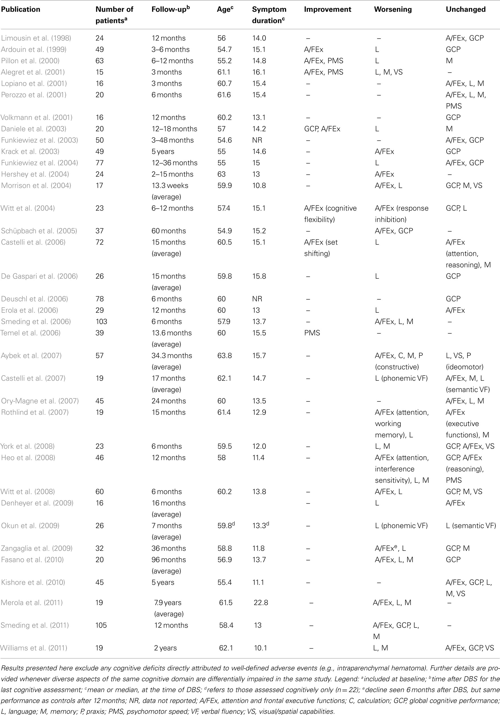
Table 1. Publications concerning cognitive functioning after deep brain stimulation of the subthalamic nucleus in Parkinson’s disease, at the time of last cognitive assessment.
Several studies have found a significant decline in phonemic (letter) VF, while semantic VF seemed to remain relatively spared after DBS (Castelli et al., 2007; Okun et al., 2009; Zangaglia et al., 2009), but others have observed significant declines in both VF modalities (Witt et al., 2008). Some studies convey information of special interest, due to the fact that a control group has also been included. Okun et al. (2009) have found interesting results, as phonemic VF declined after unilateral STN DBS, regardless of the active contact used and even with the stimulation turned off. This suggests that such an impairment might arise even after unilateral procedures and could result from microlesion or insertion effects. Witt and collaborators have conducted a multicenter controlled study enrolling patients for either STN DBS (n = 60) or best medical therapy (n = 63). By the end of the 6-month study period following DBS, they have found that VF (both phonemic and semantic) and executive functions had declined significantly in the surgical group, but this finding was independent of the observed improvement in QOL, and global cognition remained unaffected (Witt et al., 2008). In their series of 77 consecutive PD patients undergoing subthalamic DBS Funkiewiez et al. (2004) have observed that semantic fluency was significantly worse 1 and 3 years after surgery, when compared with the preoperative score. However, the difference between 1 and 3 years after surgery is not significant, suggesting that this is an early adverse effect of the therapy. Zangaglia and coworkers have observed results somewhat different from those found by other authors. In their study, 32 patients underwent STN DBS while 33 were enrolled as controls, after having refused surgery, choosing medical treatment instead. Six months after surgery, DBS patients had shown significant declines in both phonemic VF and FEx; however, the cognitive profile had returned to the values obtained before the surgical procedure by 12 months, remaining stable 3 years after surgery (Zangaglia et al., 2009). These findings seem to be in conflict with other publications, but comparing results is a challenging task, since the few studies controlled by a medical treatment group conducted so far had shorter follow-up periods. A 12-month long study was conducted by Smeding et al., who enrolled 105 STN DBS patients and 40 medically treated controls. In the surgical group a decline in cognitive performance has been noted in 36% percent of patients; significant changes were seen in global cognitive performance, VF, verbal memory, and executive functions. The authors argue that this does not seem to be a transient consequence, since effect sizes of most cognitive changes had become even larger from 6 to 12 months. Despite experiencing cognitive decline, 9% of these patients reported improvements in QOL, suggesting that cognitive decline does not necessarily mean a loss of clinical benefits gained from the surgical procedure (Smeding et al., 2011). The study by Williams et al. (2011) included 19 patients undergoing STN DBS and 18 medically treated PD controls; the final cognitive assessment was performed 2 years after surgery. Patients undergoing DBS displayed significant decline of phonemic and semantic VF, as well as non-verbal recall and information processing speed.
An array of data regarding FEx following STN DBS has been published, but conclusions are considerably harder to analyze than for VF. Several studies have reported that FEx worsen after STN DBS, whereas an approximately equal number has found unchanged scores after the procedure (see Table 1 for details). Of course, there are methodological differences between studies, and this might contribute for the disparities, but one must keep in mind that research groups have used widely accepted criteria for surgical patient selection, and the tests used for cognitive assessment are similar in many studies. Also, mean age at time of DBS and disease duration do not seem to differ significantly amongst most studies (Table 1). Hence, other factors probably account for the differences. FEx are particularly difficult to define and assess, as several different processes are encompassed under this umbrella term (Funahashi, 2001; Godefroy, 2003; Miller and Cummings, 2007; Stuss and Alexander, 2007). Therefore, strict definitions and assessment uniformization would clearly be useful in order to allow effective comparison of data published by the different study groups and eventually produce future meta-analyses of results. Other cognitive domains have been assessed, and Table 1 qualitatively summarizes the findings from each study.
Longer term data has also been published. Fasano et al. have reported their findings on a group of 20 STN DBS patients followed up for 8 years after surgery. The authors have found that part of the motor benefit had been lost since the previous assessment 3 years before, due to levodopa- and stimulation-resistant symptoms. On the other hand, they have found significant declines in VF, episodic memory, and executive functions, but only memory had significantly declined from 5 to 8 years of follow-up. Executive functions correlated significantly with postural instability (PI), which is not a surprising result, as both cognitive deterioration and PI are to be expected along disease progression – however, questions remain regarding putative common pathophysiological mechanisms or interactions. One patient had developed dementia at 5 years after DBS, with further progression at 8 years (Fasano et al., 2010).
The mechanisms leading to cognitive changes following STN DBS remain both intriguing and enigmatic. The STN seems to play an important role not only in motor function, but also in limbic and associative neural networks (Mallet et al., 2007; Rodriguez-Oroz et al., 2009; Volkmann et al., 2010). This nucleus incorporates the basal ganglia circuitry, and has traditionally been included in the so-called “indirect-pathway” inhibiting thalamocortical excitability (Obeso et al., 2008). It has been recognized that the dorsolateral portion of the STN is involved in motor function, while the intermediate part is important for cognitive processes and the anteromedial portion seems to be implicated with emotion (Mallet et al., 2007; Rodriguez-Oroz et al., 2009; Volkmann et al., 2010). Conceivably, although electrical stimulation in PD is meant to modulate motor circuitry, the energy pulse could extend to nearby non-motor regions in the STN and affect both emotion and cognitive performance, including VF. This also implies that the STN must play an important role with regard to cognitive processes, with a special clinical relevance for those involved in VF. This has also been suggested by functional neuroimaging research, namely PET scan, such as the work from Schroeder et al. (2003), who demonstrated that regional cerebral blood flow (rCBF) of the right orbitofrontal cortex and VF-associated rCBF within left-sided frontotemporal regions became notoriously reduced during STN stimulation compared with the off stimulation condition, which suggests that STN stimulation reduces the activation of a VF-related frontotemporal network.
In any case, the mechanisms underlying postsurgical cognitive decline remain obscure, and could be multifactorial, with the possible implication of individual and therapy-related risk factors. For example, it is currently unknown whether the trajectory of the electrodes, which are inserted through the frontal lobes and often cross the caudate nucleus, influences the cognitive outcome (Volkmann et al., 2010).
Dementia Following Subthalamic DBS in PD Patients
It would be important to know the incidence rate of dementia after DBS and whether this is in consonance with the natural history of the disease or if, on the other hand, there is an influence exerted by the surgical procedure itself or the stimulation. So far, data have not been conclusive, as there is a paucity of controlled and long-term studies aimed at looking into this issue. The studies considered here have demonstrated significant benefits with regard to motor symptoms and QOL, in accordance to what has been described by other authors.
The experienced Grenoble group has analyzed outcomes of STN DBS in 49 PD patients 5 years after surgery, and has found that three patients developed dementia (according to the Diagnostic and Statistical Manual of Mental Disorders fourth edition criteria) 3 years after surgery, which corresponds to a prevalence of 6% (Krack et al., 2003). Importantly, two of these patients had become demented within 3 months after surgery, which is a matter of concern, since neuropsychological testing and clinical observation had been rigorously carried out preoperatively.
Castelli et al. (2007) have published their findings from 19 patients submitted to STN DBS, assessed at baseline and at a mean of 17 months after surgery. None of the patients developed dementia in this series.
York and coworkers designed a 6-month controlled study, and included 23 PD patients who underwent STN DBS, as well as 28 medically treated PD controls. The authors have found that one patient (4%) had developed dementia 6 months after surgery, and two others had significant cognitive decline but did not fulfill criteria for dementia (York et al., 2008). The former patient was 14 years older than the average age in the series, had several vascular risk factors, and preoperative MRI had shown small vessel subcortical ischemic lesions. However, his cognitive, clinical, and psychiatric assessments did not contraindicate DBS. Postoperative CT scan has shown a small intraventricular hemorrhage and the patient suffered from transient postoperative confusion.
The study by Ory-Magne et al. (2007) has shown that 3 out of 45 patients developed dementia after a follow-up period of 24 months.
From Switzerland, Aybek et al. (2007) have obtained quite different results, as they prospectively assessed 57 patients submitted to STN DBS over 3 years. In this series, 24.5% of patients developed dementia over 3 years, whereas the remainder maintained stable cognitive scores. Those who became demented were on average older, displayed poorer executive performance, and a higher frequency of hallucinations. The average age of this series is somewhat higher than what can been observed in others, where lower incidence rates of dementia have been found. The authors argue that the incidence rate of dementia in this cohort is similar to what has been observed in medically treated patients, thus in keeping with the natural history of PD. Nonetheless, they agree that further studies should be conducted in order to define risk factors for developing dementia after DBS, especially because 36% of patients developing dementia did so within 6 months after surgery, suggesting a triggering effect of the procedure or the stimulation. From the same group, a longer observation period which included additional subjects has shown that 20% of patients (14/70) developed dementia on average 25 months after surgery (Aybek et al., 2009).
Zangaglia and collaborators where able to conduct a 3-year prospective study of STN DBS patients (n = 32) and medically treated controls who declined surgery (n = 33); at the time of last assessment one patient had become demented and one other had developed mild cognitive impairment in the surgery group only. The former patient had long disease duration (>21 years) at the time of DBS, when she was 60. Her MMSE score was at the lower limits of normal (24/30), and her preoperative L-dopa test had shown improvement of 56–36 in the Unified PD Rating Scale part III score (Zangaglia et al., 2009).
In the above mentioned series by Fasano et al. (2010) which included 20 patients with a follow-up of 8 years, only one patient developed dementia.
Kishore and colleagues have published results on 49 patients, 29 of them assessed at 5 years. At this time point five patients had become clinically demented; all of these had shown mild cognitive changes in baseline neuropsychological testing (Kishore et al., 2010).
The study by Castrioto et al. reveals data extracted during longer follow-up, as 18 patients were assessed 10 years after surgery. The authors have shown that motor benefits are still significant by this time as compared to baseline, although progressive decline has been observed, especially with regard to axial features. Three patients developed dementia in this series (Castrioto et al., 2011).
Williams et al. (2011) conducted a 2-year long controlled study, which enrolled 19 STN DBS patients and 18 medically treated PD controls. Two patients fulfilled criteria for dementia 6 months after surgery, and six patients at 2 years, twice as much as in the control group, but based on frequencies this was statistically not significant (p = 0.21). In addition, the authors have analyzed MCI frequency in this study, defined by a deficit of at least 2 SDs below the age corrected mean in one of the four cognitive domains identified in a recent expert consensus (Emre et al., 2007). Baseline MCI frequencies were similar in both groups. Three of the five stimulated patients who met criteria for MCI at baseline, had developed dementia, whereas the remaining two still fulfilled criteria for MCI at 2 years. In the control group all three PD patients with MCI at baseline had developed dementia by the time of the 2-year postsurgical evaluation. Moreover, at 2 years, four additional STN DBS patients fulfilled criteria for MCI, compared to three controls. When combining MCI and dementia patients, a trend has been observed toward higher cognitive deterioration in the surgery group (p = 0.06).
In summary, data coming from these studies seems somewhat divergent, but one must keep in mind methodological differences. Some studies suggest that cognitive decline follows the natural history of PD, whereas others have shown that a significant number of patients develop dementia in the first few months after implantation. This is an intriguing finding, and one may wonder if certain patients have some sort of specific vulnerability to accelerated postsurgical cognitive decline.
Cognitive Outcome Following DBS of the Subthalamic Nucleus: Predictive Factors
A few studies have tried to delineate predictors of cognitive decline. However, the evidence published on this matter is limited.
Funkiewiez et al. (2004) have studied 77 PD patients up to three 3 years after surgery; category VF was found to have significantly declined in this series. Patient preoperative age correlated significantly with the decay of a previously described frontal lobe score (Pillon et al., 1986) and the initiation subtest of MDRS.
The study by Ory-Magne and coworkers explored a possible role of age on clinical outcome following subthalamic DBS; 45 patients with a mean age of 60 years (range 40–73) were enrolled, 43 were reassessed at 12 months, and 39 at 24 months. The authors have found that cognitive and motor outcomes were unrelated to patient age at the time of DBS, but younger patients sustained greater improvements in QOL (Ory-Magne et al., 2007).
The study carried out by Rothlind et al. (2007) has failed to demonstrate any relation between postoperative cognitive changes and age or reductions in levodopa equivalent dose after surgery.
Heo and colleagues studied cognitive changes after STN DBS in 46 PD patients, who were assessed at 6 months and 1 year after surgery. At these time points VF had declined; mild declines have also been found in memory and executive functions. Higher formal education, higher levodopa equivalent dose, and younger age at onset correlated with cognitive worsening, but age at the time of DBS has not been found to be a predictor of decline (Heo et al., 2008).
Aybek and coworkers have found that, in their series of 70 PD patients, 14 developed dementia after an average of 25 months postsurgery. These were compared with 14 controls and the authors have found that hippocampal atrophy is a predictor of dementia in PD patients converting to dementia after subthalamic DBS (Aybek et al., 2009).
The above mentioned study by Smeding et al. also approached the issue of predictors of postsurgical decline. This controlled study has found that patients with advanced age, impaired baseline attention, and poorer levodopa response are at greater risk for postsurgical cognitive decline; importantly, the correlation between these factors was low, and multicollinearity was not significant, suggesting that their correlation with postsurgical cognitive deterioration is probably independent (Smeding et al., 2011).
In summary, the amount of evidence specifically concerning the issue of predictors of cognitive decline following DBS is somewhat small. Keeping in mind the natural history of PD (Lim et al., 2009; Rajput et al., 2009; Hawkes et al., 2010), one could postulate that certain factors would eventually predict cognitive decline following STN DBS, such as advanced age, axial signs, levodopa resistant symptoms, visual hallucinations, vascular lesion load, and poor baseline cognitive performance. Although the intuitive notion that, for instance, age would predict cognitive decline seems logical, the findings from the several studies published so far have not settled this question. This is also valid for other variables; importantly, many of them have not even been tested in the published research literature.
Psychiatric and Behavioral Effects of DBS in PD
Deep brain stimulation in PD has been frequently associated with behavioral and psychiatric symptoms, which have been mostly reported in association with STN DBS (Voon et al., 2006; Volkmann et al., 2010). Although this is not the main focus of the present manuscript, this matter should be here briefly mentioned, due to its clinical relevance.
Apathy is frequently diagnosed in PD patients, but the overlap and confusion with depression and cognitive impairment (including dementia) are common (Starkstein, 2012). Data on postsurgical apathy is quite variable, but this seems to be a frequent adverse event following STN DBS (Voon et al., 2006; Volkmann et al., 2010; Starkstein, 2012).
Elevation of mood has been reported following STN DBS; euphoria; or hypomania have been described in up to 15% of patients, whereas mania probably occurs in less than 2% of cases (Voon et al., 2006; Volkmann et al., 2010). A decrease in stimulation levels or dopaminergic drugs may be necessary for symptom remission; alternatively, switching the active contact to a more dorsal position could be tried. Depression has been reported in up to 25% of patients during postsurgical follow-up (Voon et al., 2006; Volkmann et al., 2010). Results from the COMPARE trial have suggested that more ventral contacts carry the ability to induce depressive feelings, as compared to more dorsal contacts (Okun et al., 2009). The recent randomized trial conducted by the CSP 468 Study Group has shown that depression worsened after STN stimulation, whereas it improved after pallidal stimulation (p = 0.02), despite the fact the both groups of patients improved similarly regarding motor symptoms and self-reported function (Follett et al., 2010). Interestingly, mood disorders tend to occur in the first few months after surgery (Voon et al., 2006; Volkmann et al., 2010).
Postsurgical suicide is a leading concern in the setting of STN DBS. A large international multicentre study involving more than 5000 patients has shown that the attempted and completed suicide rates can be estimated at 0.90 and 0.45%, respectively (Voon et al., 2008). Suicide rates were higher in the first postoperative year and remained high in the fourth year, as compared to the adjusted World Health Organization suicide rates. The excess death number was 13 in the first year, declining to one in the fourth. Attempted suicide risk has been related to postoperative depression, being single, a previous history of impulse control disorders, or compulsive medication use, being younger, younger PD onset, and a previous suicide attempt. Completed suicides were associated with postoperative depression, which remained a significant factor associated with attempted and completed suicides even after statistical correction (Voon et al., 2008). Also, it has been shown that impulsivity scores following STN DBS increase (Hälbig et al., 2009). However, the implications for suicide have not been adequately established, although one might speculate that this could cause greater propensity for impetuous self-destructive behaviors.
A range of impulsive compulsive behaviors (ICBs) have been described in PD, in association with dopaminergic drug therapy (Evans et al., 2009; Djamshidian et al., 2011). One would expect that STN DBS could improve ICBs by facilitating a decrease of postoperative dopaminergic medication. Unfortunately, the available evidence concerning this issue is not robust, and the outcomes regarding the effect of DBS on ICBs are conflicting, as published data have disclosed mixed results (Broen et al., 2011).
In most studies a decrease in anxiety levels has been reported (Volkmann et al., 2010).
It remains unclear which mechanisms underlie, and which risk factors associate with, the behavioral and psychiatric disorders observed after STN DBS. For instance, it has been suggested that the common postoperative reduction of dopaminergic drugs might play a role in the case of apathy or depression, but surgery itself or electrical stimulation could also be involved (Voon et al., 2006; Volkmann et al., 2010). On the other hand, an array of factors such as previous psychiatric disorders, personality traits, and psychological and psychosocial aspects might also play an important role.
Psychiatric symptoms warrant dedicated management before and after surgery, and their nature or severity may even advise against STN DBS in PD. Useful recommendations regarding management have been issued by experts in the field (Lang et al., 2006; Voon et al., 2006). This is an area of major concern and it has become clear that patient selection for DBS should be carried out by incorporating also psychiatric symptoms as important variables. In any event, patients should be informed beforehand of the expected risks in association with the procedure, especially suicide. As mentioned above, the choice of brain target for stimulation should probably be considered on a tailored perspective.
Conclusion
From the evidence collected so far, it seems reasonable to consider that DBS is generally safe from the cognitive standpoint in well selected PD patients, especially when looking at measures of global cognition. Nonetheless, there is a clear risk of postsurgical cognitive decline, which seems greater whenever the STN is used, although data concerning other targets is scanter. On the other hand, robust evidence based data is not prolific, with only one large randomized, double-blind trial conducted thus far, which has focused mainly on motor efficacy issues of STN DBS versus GPi DBS (Follett et al., 2010).
Postsurgical decline in VF has been the most consistently reported cognitive adverse effect in patients undergoing subthalamic DBS. Interestingly, our experience over the last decade, after around 200 PD patients already treated with DBS, suggests that patients are willing to accept such a tradeoff, as the motor benefits gained from the procedure seem, from their subjective point of view, to compensate for the VF changes observed. It would probably also be pertinent to systematically and objectively collect the opinions from families and caregivers on this matter, in order to confirm this impression, as frontal lobe dysfunction could bias patient self-assessment.
Several questions remain unanswered. First, it is difficult to demonstrate long-term effects of the surgical procedure or stimulation, and to differentiate these from the natural progression of the disease, as well as other confounding variables (e.g., effects of drug therapy, brain vascular lesions, PD progression, and concurrent degenerative pathology). Short-term clear cut changes (e.g., 6–12 months after surgery) are most probably due to the surgical procedure itself and/or the electrical stimulation – some small controlled studies, above mentioned, have suggested this. On the other hand, available data suggests that some aspects of cognitive functioning remain unchanged or even improve. Finally, so far it has not been explored what these postsurgical cognitive changes imply in terms of QOL and daily functioning. This seems to be an important issue, since medical decisions, as well as presurgical patient information and choices would largely benefit from this kind of evidence.
In some studies, dementia cases have been detected a few months after the surgical procedure, which is an intriguing and disturbing fact. Nonetheless, there is large heterogeneity between study results concerning this matter. Dementia cases should be systematically recorded and published, using well recognized diagnostic criteria – currently, the proposal by Emre et al. (2007) seems the most comprehensive one, and conveys the first diagnostic recommendations aimed specifically at PD dementia, although, to the best of our knowledge, prospective validation in large cohorts has not been reported so far.
In addition, research on predictive factors of postsurgical decline remains unsatisfactory, as this topic has not been approached in detail, and even at all, in most studies. Anyhow, the identification of predictive factors of outcome would be of great help, since it would allow better patient selection and information concerning the risk of poor cognitive outcome after DBS. Emphasis should probably be placed in risk factors for cognitive decline in PD (Williams-Gray et al., 2007; Aarsland and Kurz, 2010) but maybe also for dementia, broadly speaking (Korczyn and Vakhapova, 2007; Qiu et al., 2007). Notably, age, years of formal education, PD duration, disease phenotype, axial symptoms, levodopa responsiveness, hallucinations, and baseline cognitive performance seem good research candidates. One wonders if certain genetic factors may also play a role, such as apolipoprotein E polymorphisms or glucocerebrosidase mutations, and it would be very exciting to explore this matter through large multicenter collaborative research initiatives. Preoperative imaging markers such as vascular lesion load and atrophy of specific brain regions or whole brain volume would also probably deserve further exploration. It would certainly be an interesting and useful achievement to be able to stratify patients according to their risk based on a number of features, eventually even using objective mathematical and statistical models.
A large international agreement is clearly needed concerning detailed cognitive assessment methodology and cutoff scores in the setting of DBS for PD, for it seems difficult to devise strict recommendations based solely on the currently available data. It seems that expert analysis and common sense are still paramount at this point. Ideally, well founded consensus guidelines should be issued and prospectively assessed in large well designed multicenter trials.
Conflict of Interest Statement
João Massano has received educational support from Medtronic. He has acted as an advisor and received honoraria and financial support to speak or attend meetings from Bial, Grünenthal, Lundbeck, Novartis, and Tecnifar companies. Carolina Garrett has acted as an advisor and received honoraria and financial support to speak or attend meetings from Bial, Grünenthal, Janssen, Lundbeck, Novartis, and Pfizer companies.
Acknowledgments
The authors thank their colleagues at the Movement Disorders and Functional Surgery Unit, for keeping an excellent and mind stimulating discussion forum. This work has been supported by a research grant from Centro Hospitalar de São João, Porto, Portugal.
References
Aarsland, D., and Kurz, M. W. (2010). The epidemiology of dementia associated with Parkinson disease. J. Neurol. Sci. 289, 18–22.
Alegret, M., Junqué, C., Valldeoriola, F., Vendrell, P., Pilleri, M., Rumià, J., and Tolosa, E. (2001). Effects of bilateral subthalamic stimulation on cognitive function in Parkinson disease. Arch. Neurol. 58, 1223–1227.
Anderson, V. C., Burchiel, K. J., Hogarth, P., Favre, J., and Hammerstad, J. P. (2005). Pallidal vs subthalamic nucleus deep brain stimulation in Parkinson disease. Arch. Neurol. 62, 554–560.
Ardouin, C., Pillon, B., Peiffer, E., Bejjani, P., Limousin, P., Damier, P., Arnulf, I., Benabid, A., Agid, Y., and Pollak, P. (1999). Bilateral subthalamic or pallidal stimulation for Parkinson’s disease affects neither memory nor executive functions: a consecutive series of 62 patients. Ann. Neurol. 46, 217–223.
Aybek, S., Gronchi-Perrin, A., Berney, A., Chiuvé, S. C., Villemure, J.-G., Burkhard, P. R., and Vingerhoets, F. J. G. (2007). Long-term cognitive profile and incidence of dementia after STN-DBS in Parkinson’s disease. Mov. Disord. 22, 974–981.
Aybek, S., Lazeyras, F., Gronchi-Perrin, A., Burkhard, P. R., Villemure, J.-G., and Vingerhoets, F. J. G. (2009). Hippocampal atrophy predicts conversion to dementia after STN-DBS in Parkinson’s disease. Parkinsonism Relat. Disord. 15, 521–524.
Ballard, C., Gauthier, S., Corbett, A., Brayne, C., Aarsland, D., and Jones, E. (2011). Alzheimer’s disease. Lancet 377, 1019–1031.
Barone, P., Aarsland, D., Burn, D., Emre, M., Kulisevsky, J., and Weintraub, D. (2011). Cognitive impairment in nondemented Parkinson’s disease. Mov. Disord. 26, 2483–2495.
Bell, E., Maxwell, B., McAndrews, M. P., Sadikot, A., and Racine, E. (2011). Deep brain stimulation and ethics: perspectives from a multisite qualitative study of Canadian neurosurgical centers. World Neurosurg. 76, 537–547.
Benabid, A. L., Pollak, P., Louveau, A., Henry, S., and de Rougemont, J. (1987). Combined (thalamotomy and stimulation) stereotactic surgery of the VIM thalamic nucleus for bilateral Parkinson disease. Appl. Neurophysiol. 50, 344–346.
Broen, M., Duits, A., Visser-Vandewalle, V., Temel, Y., and Winogrodzka, A. (2011). Impulse control and related disorders in Parkinson’s disease patients treated with bilateral subthalamic nucleus stimulation: a review. Parkinsonism Relat. Disord. 17, 413–417.
Bronstein, J. M., Tagliati, M., Alterman, R. L., Lozano, A. M., Volkmann, J., Stefani, A., Horak, F. B., Okun, M. S., Foote, K. D., Krack, P., Pahwa, R., Henderson, J. M., Hariz, M. I., Bakay, R. A., Rezai, A., Marks, W. J. Jr., Moro, E., Vitek, J. L., Weaver, F. M., Gross, R. E., and DeLong, M. R. (2011). Deep brain stimulation for Parkinson disease: an expert consensus and review of key issues. Arch. Neurol. 68, 165–171.
Caparros-Lefebvre, D., Blond, S., Pecheux, N., Pasquier, F., and Petit, H. (1992). [Neuropsychological evaluation before and after thalamic stimulation in 9 patients with Parkinson disease]. Rev. Neurol. (Paris) 148, 117–122.
Castelli, L., Lanotte, M., Zibetti, M., Caglio, M., Rizzi, L., Ducati, A., Bergamasco, B., and Lopiano, L. (2007). Apathy and verbal fluency in STN-stimulated PD patients. An observational follow-up study. J. Neurol. 254, 1238–1243.
Castelli, L., Perozzo, P., Zibetti, M., Crivelli, B., Morabito, U., Lanotte, M., Cossa, F., Bergamasco, B., and Lopiano, L. (2006). Chronic deep brain stimulation of the subthalamic nucleus for Parkinson’s disease: effects on cognition, mood, anxiety and personality traits. Eur. Neurol. 55, 136–144.
Castrioto, A., Lozano, A. M., Poon, Y.-Y., Lang, A. E., Fallis, M., and Moro, E. (2011). Ten-year outcome of subthalamic stimulation in Parkinson disease: a blinded evaluation. Arch. Neurol. 68, 1550–1556.
Caviness, J. N., Driver-Dunckley, E., Connor, D. J., Sabbagh, M. N., Hentz, J. G., Noble, B., Evidente, V. G. H., Shill, H. A., and Adler, C. H. (2007). Defining mild cognitive impairment in Parkinson’s disease. Mov. Disord. 22, 1272–1277.
Chaudhuri, K. R., and Schapira, A. H. V. (2009). Non-motor symptoms of Parkinson’s disease: dopaminergic pathophysiology and treatment. Lancet Neurol. 8, 464–474.
Chou, K. L., Amick, M. M., Brandt, J., Camicioli, R., Frei, K., Gitelman, D., Goldman, J., Growdon, J., Hurtig, H. I., Levin, I., Litvan, I., Marsh, L., Simuni, T., Tröster, A. I., Uc, E. Y., and Parkinson Study Group Cognitive/Psychiatric Working Group. (2010). A recommended scale for cognitive screening in clinical trials of Parkinson’s disease. Mov. Disord. 25, 2501–2507.
Clausen, J. (2010). Ethical brain stimulation – neuroethics of deep brain stimulation in research and clinical practice. Eur. J. Neurosci. 32, 1152–1162.
Daniele, A., Albanese, A., Contarino, M. F., Zinzi, P., Barbier, A., Gasparini, F., Romito, L. M., Bentivoglio, A. R., and Scerrati, M. (2003). Cognitive and behavioural effects of chronic stimulation of the subthalamic nucleus in patients with Parkinson’s disease. J. Neurol. Neurosurg. Psychiatr. 74, 175–182.
De Gaspari, D., Siri, C., Di Gioia, M., Antonini, A., Isella, V., Pizzolato, A., Landi, A., Vergani, F., Gaini, S. M., Appollonio, I. M., and Pezzoli, G. (2006). Clinical correlates and cognitive underpinnings of verbal fluency impairment after chronic subthalamic stimulation in Parkinson’s disease. Parkinsonism Relat. Disord. 12, 289–295.
Defer, G. L., Widner, H., Marié, R. M., Rémy, P., and Levivier, M. (1999). Core assessment program for surgical interventional therapies in Parkinson’s disease (CAPSIT-PD). Mov. Disord. 14, 572–584.
Denheyer, M., Kiss, Z. H., and Haffenden, A. M. (2009). Behavioral effects of subthalamic deep brain stimulation in Parkinson’s disease. Neuropsychologia 47, 3203–3209.
Deuschl, G., Schade-Brittinger, C., Krack, P., Volkmann, J., Schäfer, H., Bötzel, K., Daniels, C., Deutschländer, A., Dillmann, U., Eisner, W., Gruber, D., Hamel, W., Herzog, J., Hilker, R., Klebe, S., Kloss, M., Koy, J., Krause, M., Kupsch, A., Lorenz, D., Lorenzl, S., Mehdorn, H. M., Moringlane, J. R., Oertel, W., Pinsker, M. O., Reichmann, H., Reuss, A., Schneider, G. H., Schnitzler, A., Steude, U., Sturm, V., Timmermann, L., Tronnier, V., Trottenberg, T., Wojtecki, L., Wolf, E., Poewe, W., Voges, J., and German Parkinson Study Group, Neurostimulation Section. (2006). A randomized trial of deep-brain stimulation for Parkinson’s disease. N. Engl. J. Med. 355, 896–908.
Djamshidian, A., Averbeck, B. B., Lees, A. J., and O’Sullivan, S. S. (2011). Clinical aspects of impulsive compulsive behaviours in Parkinson’s disease. J. Neurol. Sci. 310, 183–188.
Domellöf, M. E., Elgh, E., and Forsgren, L. (2011). The relation between cognition and motor dysfunction in drug-naive newly diagnosed patients with Parkinson’s disease. Mov. Disord. 26, 2183–2189.
Dorsey, E. R., Constantinescu, R., Thompson, J. P., Biglan, K. M., Holloway, R. G., Kieburtz, K., Marshall, F. J., Ravina, B. M., Schifitto, G., Siderowf, A., and Tanner, C. M. (2007). Projected number of people with Parkinson disease in the most populous nations, 2005 through 2030. Neurology 68, 384–386.
Emre, M., Aarsland, D., Brown, R., Burn, D. J., Duyckaerts, C., Mizuno, Y., Broe, G. A., Cummings, J., Dickson, D. W., Gauthier, S., Goldman, J., Goetz, C., Korczyn, A., Lees, A., Levy, R., Litvan, I., McKeith, I., Olanow, W., Poewe, W., Quinn, N., Sampaio, C., Tolosa, E., and Dubois, B. (2007). Clinical diagnostic criteria for dementia associated with Parkinson’s disease. Mov. Disord. 22, 1689–1707.
Erola, T., Heikkinen, E. R., Haapaniemi, T., Tuominen, J., Juolasmaa, A., and Myllylä, V. V. (2006). Efficacy of bilateral subthalamic nucleus (STN) stimulation in Parkinson’s disease. Acta Neurochir. (Wien) 148, 389–394.
Evans, A. H., Strafella, A. P., Weintraub, D., and Stacy, M. (2009). Impulsive and compulsive behaviors in Parkinson’s disease. Mov. Disord. 24, 1561–1570.
Fasano, A., Romito, L. M., Daniele, A., Piano, C., Zinno, M., Bentivoglio, A. R., and Albanese, A. (2010). Motor and cognitive outcome in patients with Parkinson’s disease 8 years after subthalamic implants. Brain 133, 2664–2676.
Follett, K., Weaver, F., and Stern, M. (2010). Pallidal versus subthalamic deep-brain stimulation for Parkinson’s disease. N. Engl. J. Med. 362, 2077–2091.
Funahashi, S. (2001). Neuronal mechanisms of executive control by the prefrontal cortex. Neurosci. Res. 39, 147–165.
Funkiewiez, A., Ardouin, C., Caputo, E., Krack, P., Fraix, V., Klinger, H., Chabardes, S., Foote, K., Benabid, A. L., and Pollak, P. (2004). Long term effects of bilateral subthalamic nucleus stimulation on cognitive function, mood, and behaviour in Parkinson’s disease. J. Neurol. Neurosurg. Psychiatr. 75, 834–839.
Funkiewiez, A., Ardouin, C., Krack, P., Fraix, V., Van Blercom, N., Xie, J., Moro, E., Benabid, A.-L., and Pollak, P. (2003). Acute psychotropic effects of bilateral subthalamic nucleus stimulation and levodopa in Parkinson’s disease. Mov. Disord. 18, 524–530.
Gallagher, D. A., Lees, A. J., and Schrag, A. (2010). What are the most important nonmotor symptoms in patients with Parkinson’s disease and are we missing them? Mov. Disord. 25, 2493–2500.
Goetz, C. G., Emre, M., and Dubois, B. (2008). Parkinson’s disease dementia: definitions, guidelines, and research perspectives in diagnosis. Ann. Neurol. 64, S81–S92.
Hälbig, T. D., Tse, W., Frisina, P. G., Baker, B. R., Hollander, E., Shapiro, H., Tagliati, M., Koller, W. C., and Olanow, C. W. (2009). Subthalamic deep brain stimulation and impulse control in Parkinson’s disease. Eur. J. Neurol. 16, 493–497.
Hariz, M. I., Rehncrona, S., Quinn, N. P., Speelman, J. D., and Wensing, C. (2008). Multicenter study on deep brain stimulation in Parkinson’s disease: an independent assessment of reported adverse events at 4 years. Mov. Disord. 23, 416–421.
Hawkes, C. H., Del Tredici, K., and Braak, H. (2010). A timeline for Parkinson’s disease. Parkinsonism Relat. Disord. 16, 79–84.
Heo, J.-H., Lee, K.-M., Paek, S. H., Kim, M.-J., Lee, J.-Y., Kim, J.-Y., Cho, S.-Y., Lim, Y. H., Kim, M.-R., Jeong, S. Y., and Jeon, B. S. (2008). The effects of bilateral subthalamic nucleus deep brain stimulation (STN DBS) on cognition in Parkinson disease. J. Neurol. Sci. 273, 19–24.
Hershey, T., Revilla, F., Wernle, A., Gibson, P. S., Dowling, J., and Perlmutter, J. (2004). Stimulation of STN impairs aspects of cognitive control in PD. Neurology 62, 1110–1114.
Hoops, S., Nazem, S., Siderowf, A. D., Duda, J. E., Xie, S. X., Stern, M. B., and Weintraub, D. (2009). Validity of the MoCA and MMSE in the detection of MCI and dementia in Parkinson disease. Neurology 73, 1738–1745.
Jellinger, K. A., and Attems, J. (2008). Cerebral amyloid angiopathy in Lewy body disease. J. Neural Transm. 115, 473–482.
Kaszás, B., Kovács, N., Balás, I., Kállai, J., Aschermann, Z., Kerekes, Z., Komoly, S., Nagy, F., Janszky, J., Lucza, T., and Karádi, K. (2012). Sensitivity and specificity of Addenbrooke’s Cognitive Examination, Mattis Dementia Rating Scale, Frontal Assessment Battery and Mini Mental State Examination for diagnosing dementia in Parkinson’s disease. Parkinsonism Relat. Disord. PMID: 22405839. [Epub ahead of print].
Kishore, A., Rao, R., Krishnan, S., Panikar, D., Sarma, G., Sivasanakaran, M. P., and Sarma, S. (2010). Long-term stability of effects of subthalamic stimulation in Parkinson’s disease: Indian experience. Mov. Disord. 25, 2438–2444.
Kleiner-Fisman, G., Herzog, J., Fisman, D. N., Tamma, F., Lyons, K. E., Pahwa, R., Lang, A. E., and Deuschl, G. (2006). Subthalamic nucleus deep brain stimulation: summary and meta-analysis of outcomes. Mov. Disord. 21(Suppl. 1), S290–S304.
Korczyn, A. D., and Vakhapova, V. (2007). The prevention of the dementia epidemic. J. Neurol. Sci. 257, 2–4.
Kovacs, G. G., Alafuzoff, I., Al-Sarraj, S., Arzberger, T., Bogdanovic, N., Capellari, S., Ferrer, I., Gelpi, E., Kövari, V., Kretzschmar, H., Nagy, Z., Parchi, P., Seilhean, D., Soininen, H., Troakes, C., and Budka, H. (2008). Mixed brain pathologies in dementia: the BrainNet Europe consortium experience. Dement. Geriatr. Cogn. Disord. 26, 343–350.
Krack, P., Batir, A., Van Blercom, N., Chabardes, S., Fraix, V., Ardouin, C., Koudsie, A., Limousin, P. D., Benazzouz, A., LeBas, J. F., Benabid, A. L., and Pollak, P. (2003). Five-year follow-up of bilateral stimulation of the subthalamic nucleus in advanced Parkinson’s disease. N. Eng. J. Med. 349, 1925–1934.
Kulisevsky, J., and Pagonabarraga, J. (2009). Cognitive impairment in Parkinson’s disease: tools for diagnosis and assessment. Mov. Disord. 24, 1103–1110.
Lang, A. E. (2009). When and how should treatment be started in Parkinson disease? Neurology 72, S39–S43.
Lang, A. E., Houeto, J.-L., Krack, P., Kubu, C., Lyons, K. E., Moro, E., Ondo, W., Pahwa, R., Poewe, W., Tröster, A. I., Uitti, R., and Voon, V. (2006). Deep brain stimulation: preoperative issues. Mov. Disord. 21(Suppl. 1), S171–S196.
Lim, S.-Y., Fox, S. H., and Lang, A. E. (2009). Overview of the extranigral aspects of Parkinson disease. Arch. Neurol. 66, 167–172.
Limousin, P., Krack, P., and Pollak, P. (1998). Electrical stimulation of the subthalamic nucleus in advanced Parkinson’s disease. N. Engl. J. Med. 339, 1105–1111.
Loher, T. J., Gutbrod, K., Fravi, N. L., Pohle, T., Burgunder, J.-M., and Krauss, J. K. (2003). Thalamic stimulation for tremor. Subtle changes in episodic memory are related to stimulation per se and not to a microthalamotomy effect. J. Neurol. 250, 707–713.
Lopiano, L., Rizzone, M., Bergamasco, B., Tavella, A., Torre, E., Perozzo, P., Valentini, M., and Lanotte, M. (2001). Deep brain stimulation of the subthalamic nucleus: clinical effectiveness and safety. Neurology 56, 552–554.
Mallet, L., Schüpbach, M., N’Diaye, K., Remy, P., Bardinet, E., Czernecki, V., Welter, M.-L., Pelissolo, A., Ruberg, M., Agid, Y., and Yelnik, J. (2007). Stimulation of subterritories of the subthalamic nucleus reveals its role in the integration of the emotional and motor aspects of behavior. Proc. Natl. Acad. Sci. U.S.A. 104, 10661–10666.
Massano, J., and Bhatia, K. P. (2012). Clinical approach to Parkinson’s disease: features, diagnosis, and principles of management. Cold Spring Harb. Perspect. Med. doi:10.1101/cshperspect.000870
McKhann, G. M., Knopman, D. S., Chertkow, H., Hyman, B. T., Jack, C. R., Kawas, C. H., Klunk, W. E., Koroshetz, W. J., Manly, J. J., Mayeux, R., Mohs, R. C., Morris, J. C., Rossor, M. N., Scheltens, P., Carrillo, M. C., Thies, B., Weintraub, S., and Phelps, C. H. (2011). The diagnosis of dementia due to Alzheimer’s disease: recommendations from the National Institute on Aging-Alzheimer’s Association workgroups on diagnostic guidelines for Alzheimer’s disease. Alzheimers Dement. 7, 263–269.
Merola, A., Zibetti, M., Angrisano, S., Rizzi, L., Ricchi, V., Artusi, C. A., Lanotte, M., Rizzone, M. G., and Lopiano, L. (2011). Parkinson’s disease progression at 30 years: a study of subthalamic deep brain-stimulated patients. Brain 134, 2074–2084.
Miller, B. L., and Cummings, J. L. (2007). The Human Frontal Lobes: Functions and Disorders, 2nd Edn. New York: The Guilford Press.
Moro, E., and Lang, A. E. (2006). Criteria for deep-brain stimulation in Parkinson’s disease: review and analysis. Expert Rev. Neurother. 6, 1695–1705.
Moro, E., Lozano, A. M., Pollak, P., Agid, Y., Rehncrona, S., Volkmann, J., Kulisevsky, J., Obeso, J. A., Albanese, A., Hariz, M. I., Quinn, N. P., Speelman, J. D., Benabid, A. L., Fraix, V., Mendes, A., Welter, M. L., Houeto, J. L., Cornu, P., Dormont, D., Tornqvist, A. L., Ekberg, R., Schnitzler, A., Timmermann, L., Wojtecki, L., Gironell, A., Rodriguez-Oroz, M. C., Guridi, J., Bentivoglio, A. R., Contarino, M. F., Romito, L., Scerrati, M., Janssens, M., and Lang, A. E. (2010). Long-term results of a multicenter study on subthalamic and pallidal stimulation in Parkinson’s disease. Mov. Disord. 25, 578–586.
Morrison, C. E., Borod, J. C., Perrine, K., Beric, A., Brin, M. F., Rezai, A., Kelly, P., Sterio, D., Germano, I., Weisz, D., and Olanow, C. W. (2004). Neuropsychological functioning following bilateral subthalamic nucleus stimulation in Parkinson’s disease. Arch. Clin. Neuropsychol. 19, 165–181.
Obeso, J. A., Marin, C., Rodriguez-Oroz, C., Blesa, J., Benitez-Temiño, B., Mena-Segovia, J., Rodríguez, M., and Olanow, C. W. (2008). The basal ganglia in Parkinson’s disease: current concepts and unexplained observations. Ann. Neurol. 64(Suppl. 2), S30–S46.
Okun, M. S., Fernandez, H. H., Wu, S. S., Kirsch-Darrow, L., Bowers, D., Bova, F., Suelter, M., Jacobson, C. E., Wang, X., Gordon, C. W., Zeilman, P., Romrell, J., Martin, P., Ward, H., Rodriguez, R. L., and Foote, K. D. (2009). Cognition and mood in Parkinson’s disease in subthalamic nucleus versus globus pallidus interna deep brain stimulation: the COMPARE trial. Ann. Neurol. 65, 586–595.
Okun, M. S., Rodriguez, R. L., Mikos, A., Miller, K., Kellison, I., Kirsch-Darrow, L., Wint, D. P., Springer, U., Fernandez, H. H., Foote, K. D., Crucian, G., and Bowers, D. (2007). Deep brain stimulation and the role of the neuropsychologist. Clin. Neuropsychol. 21, 162–189.
Ory-Magne, F., Brefel-Courbon, C., Simonetta-Moreau, M., Fabre, N., Lotterie, J. A., Chaynes, P., Berry, I., Lazorthes, Y., and Rascol, O. (2007). Does ageing influence deep brain stimulation outcomes in Parkinson’s disease? Mov. Disord. 22, 1457–1463.
Pahwa, R., Lyons, K. E., Wilkinson, S. B., Simpson, R. K., Ondo, W. G., Tarsy, D., Norregaard, T., Hubble, J. P., Smith, D. A., Hauser, R. A., and Jankovic, J. (2006). Long-term evaluation of deep brain stimulation of the thalamus. J. Neurosurg. 104, 506–512.
Parsons, T. D., Rogers, S. A., Braaten, A. J., Woods, S. P., and Tröster, A. I. (2006). Cognitive sequelae of subthalamic nucleus deep brain stimulation in Parkinson’s disease: a meta-analysis. Lancet Neurol. 5, 578–588.
Perozzo, P., Rizzone, M., Bergamasco, B., Castelli, L., Lanotte, M., Tavella, A., Torre, E., and Lopiano, L. (2001). Deep brain stimulation of the subthalamic nucleus in Parkinson’s disease: comparison of pre-and postoperative neuropsychological evaluation. J. Neurol. Sci. 192, 9–15.
Pillon, B. (2002). Neuropsychological assessment for management of patients with deep brain stimulation. Mov. Disord. 17, S116–S122.
Pillon, B., Ardouin, C., Damier, P., Krack, P., Houeto, J., Klinger, H., Bonnet, A., Pollak, P., Benabid, A., and Agid, Y. (2000). Neuropsychological changes between “off” and “on” STN or GPi stimulation in Parkinson’s disease. Neurology 55, 411–418.
Pillon, B., Dubois, B., Lhermitte, F., and Agid, Y. (1986). Heterogeneity of cognitive impairment in progressive supranuclear palsy, Parkinson’s disease, and Alzheimer’s disease. Neurology 36, 1179–1185.
Qiu, C., De Ronchi, D., and Fratiglioni, L. (2007). The epidemiology of the dementias: an update. Curr. Opin. Psychiatry 20, 380–385.
Rajput, A. H., Voll, A., Rajput, M. L., Robinson, C. A., and Rajput, A. (2009). Course in Parkinson disease subtypes: a 39-year clinicopathologic study. Neurology 73, 206–212.
Rodriguez, R. L., Fernandez, H. H., Haq, I., and Okun, M. S. (2007). Pearls in patient selection for deep brain stimulation. Neurologist 13, 253–260.
Rodriguez-Oroz, M. C., Jahanshahi, M., Krack, P., Litvan, I., Macias, R., Bezard, E., and Obeso, J. A. (2009). Initial clinical manifestations of Parkinson’s disease: features and pathophysiological mechanisms. Lancet Neurol. 8, 1128–1139.
Rothlind, J., Cockshott, R., Starr, P., and Marks, W. (2007). Neuropsychological performance following staged bilateral pallidal or subthalamic nucleus deep brain stimulation for Parkinson’s disease. J. Int. Neuropsychol. Soc. 13, 68–79.
Rouaud, T., Dondaine, T., Drapier, S., Haegelen, C., Lallement, F., Péron, J., Raoul, S., Sauleau, P., and Vérin, M. (2010). Pallidal stimulation in advanced Parkinson’s patients with contraindications for subthalamic stimulation. Mov. Disord. 25, 1839–1846.
Schapira, A. H. V. (2009). Neurobiology and treatment of Parkinson’s disease. Trends Pharmacol. Sci. 30, 41–47.
Schermer, M. (2011). Ethical issues in deep brain stimulation. Front. Integr. Neurosci. 5:17. doi:10.3389/fnint.2011.00017
Schroeder, U., Kuehler, A., Lange, K. W., Haslinger, B., Tronnier, V. M., Krause, M., Pfister, R., Boecker, H., and Ceballos-Baumann, A. O. (2003). Subthalamic nucleus stimulation affects a frontotemporal network: a PET study. Ann. Neurol. 54, 445–450.
Schüpbach, W. M. M., Chastan, N., Welter, M. L., Houeto, J. L., Mesnage, V., Bonnet, A. M., Czernecki, V., Maltête, D., Hartmann, A., Mallet, L., Pidoux, B., Dormont, D., Navarro, S., Cornu, P., Mallet, A., and Agid, Y. (2005). Stimulation of the subthalamic nucleus in Parkinson’s disease: a 5 year follow up. J. Neurol. Neurosurg. Psychiatr. 76, 1640–1644.
Smeding, H. M. M., Speelman, J. D., Huizenga, H. M., Schuurman, P. R., and Schmand, B. (2011). Predictors of cognitive and psychosocial outcome after STN DBS in Parkinson’s disease. J. Neurol. Neurosurg. Psychiatr. 82, 754–760.
Smeding, H. M. M., Speelman, J. D., Koning-Haanstra, M., Schuurman, P. R., Nijssen, P., van Laar, T., and Schmand, B. (2006). Neuropsychological effects of bilateral STN stimulation in Parkinson disease: a controlled study. Neurology 66, 1830–1836.
Starkstein, S. E. (2012). Apathy in Parkinson’s disease: diagnostic and etiological dilemmas. Mov. Disord. 27, 174–178.
Stuss, D. T., and Alexander, M. P. (2007). Is there a dysexecutive syndrome? Philos. Trans. R. Soc. Lond. B Biol. Sci. 362, 901–915.
Temel, Y., Blokland, A., Ackermans, L., Boon, P., van Kranen-Mastenbroek, V. H. J. M., Beuls, E. A. M., Spincemaille, G. H., and Visser-Vandewalle, V. (2006). Differential effects of subthalamic nucleus stimulation in advanced Parkinson disease on reaction time performance. Exp. Brain Res. 169, 389–399.
Temel, Y., Blokland, A., Steinbusch, H. W. M., and Visser-Vandewalle, V. (2005). The functional role of the subthalamic nucleus in cognitive and limbic circuits. Prog. Neurobiol. 76, 393–413.
Tröster, A. I., Wilkinson, S. B., Fields, J. A., Miyawaki, K., and Koller, W. C. (1998). Chronic electrical stimulation of the left ventrointermediate (Vim) thalamic nucleus for the treatment of pharmacotherapy-resistant Parkinson’s disease: a differential impact on access to semantic and episodic memory? Brain Cogn. 38, 125–149.
Utter, A. A., and Basso, M. A. (2008). The basal ganglia: an overview of circuits and function. Neurosci. Biobehav. Rev. 32, 333–342.
Volkmann, J., Allert, N., Voges, J., Weiss, P., Freund, H. J., and Sturm, V. (2001). Safety and efficacy of pallidal or subthalamic nucleus stimulation in advanced PD. Neurology 56, 548–551.
Volkmann, J., Daniels, C., and Witt, K. (2010). Neuropsychiatric effects of subthalamic neurostimulation in Parkinson disease. Nat. Rev. Neurol. 6, 487–498.
Volkmann, J., Moro, E., and Pahwa, R. (2006). Basic algorithms for the programming of deep brain stimulation in Parkinson’s disease. Mov. Disord. 21(Suppl. 1), S284–S289.
Voon, V., Krack, P., Lang, A. E., Lozano, A. M., Dujardin, K., Schüpbach, M., D’Ambrosia, J., Thobois, S., Tamma, F., Herzog, J., Speelman, J. D., Samanta, J., Kubu, C., Rossignol, H., Poon, Y. Y., Saint-Cyr, J. A., Ardouin, C., and Moro, E. (2008). A multicentre study on suicide outcomes following subthalamic stimulation for Parkinson’s disease. Brain 131, 2720–2728.
Voon, V., Kubu, C., Krack, P., Houeto, J.-L., and Tröster, A. I. (2006). Deep brain stimulation: neuropsychological and neuropsychiatric issues. Mov. Disord. 21(Suppl. 1), S305–S327.
Walter, B. L., and Vitek, J. L. (2004). Surgical treatment for Parkinson’s disease. Lancet Neurol. 3, 719–728.
Weaver, F. M., Follett, K., Stern, M., Hur, K., Harris, C., Marks, W. J., Rothlind, J., Sagher, O., Reda, D., Moy, C. S., Pahwa, R., Burchiel, K., Hogarth, P., Lai, E. C., Duda, J. E., Holloway, K., Samii, A., Horn, S., Bronstein, J., Stoner, G., Heemskerk, J., Huang, G. D., and CSP 468 Study Group. (2009). Bilateral deep brain stimulation vs best medical therapy for patients with advanced Parkinson disease: a randomized controlled trial. JAMA 301, 63–73.
Weintraub, D., Dietz, N., Duda, J. E., Wolk, D. A., Doshi, J., Xie, S. X., Davatzikos, C., Clark, C. M., and Siderowf, A. (2012). Alzheimer’s disease pattern of brain atrophy predicts cognitive decline in Parkinson’s disease. Brain 135, 170–180.
Williams, A., Gill, S., Varma, T., Jenkinson, C., Quinn, N., Mitchell, R., Scott, R., Ives, N., Rick, C., Daniels, J., Patel, S., Wheatley, K., and PD SURG Collaborative Group. (2010). Deep brain stimulation plus best medical therapy versus best medical therapy alone for advanced Parkinson’s disease (PD SURG trial): a randomised, open-label trial. Lancet Neurol. 9, 581–591.
Williams, A. E., Arzola, G. M., Strutt, A. M., Simpson, R., Jankovic, J., and York, M. K. (2011). Cognitive outcome and reliable change indices two years following bilateral subthalamic nucleus deep brain stimulation. Parkinsonism Relat. Disord. 17, 321–327.
Williams-Gray, C. H., Evans, J. R., Goris, A., Foltynie, T., Ban, M., Robbins, T. W., Brayne, C., Kolachana, B. S., Weinberger, D. R., Sawcer, S. J., and Barker, R. A. (2009). The distinct cognitive syndromes of Parkinson’s disease: 5 year follow-up of the CamPaIGN cohort. Brain 132, 2958–2969.
Williams-Gray, C. H., Foltynie, T., Brayne, C. E. G., Robbins, T. W., and Barker, R. A. (2007). Evolution of cognitive dysfunction in an incident Parkinson’s disease cohort. Brain 130, 1787–1798.
Witt, K., Daniels, C., Reiff, J., Krack, P., Volkmann, J., Pinsker, M. O., Krause, M., Tronnier, V., Kloss, M., Schnitzler, A., Wojtecki, L., Bötzel, K., Danek, A., Hilker, R., Sturm, V., Kupsch, A., Karner, E., and Deuschl, G. (2008). Neuropsychological and psychiatric changes after deep brain stimulation for Parkinson’s disease: a randomised, multicentre study. Lancet Neurol. 7, 605–614.
Witt, K., Pulkowski, U., Herzog, J., Lorenz, D., Hamel, W., Deuschl, G., and Krack, P. (2004). Deep brain stimulation of the subthalamic nucleus improves cognitive flexibility but impairs response inhibition in Parkinson disease. Arch. Neurol. 61, 697–700.
Woods, S., Fields, J., Lyons, K., Koller, W., Wilkinson, S., Pahwa, R., and Tröster, A. (2001). Neuropsychological and quality of life changes following unilateral thalamic deep brain stimulation in Parkinson’s disease: a one-year follow-up. Acta Neurochir. (Wien) 143, 1273–1278.
York, M. K., Dulay, M., Macias, A., Levin, H. S., Grossman, R., Simpson, R., and Jankovic, J. (2008). Cognitive declines following bilateral subthalamic nucleus deep brain stimulation for the treatment of Parkinson’s disease. J. Neurol. Neurosurg. Psychiatr. 79, 789–795.
Keywords: Parkinson’s disease, cognitive impairment, deep brain stimulation, dementia, functional impairment, globus pallidus interna, quality of life, subthalamic nucleus
Citation: Massano J and Garrett C (2012) Deep brain stimulation and cognitive decline in Parkinson’s disease: a clinical review. Front. Neur. 3:66. doi: 10.3389/fneur.2012.00066
Received: 01 February 2012; Accepted: 09 April 2012;
Published online: 26 April 2012.
Edited by:
Oscar López, University of Pittsburgh School of Medicine, USAReviewed by:
Oscar López, University of Pittsburgh School of Medicine, USAMarcelo L. Berthier, University of Malaga, Spain
Copyright: © 2012 Massano and Garrett. This is an open-access article distributed under the terms of the Creative Commons Attribution Non Commercial License, which permits non-commercial use, distribution, and reproduction in other forums, provided the original authors and source are credited.
*Correspondence: João Massano, Movement Disorders and Functional Surgery Unit, Department of Neurology, Centro Hospitalar de São João, Alameda Prof. Hernâni Monteiro, 4200-319 Porto, Portugal. e-mail: jmassano@med.up.pt