
Association between Arousals during Sleep and Subclinical Coronary Atherosclerosis in Patients with Obstructive Sleep Apnea


1
Department of Otolaryngology-Head and Neck Surgery, Beijing Tongren Hospital, Capital Medical University, Beijing 100005, China
2
Department of Radiology, Beijing Anzhen Hospital, Capital Medical University, Beijing 100029, China
3
Department of Radiology, Beijing Hospital of Traditional Chinese Medicine, Capital Medical University, Beijing 100010, China
*
Authors to whom correspondence should be addressed.
Brain Sci. 2022, 12(10), 1362; https://doi.org/10.3390/brainsci12101362
Submission received: 6 September 2022
/
Revised: 3 October 2022
/
Accepted: 6 October 2022
/
Published: 8 October 2022
(This article belongs to the Section Developmental Neuroscience)
Abstract
:(1) Aim: We aim to evaluate the association between arousals during sleep and subclinical coronary atherosclerosis detected by coronary computed tomography angiography (CTA) in patients with obstructive sleep apnea (OSA). (2) Methods: This was a cross-sectional study. Consecutive newly diagnosed OSA patients, who underwent coronary CTA examinations within 3 months of the sleep study, were eligible. We used the arousal index (ArI) derived from polysomnography to assess arousals during sleep and a semi-automated plaque quantification software to characterize and quantify the subclinical coronary atherosclerosis. Multiple regression models were used to evaluate the associations of the ArI with the coronary atherosclerotic plaque presence, volume, and composition. (3) Results: A total of 99 patients with OSA were included in the study. In the multivariable models, patients with a high ArI (ArI > 32.2 events/h) were more likely to have coronary plaques compared to those with a low ArI (ArI ≤ 32.2 events/h) (OR: 3.29 [95% CI: 1.284 to 8.427], p = 0.013). Furthermore, the ArI exhibited significant associations with total (β = 0.015), noncalcified (β = 0.015), and low-attenuation (β = 0.012) coronary plaque volume after accounting for established risk factors (p = 0.008, 0.004, and 0.002, respectively). However, no association between the ArI and calcified plaque volume was found. (4) Conclusion: Repetitive arousals during sleep are associated with an increased coronary plaque burden in patients with OSA, which remained robust after adjusting for multiple established cardiovascular risk factors.
1. Introduction
Obstructive sleep apnea (OSA) affects nearly 1 billion individuals worldwide and is characterized by repetitive collapse of the upper airway during sleep, resulting in intermittent hypoxia and fragmented sleep [1]. Untreated OSA has been linked to coronary atherosclerotic plaque, myocardial infarction (MI), and metabolic disorders [2,3,4,5]. Although the underlying mechanisms are complicated and multifaceted, sympathetic activation is likely to be implicated in the increased risk of coronary events seen in OSA [6]. Recent studies demonstrated that sleep fragmentation, rather than recurrent hypoxia and reoxygenation, is the primary stimulus to sympathetic activation in OSA patients since sympathetic overactivity has a strong relationship with the arousal index (ArI) but not with the apnea–hypopnea index (AHI) or desaturation index [7,8,9]. Therefore, arousals during sleep may play a more important role in the initiation and progression of OSA-related coronary events than usually believed.
Studies exploring associations between arousals during sleep and subclinical atherosclerosis are scarce. Two existing studies found that the ArI was independently correlated with the presence of carotid plaque and a high coronary artery calcification burden, respectively [10,11]. No study has yet examined the effect of arousal frequency or other objective sleep metrics (e.g., slow-wave sleep percentage) on coronary plaque volume and composition. However, it is important to note that plaques causing MI or acute coronary syndromes (ACS) have specific compositional properties on coronary computed tomography angiography (CTA). Recent clinical studies found that low-attenuation plaque identified by coronary CTA had an excellent correlation with histologically confirmed inflamed plaque, which was the strongest predictor of subsequent MI [12,13]. Moreover, the noncalcified plaque burden predicted an MI better than the calcification score and stenosis [14]. All the above studies highlighted the relevance of plaque volume and composition in MI risk prediction.
Therefore, we aim to investigate whether repetitive arousals during sleep are associated with an increase in the presence, volume, and composition of coronary plaque in patients with OSA, independent of traditional cardiovascular risk factors, AHI, and nocturnal hypoxia.
2. Materials and Methods
2.1. Study Population
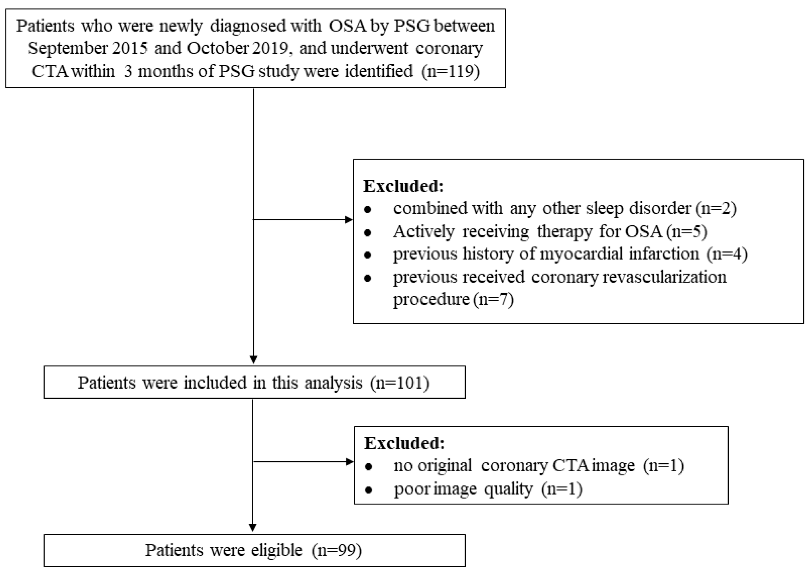
Consecutive patients who were newly diagnosed with OSA by polysomnography at Beijing Anzhen Hospital between September 2015 and October 2019 and who also underwent coronary CTAs within 3 months of the sleep studies were eligible for the current study. The patients were excluded if (1) they received OSA treatment during these 3 months; (2) they had other sleep disorders (e.g., insomnia, central sleep apnea); (3) they had a psychiatric condition (e.g., depression), and/or used antipsychotics in the last 3 months; (4) they had an MI history and/or underwent coronary revascularization; or (5) the original coronary CTA images were not available, or the quality of the image was poor.
Anthropometric measurements, medical history, and medication usage were obtained from a self-reported questionnaire and medical records. Body mass index (BMI) was calculated by dividing the body weight in kilograms by the square of height in meters. There were three categories of smoking status: never, former, and current smokers. The definitions of morbidities have been published in detail [2]. This study was approved by the Ethics Review Committee of Beijing Anzhen Hospital (No. 2017005), and written informed consent was obtained from all patients.
2.2. Sleep Study
Sleep recordings were performed using polysomnography (Grael, Compumedics, Abbotsford, VIC, Australia) at our sleep center. The sensors and recording montages mainly included electroencephalogram, bilateral electrooculograms, chin muscle electromyogram, electrocardiogram, oronasal thermistor, nasal pressure transducer/cannula, thoracic–abdominal inductance plethysmography, finger pulse oximetry, etc. Trained technicians attached the sensors and electrodes and scored the polysomnographic data according to the American Academy of Sleep Medicine scoring manual [15]. The average number of apneas and hypopneas per hour of sleep was used to determine the AHI and the patients with an AHI ≥ 5 events/h were diagnosed with OSA. Additionally, the percentage of sleep time spent in each sleep stage was also determined, namely non-rapid eye movement (NREM) stage 1 (N1%), NREM stage 2 (N2%), and NREM stage 3 (N3%, slow-wave sleep), and REM sleep (REM%). Arousal episodes were scored when there was an abrupt shift in the electroencephalogram frequencies (but not spindles) during sleep stages lasting at least 3 s, with at least 10 s of stable sleep before the change. The ArI was calculated as the number of arousals per hour of sleep. The sleep efficiency measure was the ratio between total sleep time and total bedtime.
2.3. Coronary CTA Image Acquisition
Coronary CTA was performed using prospectively electrocardiogram-triggered sequential scan techniques on a 256-row detector CT scanner (Revolution CT, GE Healthcare, Milwaukee, WI, USA) or a second-generation dual-source CT system (Somatom Definition Flash, Siemens Healthcare, Forchheim, Germany). A 50–60 mL dose of iodinated contrast agent (Ultravist, 370 mg iodine/mL; Bayer Schering Pharma, Guangzhou, China) was injected into an antecubital vein at a rate of 4–5 mL/s, followed by a 30–35 mL dose of saline solution. The scanning parameters were the gantry rotation time of 250–350 ms, a tube voltage of 100–120 kV depending on the BMI, and an automated tube current modulation. The bolus-tracking technique was used to trigger the acquisition of CT images, which started automatically after the CT value in the ascending aorta reached 120 HU. A median slice thickness of 0.625 mm and an increment of 0.5 mm were used to reconstruct the images. All procedures were performed following the Society of Cardiovascular Computed Tomography guidelines [16].
2.4. Coronary Plaque Analysis
An experienced radiologist (Z.W.) blinded to the clinical and sleep data, performed the quantitative measurement of coronary plaque using semiautomated software (Autoplaque Version 2.0, VoxelCloud Technology Co., Ltd., Suzhou, China). The overall method for plaque quantification of each coronary lesion was as follows. First, the proximal and distal limits of the plaque lesions were marked. Next, the plaque and vessel borders were automatically segmented, with manual adjustment performed if necessary. Finally, vessel volume and volume of calcified plaque, noncalcified plaque, and low-attenuation plaque (i.e., the noncalcified plaque with attenuation < 30 HU) were computed on a per-patient basis. Excellent inter-and intra-reader agreements for this software have previously been reported [17].
2.5. Statistical Analysis
Categorical variables are described as numbers with percentages and continuous variables as mean ± standard deviation (SD) or median (interquartile range) as appropriate. By dichotomizing the ArI values based on the cohort medians, we achieved low/high ArI groups (ArI ≤ 32.2 events/h and ArI > 32.2 events/h). Data between groups were compared using the independent-sample t-test, Mann–Whitney U test, χ2 test, or Fisher’s exact test, as appropriate. The Spearman’s rank correlation coefficients were obtained for the ArI with total, noncalcified, low-attenuation, and calcified coronary plaque volume, respectively. Multivariable stepwise logistic regression models were performed to test the independent associations of ArI and other sleep metrics—non-normally distributed variables—with the presence of coronary plaque in patients with OSA, by analyzing these objective sleep metrics as dichotomized variables (e.g., ArI > 32.2 events/h). The following covariates were adjusted: age, sex, BMI, smoking status, hypertension, diabetes, dyslipidemia, use of lipid-lowering medications, AHI, and lowest oxygen saturation (SpO2). Similarly, multivariable linear regression models were used to examine the independent association between ArI as the continuous variable and coronary plaque volume in patients with OSA. Since coronary plaque volumes exhibit a highly skewed distribution, they enter the linear models after logarithmic transformation. All p-values are two-sided with a significance threshold of 0.05. Statistical analyses were performed with SPSS software Version 19.0 (IBM Corp., Armonk, NY, USA). Figures were drawn using GraphPad Prism 9.
2.6. Sample Size Calculation
In this study, the coronary plaque volume was used as the outcome indicator, and multiple stepwise linear regression was performed to include 10 covariates. According to the principle of 10 outcome events per variable, the sample size should be around 100.
3. Results
3.1. Participants Characteristics
A flow diagram illustrating study inclusion is shown in Figure 1. In total, 99 patients with OSA were finally included. The mean age was 50.6 (SD, 9.4) years; 76.8% were males. As shown in Table 1, patients with high ArI were more likely to be male, to have higher BMI, neck circumference, and diastolic blood pressure, to be sleepier, to have a higher AHI, oxygen desaturation index, percentage of nighttime with oxygen saturation < 90%, and a lower lowest SpO2 and mean SpO2 than those with low ArI.
A total of 52 patients (62.6%) had at least one coronary plaque, and the median total coronary plaque volume was 56.9 (interquartile range, 0–201.5) mm3. Participant characteristics stratified by the presence of coronary plaque are shown in Table S1. In general, relative to individuals without coronary plaque, those with coronary plaques were more likely to be smokers, have a higher prevalence of hypertension, have greater usage of lipid-lowering medications, have a higher AHI and ODI, lower lowest SpO2 levels, more fragmented sleep as noted by the higher ArI, and a higher percentage of N1 and N2.
3.2. The Presence of Coronary Plaque
The prevalence of coronary plaque was higher in patients with a high ArI than in those with a low ArI, 37 (75.5%) versus 25 (50.0%), respectively (p = 0.009) (Table 1). Furthermore, we found that patients with an ArI > 32.2 events/h tended to have higher odds of having coronary plaque compared to those with a low ArI (crude OR: 3.083 [95% CI: 1.311 to 7.251], p = 0.01; adjusted OR: 3.29 [95% CI: 1.284 to 8.427], p = 0.013) (Table 2). However, there was no evidence for significant associations between adverse levels of sleep efficiency or percentage of time spent in slow-wave sleep and the presence of coronary plaque (all with p-value > 0.05).
3.3. The Volume and Composition of Coronary Plaque
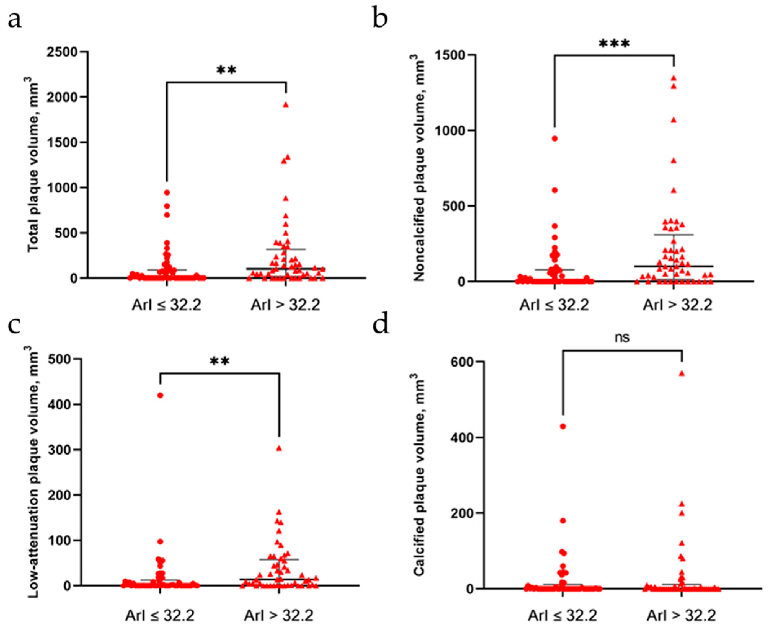
Figure 2 shows the results of the volume and distributions of coronary plaque. We observed that the total plaque volume and volumes of noncalcified low-attenuation plaque were all significantly higher among patients with a high ArI than in those with a low ArI. Spearman’s correlation analysis showed that the ArI was significantly correlated with total (rho = 0.365, p < 0.001), noncalcified (rho = 0.386, p < 0.001), and low-attenuation (rho = 0.387, p < 0.001) coronary plaque volume, but it was not significantly correlated with calcified plaque volume (rho = 0.119, p = 0.241). Furthermore, linear regression models (Table 3) demonstrated that there were significantly positive associations between the ArI and total (β = 0.019), noncalcified (β = 0.019), and low-attenuation (β = 0.015) coronary plaque volume in the unadjusted models (all with p-value ≤ 0.001), and these associations remained after adjusting for multiple cardiovascular risk factors (β = 0.015, 0.015, and 0.012, respectively, and p = 0.008, 0.004, and 0.002, respectively). Conversely, no significant associations were observed between the ArI and the calcified plaque volume. In addition, the coronary plaque volume did not seem to be associated with sleep efficiency or the percentage of time spent in slow-wave sleep.
4. Discussion
This study was the first to evaluate the relationship between arousals during sleep and coronary plaque in patients with OSA. We found that not only was coronary plaque more likely to be present in patients with a high ArI (>32.2 events/h), but the total plaque volume and volumes of noncalcified and low-attenuation plaque increased with an increase in the ArI. Importantly, adjustment for potential confounders did not substantively alter these associations. In contrast, there was no evidence of a significant association between arousals and calcified plaque. These data suggest that the ArI, which quantifies the degree of fragmented sleep, may provide unique information about arousal-related coronary events risk in patients with OSA.
Cortical arousals are normal sleep phenomena; however, if they occur frequently, they can have acute and long-term effects on cardiovascular health. A recently published study found that frequent arousals were independently linked to hypertension in patients with OSA [18]. Meanwhile, in a community study with 8001 participants, Shahrbabaki et al. demonstrated that sleep arousal burden was associated with long-term all-cause and cardiovascular mortality, particularly in women [19]. However, we know little about the relationship between frequent arousals during sleep and atherosclerotic plaque in patients with OSA. In the current study, we not only highlighted the relationship between total coronary plaque volume and increased arousal frequency but also showed a high prevalence of coronary plaque in patients with a high ArI, which may provide evidence that atherosclerosis is an intermediate step in the pathophysiology from repetitive arousals to the development of adverse cardiovascular outcomes among OSA patients. To some extent, this finding was also similar to the results of a prior study using ultrasonography to measure the severity of carotid atherosclerosis in an observational study of 83 patients with OSA, which found that the ArI was one of the independent predictors of carotid artery plaque presence [10]. A similar pattern of association between high ArI levels and carotid plaque was also demonstrated by Zhao et al. in individuals aged <68 years [20]. However, the fact that the plaque components and their relationship to arousals were not evaluated in these two studies could be potential limitations because the alteration of the plaque composition would change plaque vulnerability. In general, the plaque with a large lipid core is more prone to rupture than those with calcified components (more stable) [21].
To further explore this issue, we investigated the association between the ArI and coronary plaque composition and observed that the ArI was an independent explanatory variable for noncalcified plaque volume and low-attenuation plaque volume. Total noncalcified plaque volume predicts ACS more accurately than clinical risk profiling or conventional coronary CTA readings (e.g., calcium score, luminal stenosis, and extent of coronary artery disease) alone, which has been well documented [22]. Previous studies also showed that most ACS cases are caused by the rupture and/or erosion of coronary plaques, which are typically characterized by a lipid-rich necrotic core covered by a thin fibrous cap [23,24]. Most importantly, these specific plaque components can be detected as low-attenuation plaque on coronary CTA [13]. Therefore, our findings can be interpreted as patients with frequent arousals having a high propensity to develop unstable plaque features, which may further be related to their increased risk of subsequent coronary events. It is, however, necessary to conduct further research to confirm this hypothesis.
In the current study, arousals during sleep were not significantly associated with the volume of calcified plaques. These results appear to be inconsistent with the findings from the Multi-Ethnic Study of Atherosclerosis, which found that among patients, with a mean age of 68 years, a high coronary artery calcium score (CAC > 400) was significantly correlated with a high ArI [11]. The potential explanation is that calcification is a well-recognized hallmark for the late stage of atherosclerosis. Specifically, plaque development involves several phases, including intimal inflammation, necrosis, fibrosis, calcification, arterial remodeling, fibrous cap rupture, and thrombosis, implying that the necrotic core calcifies over time [25]. However, the patients in our study were relatively young (average age was 50.6 years), and they may still be in the early stages of atherosclerosis.
The primary mechanism underlying the link between elevated arousal frequency and coronary plaque is possibly related to sympathetic nervous system activation and vascular endothelial dysfunction. Previous studies have shown that sympathetic activation led to increased concentrations of vasoconstrictor peptides and circulating catecholamines, which directly activate platelets [26]. The endpoint effect of persistent excessive platelet activation is myocardial infarction [27]. In addition, an animal study revealed that sleep fragmentation can contribute to atherosclerosis via altering hematopoiesis [28]. Mice subjected to sleep fragmentation produced less hypocretin (a wake-promoting neuropeptide) in the lateral hypothalamus, which increased the expression of colony-stimulating factor-1, stimulated monocyte differentiation into macrophages, and ultimately accelerated atherosclerosis.
We also evaluated whether the percentage of time spent in slow-wave sleep or sleep efficiency was associated with coronary plaque volume, but no statistically significant association was observed. These results agreed with the finding from another MESA study, where they also did not find an association between sleep efficiency or slow-wave sleep and carotid plaque in the overall cohort [20].
We acknowledge several limitations in our study. First, the sample size was small. Fortunately, the study was adequately powered to demonstrate the relationship between arousals (primary exposure) and subclinical coronary plaque burden, although not powered to detect associations between other sleep metrics (e.g., sleep efficiency) and coronary plaque. Second, due to the cross-sectional nature of our study, causality between the ArI and coronary plaque cannot be inferred. Third, in the present study, the patients with a high ArI had more potential confounding factors than those with low ArI, including BMI, AHI, etc. To minimize their confounding effects, we used multivariable stepwise regression models, which included these potential risk factors as covariates for statistical analysis. Fourth, there was a selection bias in this study. Considering that some of our patients were referred to sleep medicine centers for PSG studies after cardiology visits to control for predisposing factors for coronary heart disease, our cohort may not be fully representative of typical sleep clinic patients. Fifth, the objective sleep variables were derived from single-night polysomnography recordings. As a result, night-to-night variability in these sleep variables and the first night effect may exist and affect the strength of estimates. Sixth, we did not distinguish the types of arousal (e.g., respiratory, spontaneous, and movement-related arousal) and their association with coronary plaque, since the detailed arousal data were unavailable in our study. Finally, in addition to arousal frequency, arousal duration may also contribute to sleep fragmentation and be associated with cardiovascular mortality [19,29]. However, this metric was not measured in the present study.
5. Conclusions
In sum, frequent arousals during sleep were associated with an increased burden of coronary plaque in patients with OSA. These associations remained robust after considering multiple established cardiovascular risk factors, AHI, and nocturnal hypoxemia, which suggest that repetitive arousals from sleep might be novel and independent risk factors for subclinical coronary atherosclerosis.
Supplementary Materials
The following supporting information can be downloaded at: https://www.mdpi.com/article/10.3390/brainsci12101362/s1, Table S1: Baseline characteristics of the study population according to the presence of plaque.
Author Contributions
Conceptualization, M.L. and Z.W.; methodology, M.L.; software, M.L.; validation, Z.W.; formal analysis, M.L. and Z.W.; resources, M.L. and W.Y.; data curation, M.L. and Z.W.; writing—original draft preparation, M.L.; writing—review and editing, Z.W. and Z.H.; supervision, W.Y. and Z.H.; project administration, M.L.; funding acquisition, Z.W. All authors have read and agreed to the published version of the manuscript.
Funding
This study was supported by the Beijing Municipal Administration of Hospitals Incubating Program (PX2022044). Mi Lu was financially supported by the China Scholarship Council for her visiting Ph.D. study (202108110137). The China Scholarship Council had no role in the design or conduct of this study.
Institutional Review Board Statement
The study was conducted in accordance with the Declaration of Helsinki and approved by the Ethics Review Committee of Beijing Anzhen Hospital (No. 2017005).
Informed Consent Statement
Informed consent was obtained from all subjects involved in the study.
Data Availability Statement
Not applicable.
Acknowledgments
The authors gratefully thank the staff at the Sleep Medical Center, Beijing Anzhen Hospital, China, for scoring the sleep studies according to the updated AASM scoring guidelines and the staff at the Department of Radiology, Beijing Anzhen Hospital, China, for the CCTA images scan.
Conflicts of Interest
The authors declare no conflict of interest. The funders had no role in the design of the study; in the collection, analyses, or interpretation of data; in the writing of the manuscript; or in the decision to publish the results.
References
- Benjafield, A.V.; Ayas, N.T.; Eastwood, P.R.; Heinzer, R.; Ip, M.S.M.; Morrell, M.J.; Nunez, C.M.; Patel, S.R.; Penzel, T.; Pépin, J.-L.; et al. Estimation of the global prevalence and burden of obstructive sleep apnoea: A literature-based analysis. Lancet Respir. Med. 2019, 7, 687–698. [Google Scholar] [CrossRef] [Green Version]
- Lu, M.; Fang, F.; Wang, Z.; Xu, L.; Sanderson, J.; Zhan, X.; Lianping, H.; Wu, C.; Wei, Y. Association Between OSA and Quantitative Atherosclerotic Plaque Burden: A Coronary CT Angiography Study. Chest 2021, 160, 1864–1874. [Google Scholar] [CrossRef]
- Shah, N.A.; Yaggi, H.K.; Concato, J.; Mohsenin, V. Obstructive sleep apnea as a risk factor for coronary events or cardiovascular death. Sleep Breath. 2010, 14, 131–136. [Google Scholar] [CrossRef] [PubMed]
- Yeghiazarians, Y.; Jneid, H.; Tietjens, J.R.; Redline, S.; Brown, D.L.; El-Sherif, N.; Mehra, R.; Bozkurt, B.; Ndumele, C.E.; Somers, V.K. Obstructive Sleep Apnea and Cardiovascular Disease: A Scientific Statement from the American Heart Association. Circulation 2021, 144, e56–e67. [Google Scholar] [CrossRef]
- Gabryelska, A.; Chrzanowski, J.; Sochal, M.; Kaczmarski, P.; Turkiewicz, S.; Ditmer, M.; Karuga, F.F.; Czupryniak, L.; Białasiewicz, P. Nocturnal Oxygen Saturation Parameters as Independent Risk Factors for Type 2 Diabetes Mellitus among Obstructive Sleep Apnea Patients. J. Clin. Med. 2021, 10, 3770. [Google Scholar] [CrossRef] [PubMed]
- Tietjens, J.R.; Claman, D.; Kezirian, E.J.; De Marco, T.; Mirzayan, A.; Sadroonri, B.; Goldberg, A.N.; Long, C.; Gerstenfeld, E.P.; Yeghiazarians, Y. Obstructive Sleep Apnea in Cardiovascular Disease: A Review of the Literature and Proposed Multidisciplinary Clinical Management Strategy. J. Am. Heart Assoc. 2019, 8, e010440. [Google Scholar] [CrossRef] [PubMed] [Green Version]
- Bin Kim, J.; Seo, B.S.; Kim, J.H. Effect of arousal on sympathetic overactivity in patients with obstructive sleep apnea. Sleep Med. 2019, 62, 86–91. [Google Scholar]
- Taylor, K.S.; Murai, H.; Millar, P.J.; Haruki, N.; Kimmerly, D.; Morris, B.L.; Tomlinson, G.; Bradley, T.D.; Floras, J.S. Arousal from Sleep and Sympathetic Excitation during Wakefulness. Hypertension 2016, 68, 1467–1474. [Google Scholar] [CrossRef] [PubMed]
- Ferreira, C.B.; Schoorlemmer, G.; Rocha, A.A.; Cravo, S.L. Increased sympathetic responses induced by chronic obstructive sleep apnea are caused by sleep fragmentation. J. Appl. Physiol. 2020, 129, 163–172. [Google Scholar] [CrossRef]
- Suzuki, M.; Shimamoto, K.; Sekiguchi, H.; Harada, T.; Satoya, N.; Inoue, Y.; Yamaguchi, K.; Kawana, M. Arousal index as a marker of carotid artery atherosclerosis in patients with obstructive sleep apnea syndrome. Sleep Breath. 2019, 23, 87–94. [Google Scholar] [CrossRef]
- Lutsey, P.L.; McClelland, R.L.; Duprez, D.; Shea, S.; Shahar, E.; Nagayoshi, M.; Budoff, M.; Kaufman, J.; Redline, S. Objectively measured sleep characteristics and prevalence of coronary artery calcification: The Multi-Ethnic Study of Atherosclerosis Sleep study. Thorax 2015, 70, 880–887. [Google Scholar] [CrossRef] [PubMed]
- Yuan, M.; Wu, H.; Li, R.; Yu, M.; Dai, X.; Zhang, J. The value of quantified plaque analysis by dual-source coronary CT angiography to detect vulnerable plaques: A comparison study with intravascular ultrasound. Quant. Imaging Med. Surg. 2020, 10, 668–677. [Google Scholar] [CrossRef] [PubMed]
- Williams, M.C.; Kwiecinski, J.; Doris, M.; McElhinney, P.; D’Souza, M.S.; Cadet, S.; Adamson, P.D.; Moss, A.J.; Alam, S.; Hunter, A.; et al. Low-Attenuation Noncalcified Plaque on Coronary Computed Tomography Angiography Predicts Myocardial Infarction: Results from the Multicenter SCOT-HEART Trial (Scottish Computed Tomography of the HEART). Circulation 2020, 141, 1452–1462. [Google Scholar] [CrossRef] [PubMed] [Green Version]
- Bauer, R.W.; Thilo, C.; Chiaramida, S.A.; Vogl, T.J.; Costello, P.; Schoepf, U.J. Noncalcified Atherosclerotic Plaque Burden at Coronary CT Angiography: A Better Predictor of Ischemia at Stress Myocardial Perfusion Imaging Than Calcium Score and Stenosis Severity. Am. J. Roentgenol. 2009, 193, 410–418. [Google Scholar] [CrossRef] [PubMed] [Green Version]
- Berry, R.B.; Budhiraja, R.; Gottlieb, D.J.; Gozal, D.; Iber, C.; Kapur, V.K.; Marcus, C.L.; Mehra, R.; Parthasarathy, S.; Quan, S.F.; et al. Rules for Scoring Respiratory Events in Sleep: Update of the 2007 AASM Manual for the Scoring of Sleep and Associated Events. Deliberations of the Sleep Apnea Definitions Task Force of the American Academy of Sleep Medicine. J. Clin. Sleep Med. 2012, 8, 597–619. [Google Scholar] [CrossRef] [Green Version]
- Weigold, W.G.; Abbara, S.; Achenbach, S.; Arbab-Zadeh, A.; Berman, D.; Carr, J.; Cury, R.C.; Halliburton, S.S.; McCollough, C.H.; Taylor, A.J. Standardized medical terminology for cardiac computed tomography: A report of the Society of Cardiovascular Computed Tomography. J. Cardiovasc. Comput. Tomogr. 2011, 5, 136–144. [Google Scholar] [CrossRef] [PubMed]
- Øvrehus, K.A.; Schuhbaeck, A.; Marwan, M.; Achenbach, S.; Nørgaard, B.L.; Bøtker, H.E.; Dey, D. Reproducibility of semi-automatic coronary plaque quantification in coronary CT angiography with sub-mSv radiation dose. J. Cardiovasc. Comput. Tomogr. 2016, 10, 114–120. [Google Scholar] [CrossRef] [PubMed]
- Ren, R.; Zhang, Y.; Yang, L.; Somers, V.K.; Covassin, N.; Tang, X. Association Between Arousals during Sleep and Hypertension Among Patients with Obstructive Sleep Apnea. J. Am. Heart Assoc. 2022, 11, e022141. [Google Scholar] [CrossRef] [PubMed]
- Shahrbabaki, S.S.; Linz, D.; Hartmann, S.; Redline, S.; Baumert, M. Sleep arousal burden is associated with long-term all-cause and cardiovascular mortality in 8001 community-dwelling older men and women. Eur. Heart J. 2021, 42, 2088–2099. [Google Scholar] [CrossRef] [PubMed]
- Zhao, Y.Y.; Javaheri, S.; Wang, R.; Guo, N.; Koo, B.B.; Stein, J.H.; Korcarz, C.E.; Redline, S. Associations Between Sleep Apnea and Subclinical Carotid Atherosclerosis: The Multi-Ethnic Study of Atherosclerosis. Stroke 2019, 50, 3340–3346. [Google Scholar] [CrossRef] [PubMed]
- Babaniamansour, P.; Mohammadi, M.; Babaniamansour, S.; Aliniagerdroudbari, E. The Relation between Atherosclerosis Plaque Composition and Plaque Rupture. J. Med. Signals Sens. 2020, 10, 267–273. [Google Scholar] [PubMed]
- Versteylen, M.O.; Kietselaer, B.L.; Dagnelie, P.C.; Joosen, I.A.; Dedic, A.; Raaijmakers, R.H.; Wildberger, J.E.; Nieman, K.; Crijns, H.J.; Niessen, W.J.; et al. Additive Value of Semiautomated Quantification of Coronary Artery Disease Using Cardiac Computed Tomographic Angiography to Predict Future Acute Coronary Syndrome. J. Am. Coll. Cardiol. 2013, 61, 2296–2305. [Google Scholar] [CrossRef] [PubMed]
- Naghavi, M.; Libby, P.; Falk, E.; Casscells, S.W.; Litovsky, S.; Rumberger, J.; Badimon, J.J.; Stefanadis, C.; Moreno, P.; Pasterkamp, G.; et al. From vulnerable plaque to vulnerable patient: A call for new definitions and risk assessment strategies: Part I. Circulation 2003, 108, 1664–1672. [Google Scholar] [CrossRef] [PubMed]
- Naghavi, M.; Libby, P.; Falk, E.; Casscells, S.W.; Litovsky, S.; Rumberger, J.A.; Badimón, J.J.; Stefanadis, C.; Moreno, P.R.; Pasterkamp, G.; et al. From vulnerable plaque to vulnerable patient: A call for new definitions and risk assessment strategies: Part II. Circulation 2003, 108, 1772–1778. [Google Scholar] [CrossRef] [PubMed] [Green Version]
- Bentzon, J.F.; Otsuka, F.; Virmani, R.; Falk, E. Mechanisms of Plaque Formation and Rupture. Circ. Res. 2014, 114, 1852–1866. [Google Scholar] [CrossRef] [PubMed]
- Olson, L.J.; Olson, E.J.; Somers, V.K. Obstructive sleep apnea and platelet activation: Another potential link between sleep-disordered breathing and cardiovascular disease. Chest 2004, 126, 339–341. [Google Scholar] [CrossRef] [PubMed] [Green Version]
- Gabryelska, A.; Łukasik, Z.M.; Makowska, J.S.; Bialasiewicz, P. Obstructive Sleep Apnea: From Intermittent Hypoxia to Cardiovascular Complications via Blood Platelets. Front. Neurol. 2018, 9, 635. [Google Scholar] [CrossRef] [PubMed]
- McAlpine, C.S.; Kiss, M.G.; Rattik, S.; He, S.; Vassalli, A.; Valet, C.; Anzai, A.; Chan, C.T.; Mindur, J.; Kahles, F.; et al. Sleep modulates haematopoiesis and protects against atherosclerosis. Nature 2019, 566, 383–387. [Google Scholar] [CrossRef]
- Trinder, J.; Allen, N.; Kleiman, J.; Kralevski, V.; Kleverlaan, D.; Anson, K.; Kim, Y. On the nature of cardiovascular activation at an arousal from sleep. Sleep 2003, 26, 543–551. [Google Scholar]
Figure 1.
Flow diagram of the study. CTA = computed tomography angiography; OSA = obstructive sleep apnea; PSG = polysomnography.
Figure 1.
Flow diagram of the study. CTA = computed tomography angiography; OSA = obstructive sleep apnea; PSG = polysomnography.

Figure 2.
Volume and composition of coronary plaque between patients with a low ArI and patients with a high ArI. (a) Total plaque volume; (b) noncalcified plaque volume; (c) low-attenuation plaque volume; (d) calcified plaque volume. ** indicate p-Value <0.01; *** indicate p-Value <0.001. ArI = arousal index.


Table 1.
Baseline characteristics of the study population.
| Variables | Overall (n = 99) | ArI ≤ 32.2 (n = 50) | ArI > 32.2 (n = 49) | p-Value |
|---|---|---|---|---|
| Demographic and clinical characteristics | ||||
| Age, years | 50.6 ± 9.4 | 51.52 ± 10.1 | 49.7 ± 8.8 | 0.344 |
| Male, n (%) | 76 (76.8) | 32 (64.0) | 44 (89.8) | 0.002 |
| Body mass index, kg/m2 | 27.7 (25.8, 30.8) | 26.4 (24.8, 27.8) | 30.1(27.6, 32.3) | <0.001 |
| Neck circumference, cm | 40.9 ± 3.5 | 39.6 ± 3.3 | 42.4 ± 3.2 | <0.001 |
| Smoking status | 0.541 | |||
| Never, n (%) | 52 (52.5) | 29 (58.0) | 23 (46.9) | |
| Former, n (%) | 13 (13.1) | 6 (12.0) | 7 (14.3) | |
| Current, n (%) | 34 (34.3) | 15 (30.0) | 19 (38.8) | |
| Systolic blood pressure, mmHg | 133 (125, 147) | 132(122.5, 145.5) | 135 (128.5, 150) | 0.173 |
| Diastolic blood pressure, mmHg | 83.2 ± 10.6 | 80.0 ± 9.9 | 86.4 ± 10.4 | 0.002 |
| Hypertension, n (%) | 56 (56.6) | 28 (56.0) | 28 (57.1) | 0.909 |
| Diabetes mellitus, n (%) | 14 (14.1) | 6 (12.0) | 8 (16.3) | 0.537 |
| Hyperlipidemia, n (%) | 35 (35.4) | 19 (38.0) | 16 (32.7) | 0.578 |
| Lipid-lowering medication use, n (%) | 19 (19.2) | 9 (18.0) | 10 (20.4) | 0.761 |
| Prior history of CAD, n (%) | 6 (6.1) | 3 (6.0) | 3 (6.1) | 0.100 |
| ESS | 11 (8, 15) | 9.0 (6.0, 12.5) | 13 (9, 15) | 0.006 |
| Polysomnography parameters | ||||
| AHI, /h | 36.5 (16.5, 61.4) | 18.0 (10.8, 29.8) | 60.0 (39.3, 72.3) | <0.001 |
| ODI, /h | 33.4 (16.5, 59.9) | 17.5 (7.8, 28.8) | 59.2 (41.5, 76.1) | <0.001 |
| T90, % | 3.5 (0.3, 13.1) | 0.9 (0.1, 4.1) | 10.8 (2.8, 24.1) | <0.001 |
| Lowest SpO2, % | 81.0 (72.0, 88.0) | 86.0 (79.0, 89.0) | 76.0 (66.0, 89.0) | <0.001 |
| Mean SpO2, % | 94.0 (91.0, 95.0) | 95.0 (93.0, 96.0) | 92.0 (89.0, 94.0) | <0.001 |
| TST, min | 409.0 (349.5, 447.5) | 409.0 (347.6, 447.9) | 409.0 (351.0, 442.8) | 0.755 |
| Sleep efficiency, % | 78.0 (68.1, 84.4) | 77.8 (68.1, 85.9) | 78.4 (68.6, 83.7) | 0.820 |
| Time in sleep stage, % per TST | ||||
| N1 | 22.9 (16.2, 34.4) | 16.3 (11.1, 20.9) | 31.9 (26.2, 44.4) | <0.001 |
| N2 | 48.0 (38.7, 52.6) | 51.4 (44.1, 55.8) | 42.2 (35.3, 48.7) | <0.001 |
| N3 | 13.2 (5.5, 17.3) | 15.6 (10.0, 21.4) | 8.7 (3.9, 14.6) | <0.001 |
| REM | 15.6 (11.3, 20.3) | 18.5 (13.1, 22.8) | 12.9 (9.1, 17.3) | <0.001 |
| Coronary CTA measures | ||||
| Presence of coronary plaque | 62 (62.6) | 25 (50.0) | 37 (75.5) | 0.009 |
| Total plaque length, mm | 13.4 (0, 28.1) | 4.7 (0, 22.3) | 20.8 (4.4, 35.8) | 0.005 |
| Total plaque volume, mm3 | 56.9 (0, 201.5) | 9.2 (0, 90.8) | 103.7 (18.1, 318.0) | 0.002 |
| Noncalcified plaque volume, mm3 | 47.8 (0, 173.3) | 9.2 (0, 78.6) | 100.8 (14.5, 309.5) | <0.001 |
| Low-attenuation plaque volume, mm3 | 5.0 (0, 30.2) | 0.3 (0, 12.4) | 13.8 (0.6, 57.8) | 0.001 |
| Calcified plaque volume, mm3 | 0 (0, 10.4) | 0 (0, 11.7) | 0 (0, 11.8) | 0.669 |
Data are presented as n (%), mean ± standard deviation, or median (interquartile range). AHI, apnea–hypopnea index; ArI, arousal index; CAD, coronary artery disease; ESS, Epworth sleepiness scale; ODI, oxygen desaturation index; REM, rapid eye movement; SpO2, oxygen saturation; TST, total sleep time; T90, percent of nighttime spent with oxygen saturation below 90%.
Table 2.
Logistic regression models assessing the association between sleep measures and the presence of coronary plaque.
Table 2.
Logistic regression models assessing the association between sleep measures and the presence of coronary plaque.
| Unadjusted Odds Ratio (95% CI) | p-Value | Adjusted Odds Ratio * (95% CI) | p-Value # | |
|---|---|---|---|---|
| Arousal index (>32.2 /h) | 3.083 (1.311, 7.251) | 0.010 | 3.290 (1.284, 8.427) | 0.013 |
| Sleep efficiency (>78.0%) | 1.494 (0.658, 3.39) | 0.337 | NA | NA |
| Total sleep time (>409.0 min) | 1.056 (0.467, 2.384) | 0.896 | NA | NA |
| Slow-wave sleep percentage (N3 > 13.2%) | 1.056 (0.467, 2.384) | 0.896 | NA | NA |
* Adjusted for age, sex, body mass index, smoking status, the presence of hypertension, diabetes, hyperlipidemia, lipid-lowering medications, AHI, and low SpO2. # Tested by multivariable backward stepwise logistic regression analyses. NA means the variable was not in the final model due to the corresponding p-Value > 0.1.
Table 3.
Linear models assessing the association between sleep measures and coronary plaque volume.
| Dependent Variables | Independent variables | Unadjusted β ± SE | p-Value | Adjusted * β ± SE | p-Value # |
|---|---|---|---|---|---|
| Total plaque volume, mm3 | |||||
| Arousal index, /h | 0.019 ± 0.06 | 0.001 | 0.015 ± 0.05 | 0.008 | |
| Sleep efficiency, % | −0.001 ± 0.009 | 0.951 | NA | NA | |
| Total sleep time, min | −0.001 ± 0.002 | 0.678 | NA | NA | |
| Slow-wave sleep (N3), % | −0.007 ± 0.015 | 0.618 | NA | NA | |
| Non-calcified plaque volume, mm3 | |||||
| Arousal index, /h | 0.019 ± 0.06 | 0.001 | 0.015 ± 0.05 | 0.004 | |
| Sleep efficiency, % | −0.001 ± 0.009 | 0.912 | NA | NA | |
| Total sleep time, min | −0.001 ± 0.002 | 0.608 | NA | NA | |
| Slow-wave sleep (N3), % | −0.007 ± 0.014 | 0.624 | NA | NA | |
| Low-attenuation plaque volume, mm3 | |||||
| Arousal index, /h | 0.015 ± 0.04 | <0.001 | 0.012 ± 0.04 | 0.002 | |
| Sleep efficiency, % | −0.002 ± 0.006 | 0.749 | NA | NA | |
| Total sleep time, min | −0.001 ± 0.001 | 0.289 | NA | NA | |
| Slow-wave sleep (N3), % | −0.007 ± 0.01 | 0.476 | NA | NA | |
| Calcified plaque volume, mm3 | |||||
| Arousal index, /h | 0.003 ± 0.004 | 0.439 | NA | NA | |
| Sleep efficiency, % | 0.006 ± 0.006 | 0.306 | NA | NA | |
| Total sleep time, min | 0.001 ± 0.001 | 0.398 | NA | NA | |
| Slow-wave sleep (N3), % | 0.002 ± 0.01 | 0.867 | NA | NA |
* Adjusted for age, sex, body mass index, smoking status, the presence of hypertension, diabetes, hyperlipidemia, lipid-lowering medications, AHI, and low SpO2. # Tested by multivariable stepwise linear regression analyses. The coronary plaque volume data were entered into the models after logarithmic transformation as log (variable +1). NA means the variable was not in the final multivariable linear model due to the corresponding p-Value > 0.1.
Publisher’s Note: MDPI stays neutral with regard to jurisdictional claims in published maps and institutional affiliations. |
© 2022 by the authors. Licensee MDPI, Basel, Switzerland. This article is an open access article distributed under the terms and conditions of the Creative Commons Attribution (CC BY) license (https://creativecommons.org/licenses/by/4.0/).
Share and Cite
MDPI and ACS Style
Lu, M.; Yu, W.; Wang, Z.; Huang, Z. Association between Arousals during Sleep and Subclinical Coronary Atherosclerosis in Patients with Obstructive Sleep Apnea. Brain Sci. 2022, 12, 1362. https://doi.org/10.3390/brainsci12101362
AMA Style
Lu M, Yu W, Wang Z, Huang Z. Association between Arousals during Sleep and Subclinical Coronary Atherosclerosis in Patients with Obstructive Sleep Apnea. Brain Sciences. 2022; 12(10):1362. https://doi.org/10.3390/brainsci12101362
Chicago/Turabian StyleLu, Mi, Wei Yu, Zhenjia Wang, and Zhigang Huang. 2022. "Association between Arousals during Sleep and Subclinical Coronary Atherosclerosis in Patients with Obstructive Sleep Apnea" Brain Sciences 12, no. 10: 1362. https://doi.org/10.3390/brainsci12101362
Note that from the first issue of 2016, this journal uses article numbers instead of page numbers. See further details here.