Review

Targeting the Interplay between Epithelial-to-Mesenchymal-Transition and the Immune System for Effective Immunotherapy

by
Rama Soundararajan
1,†,
Jared J. Fradette
2,†,
Jessica M. Konen
2,†,
Stacy Moulder
3,†,
Xiang Zhang
4,†,
Don L. Gibbons
2,†,
Navin Varadarajan
5,†,
Ignacio I. Wistuba
1,†,
Debasish Tripathy
3,†,
Chantale Bernatchez
6,
Lauren A. Byers
2,
Jeffrey T. Chang
7,†,
Alejandro Contreras
8,
Bora Lim
3,
Edwin Roger Parra
1,
Emily B. Roarty
2,
Jing Wang
9,
Fei Yang
1,
Michelle Barton
10,
Jeffrey M. Rosen
4,*,† and
Sendurai A. Mani
1,*,†
add Show full author list remove Hide full author list
1
Department of Translational Molecular Pathology, The University of Texas MD Anderson Cancer Center, Houston, TX 77030, USA
2
Department of Thoracic/Head-Neck Medical Oncology, The University of Texas MD Anderson Cancer Center, Houston, TX 77030, USA
3
Department of Breast Medical Oncology, The University of Texas MD Anderson Cancer Center, Houston, TX 77030, USA
4
Department of Molecular and Cellular Biology, Baylor College of Medicine, Houston, TX 77030, USA
5
Department of Chemical and Biomolecular Engineering, University of Houston, Houston, TX 77204, USA
6
Departments of Melanoma Medical Oncology–Research and Translational Molecular Pathology, The University of Texas MD Anderson Cancer Center, Houston, TX 77030, USA
7
Department of Integrative Biology and Pharmacology, The University of Texas Health Sciences Center, Houston, TX 77030, USA
8
Department of Pathology (Anatomical), The University of Texas MD Anderson Cancer Center, Houston, TX 77030, USA
9
Department of Bioinformatics and Computational Biology, The University of Texas MD Anderson Cancer Center, Houston, TX 77030, USA
10
Department of Epigenetics and Molecular Carcinogenesis, The University of Texas MD Anderson Cancer Center, Houston, TX 77030, USA
*
Authors to whom correspondence should be addressed.
These authors contributed equally to this work.
Cancers 2019, 11(5), 714; https://doi.org/10.3390/cancers11050714
Submission received: 26 February 2019 / Revised: 14 May 2019 / Accepted: 20 May 2019 / Published: 24 May 2019

Abstract

:
Over the last decade, both early diagnosis and targeted therapy have improved the survival rates of many cancer patients. Most recently, immunotherapy has revolutionized the treatment options for cancers such as melanoma. Unfortunately, a significant portion of cancers (including lung and breast cancers) do not respond to immunotherapy, and many of them develop resistance to chemotherapy. Molecular characterization of non-responsive cancers suggest that an embryonic program known as epithelial-mesenchymal transition (EMT), which is mostly latent in adults, can be activated under selective pressures, rendering these cancers resistant to chemo- and immunotherapies. EMT can also drive tumor metastases, which in turn also suppress the cancer-fighting activity of cytotoxic T cells that traffic into the tumor, causing immunotherapy to fail. In this review, we compare and contrast immunotherapy treatment options of non-small cell lung cancer (NSCLC) and triple negative breast cancer (TNBC). We discuss why, despite breakthrough progress in immunotherapy, attaining predictable outcomes in the clinic is mostly an unsolved problem for these tumors. Although these two cancer types appear different based upon their tissues of origin and molecular classification, gene expression indicate that they possess many similarities. Patient tumors exhibit activation of EMT, and resulting stem cell properties in both these cancer types associate with metastasis and resistance to existing cancer therapies. In addition, the EMT transition in both these cancers plays a crucial role in immunosuppression, which exacerbates treatment resistance. To improve cancer-related survival we need to understand and circumvent, the mechanisms through which these tumors become therapy resistant. In this review, we discuss new information and complementary perspectives to inform combination treatment strategies to expand and improve the anti-tumor responses of currently available clinical immune checkpoint inhibitors.

1. Rethinking Cancer Therapy Development

Over the last decade, pivotal technological and clinical advances have dramatically impacted the survival of some cancer patients. This began with rapid and efficient genomic sequencing that markedly expanded our knowledge beyond the scaffold delivered by the initial Human Genome Project into the realm of tumor-driving mutations, some of which are seen in only a small fraction of cancers. Primarily due to the advancements facilitated by these data, the majority of new oncologic agents approved today are biologically targeted as opposed to cytotoxics.
Among various cancers, non-small cell lung cancer (NSCLC) and triple-negative breast cancer (TNBC) are the first and fourth most common causes of cancer-related mortality in the U.S. [1]. These cancers possess many similarities based on molecular classification and gene expression analyses despite their distinct tissues of origin [2]. Epithelial cells are the heartiest of embryologically derived layers. Topologically, they are external-facing barrier cells that are therefore endowed with protective mechanisms including membrane transport channels, tight junctions, and built-in plasticity mechanisms for adaptive responses to numerous insults even in their benign states—this makes them formidable enemies when they undergo malignant transformation. Targeted therapies for oncogenic aberration in lung (e.g., EGFR and ALK kinase inhibitors) and breast (e.g., HER2 therapies) cancer have improved survival, but have not resulted in cures for all patients. In advanced NSCLC, responsible for the largest number of cancer-caused deaths in the U.S., it has now become standard clinical practice in metastatic disease to obtain genomic sequencing, including for EGFR or ALK gene mutations/rearrangements in order to select drugs that significantly improve survival. Assays for HER2 overexpression and/or gene amplification are standard for every breast cancer case.
As the cost of gene sequencing has dropped, methods of “deeper” sequencing with accuracy to single-cell resolution have been developed. Single-cell sequencing has revealed that tumors are composed of genomically and transcriptionally diverse cells. Clonal selection and adaptive responses lead to drug resistance, immune escape, and tumor dissemination. Single-cell sequencing using topographic spatial information in tissue sections of breast ductal carcinoma and associated metastases revealed the direct genomic lineage between in situ and invasive subpopulations, demonstrating that such diversity is an early phenomenon that allows for pre-invasive selection and likely explains the complex constellation of phenotypes that cancer cells possess from the outset [3,4,5]. Innovations in both genomics and proteomic analytic techniques, increasingly being applied pre- and post-treatment, have also revealed extensive rewiring of cellular networks associated with tumor progression, metastasis, and drug resistance. These adaptive changes can be mediated by epigenetic modifications or microRNAs (miRNAs), and other pre- and post-transcriptional, post-translational and tumor microenvironmental events [6,7,8]. Each of these processes represents therapeutic opportunities that can be tested in preclinical models and ultimately in the clinic.
Importantly, interactions of malignant cells with the tumor microenvironment, including immune cells, the vascular system, stromal cells, and stem cells of different lineages contribute to phenotypic plasticity driven by de-differentiation, increased stem-cell behavior, and cells that have undergone the epithelial-mesenchymal transition (EMT) [9]. These events can generate overlapping yet distinct functional compartments that escape natural immunity and subsequent treatment [10,11,12]. In fact, the characteristics of immunoevasion, energy reprogramming, and “collusion” with the tumor microenvironment are recognized as the next generation of the hallmarks of cancer [13,14]. These interacting traits are part of a multi-dimensional construct that necessitates creative approaches to therapeutic intervention. RECIST (Response Evaluation Criteria in Solid Tumors) is currently used for evaluating objective treatment response for the majority of clinical trials. Clearly, based upon the above discussion RECIST as the sole endpoint for clinical trials is clearly not sufficient as revealed by the extensive intratumoral and microenvironmental heterogeneity observed in both primary and metastatic disease. Instead, better criteria, including the inclusion of the expression and spatial localization of specific biomarkers, both tumor intrinsic and in the microenvironment, and the detailed evaluation of residual disease will be required to develop more efficacious cancer therapies.

2. Tumors Responsive to Immunotherapy

Though immune responses to cancer have been recognized for decades, the general consensus was that most were insufficient to eradicate established cancers due to immune suppression mediated through several mechanisms. Immune checkpoint blockade therapies (ICBT) are revolutionizing and rapidly emerging as a game-changing approach in the treatment of many cancer types. ICBTs are remarkably effective and approved for several cancer types including metastatic melanoma and non-small cell lung cancer [15,16,17]. The premise of cancer immune checkpoint therapy (ICBT) is that harnessing the patient’s natural cancer immune defense system leads these cells to selectively search out and destroy cancer cells [18]. By targeting negative regulators of T cell activity, they unleash anti-tumor immunity.
In September 2014, the anti-PD-1 antibody pembrolizumab was the first agent targeting the PD-1/PD-L1 interaction to receive FDA approval for metastatic melanoma. There are data suggesting that PD-L1 expression on the tumor may be a biomarker predicting response to this class of therapy [19,20]. PD-L1 expression was also seen in 20% of TNBC tumors suggesting that targeting PD-1 or PD-L1 may have therapeutic benefit in TNBC [21]. Single-agent trials of PD-1 or PD-L1 inhibition in TNBC have demonstrated response rates of 5–19%, with some patients experiencing prolonged, durable responses. [22,23,24]. Some of these studies required ≥1% expression of PD-L1 as an entry criterion, while others did not. Responses have been seen in tumors that lack PD-L1 expression, and thus far, the data do not definitively suggest that PD-L1 expression is required for single agent biologic activity of immune checkpoint inhibitors in TNBC.
Tumor cell killing by cytotoxic chemotherapy like anthracyclines and carboplatins can facilitate immunogenic cell death and facilitate an adaptive immune response [25]. Invigorating tumor-specific T-cell immunity in this setting by inhibiting PD-L1/PD-1 signaling may result in deeper and more durable responses compared to standard chemotherapy alone.
Supporting this hypothesis, a randomized phase III trial of nab-paclitaxel+/− atezolizumab for the first line treatment of metastatic TNBC was the first to show a benefit for immunotherapy [26]. Notably, unlike single agent checkpoint inhibitor trials, benefit was only seen in the group of tumors that expressed PD-L1 within the immune infiltrate and, as such, the combinatorial strategy has been recently FDA approved for unresectable locally advanced or metastatic TNBC patients who have PD-L1 stained tumor-infiltrating immune cells covering at least ≥1% of the tumor area (https://www.fda.gov/Drugs/InformationOnDrugs/ApprovedDrugs/ucm633065.htm).
In lung cancer, immunotherapy has rapidly become a standard part of therapy for patients with advanced cancers, with several anti-PD1 and/or anti-PDL1 drugs approved by the FDA for non-small cell lung cancer (NSCLC). Specifically, patients with metastatic NSCLC and PDL1 tumor proportion score of at least 50% can be treated with front-line single agent pembrolizumab, while patients with PDL1 levels below 50% can receive immunotherapy in combination with chemotherapy (i.e., carboplatin-pemetrexed-pembrolizumab, carboplatin-taxane-pembrlizumab combinations) in the frontline setting or one of three single-agent immunotherapy agents (pembrolizumab, nivolumab, or atezolizumab) in the second-line setting for relapsed disease [27,28,29]. However, despite the enthusiasm for immunotherapy for lung cancer, many patients do not receive clinical benefit from these agents, and even in those who do respond initially, therapeutic resistance can develop over time.
The renaissance of immunotherapy with the discovery of checkpoints and other modulators of immunity has brought on a new era in cancer therapeutics. In many types of non-epithelial malignancies, immune checkpoint blockade therapy (ICBT) achieves long-term remissions. However, ICBT is often ineffective in lung cancer and is rarely successful for breast cancer, for which this therapy remains investigational [30,31]. In addition to intrinsic resistance to ICBT, acquired resistance, defined as clinical progression after an initial response or prolonged stability, is also seen with targeted or cytotoxic therapy.

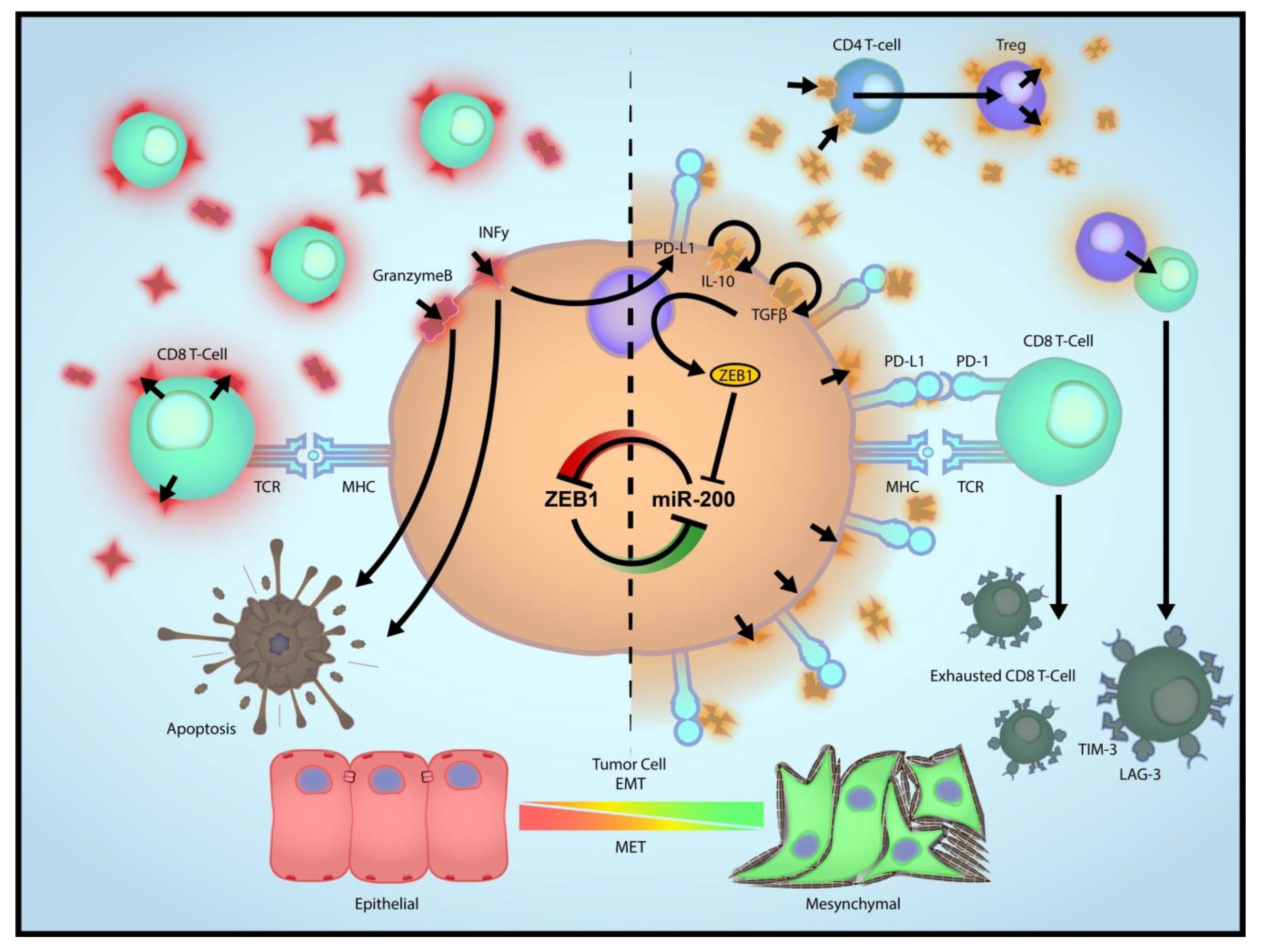
3. Role of EMT in Immune Evasion

EMT directly regulates expression of PD-L1 and is associated with several other checkpoint ligands [32,33] (Figure 1). Thus, EMT is expected to induce checkpoint-dependent resistance to anti-tumor immunity. Due to the redundancy of the multiple checkpoints, EMT may render cancer cells non-responsive to therapies targeting one or few checkpoints (e.g., anti-PD-L1 and anti-CTLA4). Due to the limited scope of this review, we are unable to discuss many articles here; however, the reader is referred some notable publications in this context [12,34,35,36,37,38]. Additionally, EMT drives the recruitment of tumor-associated macrophages, which may, in turn, mediate resistance to immunotherapies [39,40]. This may be achieved through direct regulation of cytokinome of cancer cells (e.g., CCL2). The immunosuppression by macrophages, especially the alternatively activated macrophages (M2), has been extensively studied and involves several mechanisms [41]. The tumor suppressive and tumor promoting effects of EMT shift the balance between macrophages and neutrophils. Thus, inhibition of EMT, while overcoming immunosuppression by cancer cells and macrophages, may coincidentally cause accumulation of a type of neutrophils in some tumors defined as myeloid-derived suppressor cells leading to an “escape” pathway from anti-EMT treatment. Therefore, it is important to examine the clinical correlation between EMT and the entire immune microenvironment, including the myeloid cell compartment.

4. EMT as a Driver of Immune Escape from ICBT

Despite the presence of an immune response, some tumors continue to grow, a process that has been referred to as “immune escape”. Tumor cell escape can occur through multiple different mechanisms [42]. Tumor cells themselves promote an immunosuppressive microenvironment by producing suppressive cytokines including TGF-β, VEGF, or indoleamine 2,3-dioxygenase. The tumor microenvironment also contains immune cells such as regulatory T cells and myeloid-derived suppressor cells that function to suppress the immune response. At the individual tumor cell level, alterations leading to decreased immune recognition (such as loss of tumor antigens, downregulation of major histocompatibility complex molecules, or loss of antigen processing function within the tumor cell), or increased resistance to the cytotoxic effects of immunity (such as via induction of anti-apoptotic mechanisms) can promote tumor growth. Finally, tumor cells can upregulate T cell-inhibitory molecules such as PD-L1, which is why activating the immune system for therapeutic benefit in cancer is an area of active investigation. For a more elaborate discussion on this topic, we refer readers to some recent reviews [10,43,44].
We and others have previously described epithelial-mesenchymal transition (EMT) as a frequent mechanism of de novo and acquired therapeutic resistance in mammary tumor cells, as well as all subtypes of NSCLC, including lung cancers with oncogenic driver mutations in EGFR and KRAS [45,46,47,48]. Furthermore, we and others have demonstrated that EMT upregulates expression of PD-L1 in murine and human NSCLC and directly leads to CD8+ T cell exhaustion and immunosuppression. To facilitate the study of EMT across large cohorts, our group identified a robust, platform-independent lung cancer EMT gene expression signature valid in lung cancer cell lines and NSCLC patient tumors [45]. The signature was then further refined to develop a patient-based, pan-cancer EMT signature using 1934 patient tumors from multiple solid tumor types including breast, lung, colon, and other common cancers [2]. The Lung EMT and Pan-Cancer EMT signatures are highly correlated with mesenchymal cancers from distinct tumor types showing striking similarities in their molecular profiles. Using the EMT signatures, an individual cancer cell line or tumor can be scored for the degree to which it has undergone EMT. These mRNA-based EMT signature scores are easy to measure, and are highly correlated with other established EMT markers (such as expression of E-cadherin protein) as well as with other factors known to regulate EMT (e.g., miR200 family and the transcription factor ZEB1). The applicability of EMT scores to predict lung cancer has been mirrored in breast cancer patients as well [49]. Residual breast cancers after conventional chemotherapy were shown to exhibit mesenchymal features. In addition to applying the EMT scores in our lung cancer research, we generated EMT scores for multiple TCGA cohorts, which has allowed integrated analyses of the relationships between EMT and other molecular or immune data profiles (i.e., miRNA and methylation profiles, iCLUSTER data) [32,50,51].
In addition to its broad tumor cell-autonomous impact, EMT in breast cancer also profoundly alters the microenvironment landscape, especially immune cell constituents (Figure 1). Macrophages and neutrophils are key modulators of the tumor microenvironment [52,53]. Recent evidence suggests a strong correlation between EMT and a switch from a neutrophil- enriched immune profile to a macrophage-dominant profile. Tumors exhibiting an epithelial-like phenotype tend to have local and systemic accumulation of neutrophils. In contrast, tumors with mesenchymal features are predominantly infiltrated with macrophages that are often but not always polarized to the M2 (alternatively activated) status. These two categories of tumors are defined as a neutrophil-enriched subtype (NES) and a macrophage-enriched subtype (MES), respectively. Inducible expression of miR-200, a master regulator of EMT, shifts the macrophage/neutrophil balance, supporting the causal role of EMT in determining the myeloid cell profile of the tumor microenvironment.
Mechanistically, several transcriptional suppressors regulate EMT, including the two-handed zinc-finger δEF1 family transcription factors ZEB1 and ZEB2 [54,55,56,57]. ZEB1/2 binds to E-box regions in the promoters of key epithelial differentiation genes such as E-cadherin, and transcriptionally suppress their expression [58]. ZEB1/2 also regulates the miR-200 family of miRNAs miR-141, 200a/b/c, and 429 that are broadly expressed in normal epithelial cells [59]. miR-200 is a master EMT regulator, governed by a double-negative feedback loop with the ZEB repressors [60,61,62,63,64] and regulated by multiple EMT inducers (e.g., TGFβ) [61,62]. miR-200 loss has been linked to stem-like features and chemoresistance [65,66]. Evidence from several epithelial tumor types, including lung and breast, implicates miR-200 dysregulation in disease progression [64,67,68]. Using the KP mouse model and a panel of human NSCLC cell lines, we have demonstrated that the miR-200/ZEB1 feedback loop is a critical regulatory axis that determines metastatic potential [69,70] by controlling global mRNA changes in an invasive subset of tumor cells, modulating matrix-dependent tumor activation and invasion [70,71,72,73].

5. Relationship between CD8 T Cells and EMT, and Impact of EMT on ICBT

In breast cancer, there is a strong correlation between tumor-infiltrating T and B cells and favorable prognostic outcome or therapeutic responses (standard therapies) [18,74,75,76,77]. Many standard-of-care therapies require the immune system to exert their effects. The most prominent example is anti-HER2 treatment with trastuzumab, which heavily relies on functional host immunity [78]. Furthermore, it has recently been recognized that the response of some chemotherapy regimens is achieved by their impact on immunosuppressive cells [79,80,81]. Thus, it appears that the immune system in breast cancer patients retains the potential of fighting cancer. Ongoing clinical trials suggest that although some breast cancer patients may benefit from ICBT [22,82], the percentage is disappointingly low. Possible mechanisms of resistance include low level or ineffective neoantigens [83,84] and/or the enrichment of immunosuppressive cells [85,86], and the elaboration of immunosuppressive cytokines (e.g., IL-10, TGF-β), all of which may result in a scarcity of functional cytotoxic T cells. Therefore, to improve the efficacy of immunotherapy it will be critically important to tackle these potential mechanisms. In the following section, we will focus on the latter: the immunosuppressive microenvironment that prevents anti-tumor immunity.
ICBT enhances anti-tumor responses by increasing the activity of the cytotoxic CD8+ T lymphocyte subpopulations; these cells are key players in the effector functions of adaptive immunity [87]. In addition to secreting chemokines and cytokines, tumor-specific T cells interact through T cell receptors with the major histocompatibility complex (MHC) on antigen-presenting cells, which in turn triggers a signaling cascade resulting in the death of target cells [88,89]. Currently, adoptive T cell-transfer, oncolytic viruses, cancer vaccines, T cell co-stimulatory agonists and monoclonal antibodies are used either alone or in combination with ICBT [90]. Checkpoint regulators of immune activation help maintain immune homeostasis and prevent autoimmunity. However, the immune checkpoint pathways are frequently activated in cancer to suppress the nascent anti-tumor immune response. A combination of immune checkpoint inhibitors namely PD-1 and CTLA-4 can effectively kill cancer cells because they function primarily through complementary mechanisms [91]. When CD8+ T cells recognize self-antigen on tumor cells, they fail to kill cancer cells; this immunological tolerance is a drawback of the ICBT [92]. In addition, CD8+ T cell exhaustion due to chronic exposure to antigens negatively affects the efficacy of ICBT [93].
Tumor cells evade immunosurveillance by altering their phenotype via immunoediting, and it is known that immuno-edited tumors display properties of cells that have undergone EMT [94]. Activated CD8+ T cells, macrophages, and several other immune cell types produce TGF-β, a crucial promoter of EMT [95,96,97]. We previously demonstrated that two key EMT factors that are also markers of cancer stem cells (CSCs), FOXC2 and Twist, are necessary for the process of breast carcinoma metastasis [57,98]. Early evidence suggested an antitumor response of CD8+ T cells delayed metastasis and eliminated disseminated tumor cells (DTC) of P815 mastocytoma [99]. In a melanoma mouse model, CD8+ T cells are involved in maintaining DTC dormancy in visceral organs like the lungs and the reproductive tract, thereby preventing overt metastasis and limiting disease progression [32]. CD8+ T cells are also known to inhibit tumor growth thus prolonging the survival of experimental mice by selectively targeting cancer stem cells (CSCs) [100]. CD8+ T cell suppression within the tumor microenvironment is dependent on PD-L1 regulation on tumor cells via a pathway involving the microRNA miR-200 and the transcription factor ZEB1; these molecules are the links between EMT, CD8+ T cell exhaustion, and tumor suppression [32]. Furthermore, the inhibition of breast cancer onset and progression is inhibited by CD8+ T cells and natural killer (NK) cells due to increased cytotoxic activity mediated by the protein TIPE2 [101]. Although EMT leads to up-regulation of multiple checkpoint molecules that inhibit T cell-mediated cytotoxicity, it also reduces expression of many adhesion molecules including E-cadherin, which is a known inhibitory ligand of NK cell receptor (KLRG1) [102]. Other well-established NK cell-activating molecules, such as PVR, are also upregulated upon EMT induction [103,104]. It is, therefore, not surprising that EMT may be accompanied by increased sensitivity to NK cell-mediated cytotoxicity [105]. Although the anti-tumor potential of CD8+ T cells is well accepted, the prognostic significance of their intratumoral homing is highly variable across different breast tumor subtypes [106]. The impact of CD8+ T cells in the tumor microenvironment (TME) on tumor epithelial-mesenchymal plasticity, on the interplay with other immune cells, and on associated metastatic traits in breast cancer cells are incompletely understood. Furthermore, it will be critical to clarify how the inflammatory TME and epithelial-mesenchymal plasticity influence CD8+ T cell activity and survival.

6. Impact of Immunosuppressive TME on ICBT

The intertumoral heterogeneity of the immune microenvironment dictates ICBT responses. We profiled the immune compartment in a wide variety of syngeneic triple-negative breast cancer (TNBC) models and discovered two prototypes of the immune microenvironment. In the first prototype, tumors induce systemic accumulation of neutrophils. These neutrophils overexpress multiple immunosuppressive pathways and may represent granulocytic myeloid-derived suppressor cells (gMDSCs). Macrophages co-exist but only constitute a minority of the myeloid cells in the tumor. In the second prototype, there does not appear to be an increase of neutrophils; rather, there is exclusively a local enrichment of tumor macrophages, which often (but not always) polarize toward the immunosuppressive M2 status. We have denoted these two “immunosubtypes” of mammary tumors as NES and MES, respectively. Apparently, NES and MES rely on different types of myeloid cells to escape immunosurveillance (data not shown). Indeed, when initially responsive MES tumors recurred neutrophils or gMDSCs accumulated, suggesting a conversion from MES toward NES or a switch of suppressor cell types. Depletion of neutrophils reduced this acquired ICBT resistance. Thus, immunosuppression may be exerted by different cell types in different tumor contexts.
EMT clearly contributes to the development of different immune microenvironments. Intriguingly, analyses of eight murine models representative of both NES and MES subtypes revealed that EMT contributes to the development of the dichotomous myeloid microenvironment. EMT has been linked to the recruitment of macrophages to the tumor microenvironment via chemokines like CCL2 [39,40]. Previous studies have also demonstrated that EMT drives expression of checkpoint ligands in cancer cells [51]. Taken together, the connection between EMT and cancer cell- or macrophage-mediated immunosuppression has been well established, which makes it an appealing therapeutic target to enhance immunotherapies. Inhibition of EMT, by targeting the EMT signaling pathways using TGF-β1 inhibitors while reverse immunosuppression by cancer cells and macrophages, may coincidentally cause accumulation of neutrophils, which can act as gMDSCs and lead to an alternative immunosuppressive mechanism, independent of checkpoints. Indeed, previous studies using syngeneic lung cancer models already indicate the existence of the dichotomous myeloid cell compartment in this cancer type as well [107]. Preclinical studies demonstrate that gMDSCs promote tumor progression through suppressing anti-tumor immunity [108,109] and promoting tumor-initiating cells (TIC) through the Notch pathway [109]. Interestingly, a recent study suggested that the former activity may depend on endogenous estrogen receptor alpha (ER) signaling [110], raising the possibility that endocrine deprivation therapies could be used for gMDSC elimination [111]. Endocrine deprivation therapies are standard-of-care for ER+ breast cancer.
All these findings suggest that EMT drives a switch of immunosuppression from gMDSC-mediated, checkpoint-independent mechanisms to macrophage/cancer cell-mediated, checkpoint-dependent mechanisms. Consequently, gMDSC accumulation may represent an escape pathway upon EMT inhibition that allows tumors to maintain an immunosuppressive microenvironment. Moreover, estrogen signaling and the Notch pathway mediate the pro-tumor effects of gMDSCs and provide potential therapeutic targets to eliminate these cells, a strategy that may complement anti-EMT treatment and ICBT.

7. Role of Bioinformatics

To identify potential relationships between phenotypic traits of tumors, including EMT and immune cell populations, and cancer outcomes, bioinformatics has long played a critical role. Gene expression profiles reveal the underlying biology of the tumors and can be used to dissect the features that are correlated with responses. A key observation is that signaling events lead to a transcriptional response, even if they are driven upstream by post-translational modifications. Thus, the signature of a pathway can be used to identify the molecular processes associated with clinical events.
Gene expression signatures involving biological processes have been generated using machine learning techniques. In short, the development of signatures is typically framed as a classification problem, where the goal is to identify a set of genes that can distinguish two biological states, such as epithelial or mesenchymal cells. To accomplish this, a training set is generated comprised of gene expression profiles of the two states. Then, differential expression analysis methods, such as DESeq2 [112] or EdgeR [113], are used to identify genes that can differentiate the two states. Finally, machine learning algorithms, such as SIGNATURE [114], can be used to score the expression profiles of new samples to provide a quantitative measure of the similarity to one or the other state.
Based on such approaches, an EMT signature has been linked to several outcomes including responses to chemotherapy [115], targeted therapies [116], patient survival [117,118], recurrence [119], and metastasis [120]. In addition to these outcomes, our studies and others have linked an EMT signature with markers or other evidence of immune modulation such as immune checkpoints [51] or low T cell infiltration in NSCLC [121]. Indeed, an increasing amount of evidence points to an association between EMT and the immune system in human tumors.
Independent of EMT, several bioinformatic analyses have supported the role of immune cells and clinical outcomes [122,123,124,125,126,127,128,129,130]. This demonstrates the ability of this technology to identify signatures of immune cells in bulk tumor samples. These analyses are enabled by the fact that the gene expression profiles of tumors reflect both the cancer cell and stromal/immune compartments. Although the signature of the immune cells can be seen in the bulk gene expression profiles, methods have been developed that can deconvolute the profiles into the constituent parts, enabling a more accurate quantification of the immune cell types that comprise the tumor [131,132].
One of the limitations of the prior studies is that they provide a limited resolution in quantifying the immune cell types (i.e., how many CD4+ T cells there are), and also in identifying the cell subtypes (i.e., are these Th1, Th2, or other CD4+ T cells). To address these questions, single-cell sequencing technologies that have the have the capacity to profile a range of cell types are rapidly being adopted. While powerful, these technologies have introduced a new bioinformatics challenge. The result of a single-cell RNA-Seq (scRNA-Seq) assay is the generation of transcriptional profiles of a likely heterogeneous immune cell population. Therefore, one step in the processing is to identify the cell types present in the population. While markers for immune cells have long been established for flow cytometry experiments, they are of more limited use in scRNA-Seq profiles due to issues such as drop-out, a phenomenon where a gene is not profiled due to factors including a lack of sensitivity in the assay; or ambiguities in the accepted markers. In the future, to address this, machine learning methods can be applied to identify immune cells from scRNA-Seq profiles. Nevertheless, a recent study has revealed a previously unknown range of T cell activation states within breast tumors [133]. Future studies using single-cell technologies, coupled with increasingly sophisticated bioinformatic analyses, will likely reveal new nuances in the relationships between the immune system, EMT, and cancer. Additional multiplex technologies will allow the spatial localization of these cells.

8. Conclusions

In this review, we have highlighted mechanistic vulnerabilities in mesenchymal tumor cells due to their ability to reprogram the tumor immune microenvironment. Because immunotherapy is rapidly emerging as a game-changing approach in many cancer types, including lung cancer, it will be critical to develop a greater understanding of those patients most likely to benefit and the mechanisms defining primary and acquired resistance. It will also be important to co-target EMT-related vulnerabilities along with the PD-L1/PD-1 immune checkpoint axis, given the large contribution of EMT to resistance mechanisms. Most importantly, the EMT phenotype has a distinct relationship with the immune microenvironment that can potentially be leveraged for a transformative clinical benefit for common epithelial tumors. EMT broadly up-regulates multiple immune checkpoint and inflammatory molecules to produce CD8+ T cell exhaustion, highlighting multiple potential mechanisms for the development of therapeutic resistance to immune therapy in mesenchymal tumors. EMT is a multidimensional process with different axes and these multiple parameters are exploited in malignancy under selective pressures including cytotoxic and biological therapies, hypoxia, energetics, and immune surveillance. As such, there is a pressing clinical need for targeted, biomarker-directed therapies to address both primary and acquired immunotherapy resistance. Incorporation of targeted agents (and validated biomarkers) that modulate immune suppression could expand the patient population that responds to immune checkpoint inhibitors and help address immunotherapy resistance. Studying these questions and integrating information across cancer types, instead of a single cancer, will shed light on combinatorial strategies that may be more generally applicable. Moreover, these comparative studies may also yield new insights since the treatment of cancer as evidenced in recent basket trials now is geared to understanding common vulnerabilities, e.g., mismatch repair deficiencies, as criteria for using ICBT across tumor types.

Acknowledgments

We thank NIH/NCI CCSG P30-CA01667 (L.A.B.), NIH/NCI SPORE P5-CA070907 (L.A.B.), an MD Andersen Cancer Center Physician Scientist Award (L.A.B.), LUNGevity Foundation (L.A.B.), philanthropic contributions to The University of Texas MD Anderson Cancer Center Lung Cancer Moonshot Program (L.A.B.), NIH R37 CA214609-01A1 (D.L.G.), CPRIT-MIRA-RP160652-P3 (D.L.G.), Rexanna’s Foundation for Fighting Lung Cancer (D.L.G.), P50 CA070907 from UT Lung SPORE Career Enhancement Award (J.M.K.), NIH/NCI-R01CA200970 (S.A.M.), NIH/NCI-2R01CA155243 (S.A.M.), National Science Foundation-15-597-1605817 (S.A.M.), CPRIT-MIRA-RP160710 (S.A.M., Project-3), CPRIT-IIRA-RP170172 (J.M.R., S.A.M.), Diana Helis Henry and Andrienne Helis Malvin Medical Research Foundation Grant (J.M.R., X.Z.), P50 CA140388 from the National Cancer Institute (University of Texas MD Anderson Prostate Cancer SPORE Career Enhancement Program Award) (R.S.), MD Anderson Institutional Research Grant (R.S.), CPRIT-MIRA-RP180712 (D.T., Core), CPRIT-RP180466 (N.V.), MRA-509800 (N.V.), for financial support.

Conflicts of Interest

L.A.B. serves as a consultant for AstraZeneca; AbbVie; GenMab; BergenBio; and Pharma Mar, SA, and has research support from AbbVie; AstraZeneca; GenMab; Sierra Oncology; and Tolero Pharmaceuticals. N.V. is the founder and CSO of CellChorus. All other authors declare no conflict of interest.

References

  1. Siegel, R.L.; Miller, K.D.; Jemal, A. Cancer Statistics, 2017. CA Cancer J. Clin. 2017, 67, 7–30. [Google Scholar] [CrossRef] [PubMed]
  2. Mak, M.P.; Tong, P.; Diao, L.; Cardnell, R.J.; Gibbons, D.L.; William, W.N.; Skoulidis, F.; Parra, E.R.; Rodriguez-Canales, J.; Wistuba, I.I.; et al. A Patient-Derived, Pan-Cancer EMT Signature Identifies Global Molecular Alterations and Immune Target Enrichment Following Epithelial-to-Mesenchymal Transition. Clin. Cancer Res. 2016, 22, 609–620. [Google Scholar] [CrossRef] [PubMed]
  3. Casasent, A.K.; Schalck, A.; Gao, R.; Sei, E.; Long, A.; Pangburn, W.; Casasent, T.; Meric-Bernstam, F.; Edgerton, M.E.; Navin, N.E. Multiclonal Invasion in Breast Tumors Identified by Topographic Single Cell Sequencing. Cell 2018, 172, 205–217.e212. [Google Scholar] [CrossRef] [PubMed]
  4. Hernandez, L.; Wilkerson, P.M.; Lambros, M.B.; Campion-Flora, A.; Rodrigues, D.N.; Gauthier, A.; Cabral, C.; Pawar, V.; Mackay, A.; A’Hern, R.; et al. Genomic and mutational profiling of ductal carcinomas in situ and matched adjacent invasive breast cancers reveals intra-tumour genetic heterogeneity and clonal selection. J. Pathol. 2012, 227, 42–52. [Google Scholar] [CrossRef] [Green Version]
  5. Niyomnaitham, S.; Parinyanitikul, N.; Roothumnong, E.; Jinda, W.; Samarnthai, N.; Atikankul, T.; Suktitipat, B.; Thongnoppakhun, W.; Limwongse, C.; Pithukpakorn, M. Tumor mutational profile of triple negative breast cancer patients in Thailand revealed distinctive genetic alteration in chromatin remodeling gene. PeerJ 2019, 7, e6501. [Google Scholar] [CrossRef]
  6. Fardi, M.; Alivand, M.; Baradaran, B.; Farshdousti Hagh, M.; Solali, S. The crucial role of ZEB2: From development to epithelial-to-mesenchymal transition and cancer complexity. J. Cell Physiol. 2019. [Google Scholar] [CrossRef] [PubMed]
  7. Zhang, L.; Liang, Y.; Li, S.; Zeng, F.; Meng, Y.; Chen, Z.; Liu, S.; Tao, Y.; Yu, F. The interplay of circulating tumor DNA and chromatin modification, therapeutic resistance, and metastasis. Mol. Cancer 2019, 18, 36. [Google Scholar] [CrossRef]
  8. Schwarzenbacher, D.; Balic, M.; Pichler, M. The role of microRNAs in breast cancer stem cells. Int. J. Mol. Sci. 2013, 14, 14712–14723. [Google Scholar] [CrossRef]
  9. Markiewicz, A.; Topa, J.; Nagel, A.; Skokowski, J.; Seroczynska, B.; Stokowy, T.; Welnicka-Jaskiewicz, M.; Zaczek, A.J. Spectrum of Epithelial-Mesenchymal Transition Phenotypes in Circulating Tumour Cells from Early Breast Cancer Patients. Cancers 2019, 11, 59. [Google Scholar] [CrossRef] [PubMed]
  10. Terry, S.; Savagner, P.; Ortiz-Cuaran, S.; Mahjoubi, L.; Saintigny, P.; Thiery, J.P.; Chouaib, S. New insights into the role of EMT in tumor immune escape. Mol. Oncol. 2017, 11, 824–846. [Google Scholar] [CrossRef] [Green Version]
  11. Akalay, I.; Janji, B.; Hasmim, M.; Noman, M.Z.; Andre, F.; De Cremoux, P.; Bertheau, P.; Badoual, C.; Vielh, P.; Larsen, A.K.; et al. Epithelial-to-mesenchymal transition and autophagy induction in breast carcinoma promote escape from T-cell-mediated lysis. Cancer Res. 2013, 73, 2418–2427. [Google Scholar] [CrossRef]
  12. Chouaib, S.; Janji, B.; Tittarelli, A.; Eggermont, A.; Thiery, J.P. Tumor plasticity interferes with anti-tumor immunity. Crit. Rev. Immunol. 2014, 34, 91–102. [Google Scholar] [CrossRef] [PubMed]
  13. Hanahan, D.; Weinberg, R.A. Hallmarks of cancer: The next generation. Cell 2011, 144, 646–674. [Google Scholar] [CrossRef]
  14. Terry, S.; Chouaib, S. EMT in immuno-resistance. Oncoscience 2015, 2, 841–842. [Google Scholar] [CrossRef] [PubMed]
  15. Sharma, P.; Allison, J.P. Immune checkpoint targeting in cancer therapy: Toward combination strategies with curative potential. Cell 2015, 161, 205–214. [Google Scholar] [CrossRef]
  16. Pardoll, D.M. The blockade of immune checkpoints in cancer immunotherapy. Nat. Rev. Cancer 2012, 12, 252–264. [Google Scholar] [CrossRef] [Green Version]
  17. Topalian, S.L.; Drake, C.G.; Pardoll, D.M. Immune checkpoint blockade: A common denominator approach to cancer therapy. Cancer Cell 2015, 27, 450–461. [Google Scholar] [CrossRef]
  18. Gentles, A.J.; Newman, A.M.; Liu, C.L.; Bratman, S.V.; Feng, W.; Kim, D.; Nair, V.S.; Xu, Y.; Khuong, A.; Hoang, C.D.; et al. The prognostic landscape of genes and infiltrating immune cells across human cancers. Nat. Med. 2015, 21, 938–945. [Google Scholar] [CrossRef] [PubMed]
  19. Brahmer, J.R.; Tykodi, S.S.; Chow, L.Q.; Hwu, W.J.; Topalian, S.L.; Hwu, P.; Drake, C.G.; Camacho, L.H.; Kauh, J.; Odunsi, K.; et al. Safety and activity of anti-PD-L1 antibody in patients with advanced cancer. N. Engl. J. Med. 2012, 366, 2455–2465. [Google Scholar] [CrossRef]
  20. Zhang, M.; Sun, H.; Zhao, S.; Wang, Y.; Pu, H.; Wang, Y.; Zhang, Q. Expression of PD-L1 and prognosis in breast cancer: A meta-analysis. Oncotarget 2017, 8, 31347–31354. [Google Scholar] [CrossRef] [PubMed]
  21. Mittendorf, E.A.; Philips, A.V.; Meric-Bernstam, F.; Qiao, N.; Wu, Y.; Harrington, S.; Su, X.; Wang, Y.; Gonzalez-Angulo, A.M.; Akcakanat, A.; et al. PD-L1 expression in triple-negative breast cancer. Cancer Immunol. Res. 2014, 2, 361–370. [Google Scholar] [CrossRef]
  22. Nanda, R.; Chow, L.Q.; Dees, E.C.; Berger, R.; Gupta, S.; Geva, R.; Pusztai, L.; Pathiraja, K.; Aktan, G.; Cheng, J.D.; et al. Pembrolizumab in Patients With Advanced Triple-Negative Breast Cancer: Phase Ib KEYNOTE-012 Study. J. Clin. Oncol. 2016, 34, 2460–2467. [Google Scholar] [CrossRef]
  23. Dirix, L.Y.; Takacs, I.; Jerusalem, G.; Nikolinakos, P.; Arkenau, H.T.; Forero-Torres, A.; Boccia, R.; Lippman, M.E.; Somer, R.; Smakal, M.; et al. Avelumab, an anti-PD-L1 antibody, in patients with locally advanced or metastatic breast cancer: A phase 1b JAVELIN Solid Tumor study. Breast Cancer Res. Treat. 2018, 167, 671–686. [Google Scholar] [CrossRef]
  24. Adams, S.; Schmid, P.; Rugo, H.S.; Winer, E.P.; Loirat, D.; Awada, A.; Cescon, D.W.; Iwata, H.; Campone, M.; Nanda, R.; et al. Pembrolizumab Monotherapy for Previously Treated Metastatic Triple-Negative Breast Cancer: Cohort A of the Phase 2 KEYNOTE-086 Study. Ann. Oncol. 2018. [Google Scholar] [CrossRef]
  25. Wang, Y.J.; Fletcher, R.; Yu, J.; Zhang, L. Immunogenic effects of chemotherapy-induced tumor cell death. Genes Dis. 2018, 5, 194–203. [Google Scholar] [CrossRef]
  26. Schmid, P.; Adams, S.; Rugo, H.S.; Schneeweiss, A.; Barrios, C.H.; Iwata, H.; Dieras, V.; Hegg, R.; Im, S.A.; Shaw Wright, G.; et al. Atezolizumab and Nab-Paclitaxel in Advanced Triple-Negative Breast Cancer. N. Engl. J. Med. 2018. [Google Scholar] [CrossRef]
  27. Reck, M.; Rodriguez-Abreu, D.; Robinson, A.G.; Hui, R.; Csoszi, T.; Fulop, A.; Gottfried, M.; Peled, N.; Tafreshi, A.; Cuffe, S.; et al. Pembrolizumab versus Chemotherapy for PD-L1-Positive Non-Small-Cell Lung Cancer. N. Engl. J. Med. 2016, 375, 1823–1833. [Google Scholar] [CrossRef] [PubMed]
  28. Langer, C.J.; Gadgeel, S.M.; Borghaei, H.; Papadimitrakopoulou, V.A.; Patnaik, A.; Powell, S.F.; Gentzler, R.D.; Martins, R.G.; Stevenson, J.P.; Jalal, S.I.; et al. Carboplatin and pemetrexed with or without pembrolizumab for advanced, non-squamous non-small-cell lung cancer: A randomised, phase 2 cohort of the open-label KEYNOTE-021 study. Lancet Oncol. 2016, 17, 1497–1508. [Google Scholar] [CrossRef]
  29. Rittmeyer, A.; Barlesi, F.; Waterkamp, D.; Park, K.; Ciardiello, F.; von Pawel, J.; Gadgeel, S.M.; Hida, T.; Kowalski, D.M.; Dols, M.C.; et al. Atezolizumab versus docetaxel in patients with previously treated non-small-cell lung cancer (OAK): A phase 3, open-label, multicentre randomised controlled trial. Lancet 2017, 389, 255–265. [Google Scholar] [CrossRef]
  30. Reck, M.; Rabe, K.F. Precision Diagnosis and Treatment for Advanced Non-Small-Cell Lung Cancer. N. Engl. J. Med. 2017, 377, 849–861. [Google Scholar] [CrossRef]
  31. McArthur, H.L.; Page, D.B. Immunotherapy for the treatment of breast cancer: Checkpoint blockade, cancer vaccines, and future directions in combination immunotherapy. Clin. Adv. Hematol. Oncol. 2016, 14, 922–933. [Google Scholar] [PubMed]
  32. Chen, L.; Gibbons, D.L.; Goswami, S.; Cortez, M.A.; Ahn, Y.H.; Byers, L.A.; Zhang, X.; Yi, X.; Dwyer, D.; Lin, W.; et al. Metastasis is regulated via microRNA-200/ZEB1 axis control of tumour cell PD-L1 expression and intratumoral immunosuppression. Nat. Commun. 2014, 5, 5241. [Google Scholar] [CrossRef]
  33. Loi, S.; Dushyanthen, S.; Beavis, P.A.; Salgado, R.; Denkert, C.; Savas, P.; Combs, S.; Rimm, D.L.; Giltnane, J.M.; Estrada, M.V.; et al. RAS/MAPK Activation Is Associated with Reduced Tumor-Infiltrating Lymphocytes in Triple-Negative Breast Cancer: Therapeutic Cooperation Between MEK and PD-1/PD-L1 Immune Checkpoint Inhibitors. Clin. Cancer Res. 2016, 22, 1499–1509. [Google Scholar] [CrossRef]
  34. Dong, P.; Xiong, Y.; Yue, J.; Hanley, S.J.B.; Watari, H. Tumor-Intrinsic PD-L1 Signaling in Cancer Initiation, Development and Treatment: Beyond Immune Evasion. Front. Oncol. 2018, 8, 386. [Google Scholar] [CrossRef] [Green Version]
  35. Asgarova, A.; Asgarov, K.; Godet, Y.; Peixoto, P.; Nadaradjane, A.; Boyer-Guittaut, M.; Galaine, J.; Guenat, D.; Mougey, V.; Perrard, J.; et al. PD-L1 expression is regulated by both DNA methylation and NF-kB during EMT signaling in non-small cell lung carcinoma. Oncoimmunology 2018, 7, e1423170. [Google Scholar] [CrossRef] [PubMed] [Green Version]
  36. Li, F.; Zhu, T.; Yue, Y.; Zhu, X.; Wang, J.; Liang, L. Preliminary mechanisms of regulating PDL1 expression in nonsmall cell lung cancer during the EMT process. Oncol. Rep. 2018, 40, 775–782. [Google Scholar] [CrossRef]
  37. Kumar, S.; Davra, V.; Obr, A.E.; Geng, K.; Wood, T.L.; De Lorenzo, M.S.; Birge, R.B. Crk adaptor protein promotes PD-L1 expression, EMT and immune evasion in a murine model of triple-negative breast cancer. Oncoimmunology 2017, 7, e1376155. [Google Scholar] [CrossRef] [PubMed]
  38. Kim, S.; Koh, J.; Kim, M.Y.; Kwon, D.; Go, H.; Kim, Y.A.; Jeon, Y.K.; Chung, D.H. PD-L1 expression is associated with epithelial-to-mesenchymal transition in adenocarcinoma of the lung. Hum. Pathol. 2016, 58, 7–14. [Google Scholar] [CrossRef] [PubMed]
  39. Low-Marchelli, J.M.; Ardi, V.C.; Vizcarra, E.A.; van Rooijen, N.; Quigley, J.P.; Yang, J. Twist1 induces CCL2 and recruits macrophages to promote angiogenesis. Cancer Res. 2013, 73, 662–671. [Google Scholar] [CrossRef]
  40. Hsu, D.S.; Wang, H.J.; Tai, S.K.; Chou, C.H.; Hsieh, C.H.; Chiu, P.H.; Chen, N.J.; Yang, M.H. Acetylation of snail modulates the cytokinome of cancer cells to enhance the recruitment of macrophages. Cancer Cell 2014, 26, 534–548. [Google Scholar] [CrossRef]
  41. Noy, R.; Pollard, J.W. Tumor-associated macrophages: From mechanisms to therapy. Immunity 2014, 41, 49–61. [Google Scholar] [CrossRef]
  42. Schreiber, R.D.; Old, L.J.; Smyth, M.J. Cancer immunoediting: Integrating immunity’s roles in cancer suppression and promotion. Science 2011, 331, 1565–1570. [Google Scholar] [CrossRef]
  43. Galon, J.; Bruni, D. Approaches to treat immune hot, altered and cold tumours with combination immunotherapies. Nat. Rev. Drug Discov. 2019, 18, 197–218. [Google Scholar] [CrossRef]
  44. Otsuki, Y.; Saya, H.; Arima, Y. Prospects for new lung cancer treatments that target EMT signaling. Dev. Dyn. 2018, 247, 462–472. [Google Scholar] [CrossRef]
  45. Byers, L.A.; Diao, L.; Wang, J.; Saintigny, P.; Girard, L.; Peyton, M.; Shen, L.; Fan, Y.; Giri, U.; Tumula, P.K.; et al. An epithelial-mesenchymal transition gene signature predicts resistance to EGFR and PI3K inhibitors and identifies Axl as a therapeutic target for overcoming EGFR inhibitor resistance. Clin. Cancer Res. 2013, 19, 279–290. [Google Scholar] [CrossRef] [PubMed]
  46. Camidge, D.R.; Pao, W.; Sequist, L.V. Acquired resistance to TKIs in solid tumours: Learning from lung cancer. Nat. Rev. Clin. Oncol. 2014, 11, 473–481. [Google Scholar] [CrossRef] [PubMed]
  47. Zhang, Z.; Lee, J.C.; Lin, L.; Olivas, V.; Au, V.; Laframboise, T.; Abdel-Rahman, M.; Wang, X.; Levine, A.D.; Rho, J.K.; et al. Activation of the AXL kinase causes resistance to EGFR-targeted therapy in lung cancer. Nat. Genet. 2012, 44, 852–860. [Google Scholar] [CrossRef] [PubMed]
  48. Dongre, A.; Rashidian, M.; Reinhardt, F.; Bagnato, A.; Keckesova, Z.; Ploegh, H.L.; Weinberg, R.A. Epithelial-to-Mesenchymal Transition Contributes to Immunosuppression in Breast Carcinomas. Cancer Res. 2017, 77, 3982–3989. [Google Scholar] [CrossRef] [PubMed]
  49. Creighton, C.J.; Li, X.; Landis, M.; Dixon, J.M.; Neumeister, V.M.; Sjolund, A.; Rimm, D.L.; Wong, H.; Rodriguez, A.; Herschkowitz, J.I.; et al. Residual breast cancers after conventional therapy display mesenchymal as well as tumor-initiating features. Proc. Natl. Acad. Sci. USA 2009, 106, 13820–13825. [Google Scholar] [CrossRef] [Green Version]
  50. Allison Stewart, C.; Tong, P.; Cardnell, R.J.; Sen, T.; Li, L.; Gay, C.M.; Masrorpour, F.; Fan, Y.; Bara, R.O.; Feng, Y.; et al. Dynamic variations in epithelial-to-mesenchymal transition (EMT), ATM, and SLFN11 govern response to PARP inhibitors and cisplatin in small cell lung cancer. Oncotarget 2017, 8, 28575–28587. [Google Scholar] [CrossRef] [Green Version]
  51. Lou, Y.; Diao, L.; Cuentas, E.R.; Denning, W.L.; Chen, L.; Fan, Y.H.; Byers, L.A.; Wang, J.; Papadimitrakopoulou, V.A.; Behrens, C.; et al. Epithelial-Mesenchymal Transition Is Associated with a Distinct Tumor Microenvironment Including Elevation of Inflammatory Signals and Multiple Immune Checkpoints in Lung Adenocarcinoma. Clin. Cancer Res. 2016, 22, 3630–3642. [Google Scholar] [CrossRef] [PubMed]
  52. Kim, J.; Bae, J.S. Tumor-Associated Macrophages and Neutrophils in Tumor Microenvironment. Mediat. Inflamm. 2016, 2016, 6058147. [Google Scholar] [CrossRef] [PubMed]
  53. Carron, E.C.; Homra, S.; Rosenberg, J.; Coffelt, S.B.; Kittrell, F.; Zhang, Y.; Creighton, C.J.; Fuqua, S.A.; Medina, D.; Machado, H.L. Macrophages promote the progression of premalignant mammary lesions to invasive cancer. Oncotarget 2017, 8, 50731–50746. [Google Scholar] [CrossRef] [PubMed] [Green Version]
  54. Comijn, J.; Berx, G.; Vermassen, P.; Verschueren, K.; van Grunsven, L.; Bruyneel, E.; Mareel, M.; Huylebroeck, D.; van Roy, F. The two-handed E box binding zinc finger protein SIP1 downregulates E-cadherin and induces invasion. Mol. Cell 2001, 7, 1267–1278. [Google Scholar] [CrossRef]
  55. Huber, M.A.; Kraut, N.; Beug, H. Molecular requirements for epithelial-mesenchymal transition during tumor progression. Curr. Opin. Cell Biol. 2005, 17, 548–558. [Google Scholar] [CrossRef]
  56. Peinado, H.; Olmeda, D.; Cano, A. Snail, Zeb and bHLH factors in tumour progression: An alliance against the epithelial phenotype? Nat. Rev. Cancer 2007, 7, 415–428. [Google Scholar] [CrossRef]
  57. Yang, J.; Mani, S.A.; Donaher, J.L.; Ramaswamy, S.; Itzykson, R.A.; Come, C.; Savagner, P.; Gitelman, I.; Richardson, A.; Weinberg, R.A. Twist, a master regulator of morphogenesis, plays an essential role in tumor metastasis. Cell 2004, 117, 927–939. [Google Scholar] [CrossRef]
  58. Eger, A.; Aigner, K.; Sonderegger, S.; Dampier, B.; Oehler, S.; Schreiber, M.; Berx, G.; Cano, A.; Beug, H.; Foisner, R. DeltaEF1 is a transcriptional repressor of E-cadherin and regulates epithelial plasticity in breast cancer cells. Oncogene 2005, 24, 2375–2385. [Google Scholar] [CrossRef] [Green Version]
  59. Gregory, P.A.; Bracken, C.P.; Bert, A.G.; Goodall, G.J. MicroRNAs as regulators of epithelial-mesenchymal transition. Cell Cycle 2008, 7, 3112–3117. [Google Scholar] [CrossRef]
  60. Bracken, C.P.; Gregory, P.A.; Kolesnikoff, N.; Bert, A.G.; Wang, J.; Shannon, M.F.; Goodall, G.J. A double-negative feedback loop between ZEB1-SIP1 and the microRNA-200 family regulates epithelial-mesenchymal transition. Cancer Res. 2008, 68, 7846–7854. [Google Scholar] [CrossRef] [PubMed]
  61. Burk, U.; Schubert, J.; Wellner, U.; Schmalhofer, O.; Vincan, E.; Spaderna, S.; Brabletz, T. A reciprocal repression between ZEB1 and members of the miR-200 family promotes EMT and invasion in cancer cells. EMBO Rep. 2008, 9, 582–589. [Google Scholar] [CrossRef] [Green Version]
  62. Gregory, P.A.; Bert, A.G.; Paterson, E.L.; Barry, S.C.; Tsykin, A.; Farshid, G.; Vadas, M.A.; Khew-Goodall, Y.; Goodall, G.J. The miR-200 family and miR-205 regulate epithelial to mesenchymal transition by targeting ZEB1 and SIP1. Nat. Cell Biol. 2008, 10, 593–601. [Google Scholar] [CrossRef]
  63. Korpal, M.; Lee, E.S.; Hu, G.; Kang, Y. The miR-200 family inhibits epithelial-mesenchymal transition and cancer cell migration by direct targeting of E-cadherin transcriptional repressors ZEB1 and ZEB2. J. Biol. Chem. 2008, 283, 14910–14914. [Google Scholar] [CrossRef]
  64. Park, S.M.; Gaur, A.B.; Lengyel, E.; Peter, M.E. The miR-200 family determines the epithelial phenotype of cancer cells by targeting the E-cadherin repressors ZEB1 and ZEB2. Genes Dev. 2008, 22, 894–907. [Google Scholar] [CrossRef] [Green Version]
  65. Shimono, Y.; Zabala, M.; Cho, R.W.; Lobo, N.; Dalerba, P.; Qian, D.; Diehn, M.; Liu, H.; Panula, S.P.; Chiao, E.; et al. Downregulation of miRNA-200c links breast cancer stem cells with normal stem cells. Cell 2009, 138, 592–603. [Google Scholar] [CrossRef]
  66. Wellner, U.; Schubert, J.; Burk, U.C.; Schmalhofer, O.; Zhu, F.; Sonntag, A.; Waldvogel, B.; Vannier, C.; Darling, D.; zur Hausen, A.; et al. The EMT-activator ZEB1 promotes tumorigenicity by repressing stemness-inhibiting microRNAs. Nat. Cell Biol. 2009, 11, 1487–1495. [Google Scholar] [CrossRef] [PubMed]
  67. Ceppi, P.; Mudduluru, G.; Kumarswamy, R.; Rapa, I.; Scagliotti, G.V.; Papotti, M.; Allgayer, H. Loss of miR-200c expression induces an aggressive, invasive, and chemoresistant phenotype in non-small cell lung cancer. Mol. Cancer Res. 2010, 8, 1207–1216. [Google Scholar] [CrossRef] [PubMed]
  68. Hu, X.; Schwarz, J.K.; Lewis, J.S., Jr.; Huettner, P.C.; Rader, J.S.; Deasy, J.O.; Grigsby, P.W.; Wang, X. A microRNA expression signature for cervical cancer prognosis. Cancer Res. 2010, 70, 1441–1448. [Google Scholar] [CrossRef] [PubMed]
  69. Gibbons, D.L.; Lin, W.; Creighton, C.J.; Rizvi, Z.H.; Gregory, P.A.; Goodall, G.J.; Thilaganathan, N.; Du, L.; Zhang, Y.; Pertsemlidis, A.; et al. Contextual extracellular cues promote tumor cell EMT and metastasis by regulating miR-200 family expression. Genes Dev. 2009, 23, 2140–2151. [Google Scholar] [CrossRef] [Green Version]
  70. Ahn, Y.H.; Gibbons, D.L.; Chakravarti, D.; Creighton, C.J.; Rizvi, Z.H.; Adams, H.P.; Pertsemlidis, A.; Gregory, P.A.; Wright, J.A.; Goodall, G.J.; et al. ZEB1 drives prometastatic actin cytoskeletal remodeling by downregulating miR-34a expression. J. Clin. Investig. 2012, 122, 3170–3183. [Google Scholar] [CrossRef] [PubMed] [Green Version]
  71. Yang, Y.; Ahn, Y.H.; Gibbons, D.L.; Zang, Y.; Lin, W.; Thilaganathan, N.; Alvarez, C.A.; Moreira, D.C.; Creighton, C.J.; Gregory, P.A.; et al. The Notch ligand Jagged2 promotes lung adenocarcinoma metastasis through a miR-200-dependent pathway in mice. J. Clin. Investig. 2011, 121, 1373–1385. [Google Scholar] [CrossRef] [PubMed]
  72. Ungewiss, C.; Rizvi, Z.H.; Roybal, J.D.; Peng, D.H.; Gold, K.A.; Shin, D.H.; Creighton, C.J.; Gibbons, D.L. The microRNA-200/Zeb1 axis regulates ECM-dependent beta1-integrin/FAK signaling, cancer cell invasion and metastasis through CRKL. Sci. Rep. 2016, 6, 18652. [Google Scholar] [CrossRef] [PubMed]
  73. Schliekelman, M.J.; Gibbons, D.L.; Faca, V.M.; Creighton, C.J.; Rizvi, Z.H.; Zhang, Q.; Wong, C.H.; Wang, H.; Ungewiss, C.; Ahn, Y.H.; et al. Targets of the tumor suppressor miR-200 in regulation of the epithelial-mesenchymal transition in cancer. Cancer Res. 2011, 71, 7670–7682. [Google Scholar] [CrossRef]
  74. Mahmoud, S.M.; Paish, E.C.; Powe, D.G.; Macmillan, R.D.; Grainge, M.J.; Lee, A.H.; Ellis, I.O.; Green, A.R. Tumor-infiltrating CD8+ lymphocytes predict clinical outcome in breast cancer. J. Clin. Oncol. 2011, 29, 1949–1955. [Google Scholar] [CrossRef] [PubMed]
  75. Garcia-Martinez, E.; Gil, G.L.; Benito, A.C.; Gonzalez-Billalabeitia, E.; Conesa, M.A.; Garcia Garcia, T.; Garcia-Garre, E.; Vicente, V.; Ayala de la Pena, F. Tumor-infiltrating immune cell profiles and their change after neoadjuvant chemotherapy predict response and prognosis of breast cancer. Breast Cancer Res. 2014, 16, 488. [Google Scholar] [CrossRef]
  76. Iglesia, M.D.; Vincent, B.G.; Parker, J.S.; Hoadley, K.A.; Carey, L.A.; Perou, C.M.; Serody, J.S. Prognostic B-cell signatures using mRNA-seq in patients with subtype-specific breast and ovarian cancer. Clin. Cancer Res. 2014, 20, 3818–3829. [Google Scholar] [CrossRef]
  77. Liu, S.; Lachapelle, J.; Leung, S.; Gao, D.; Foulkes, W.D.; Nielsen, T.O. CD8+ lymphocyte infiltration is an independent favorable prognostic indicator in basal-like breast cancer. Breast Cancer Res. 2012, 14, R48. [Google Scholar] [CrossRef]
  78. Hudis, C.A. Trastuzumab--mechanism of action and use in clinical practice. N. Engl. J. Med. 2007, 357, 39–51. [Google Scholar] [CrossRef]
  79. Shurin, M.R. Dual role of immunomodulation by anticancer chemotherapy. Nat. Med. 2013, 19, 20–22. [Google Scholar] [CrossRef] [Green Version]
  80. Zitvogel, L.; Galluzzi, L.; Smyth, M.J.; Kroemer, G. Mechanism of action of conventional and targeted anticancer therapies: Reinstating immunosurveillance. Immunity 2013, 39, 74–88. [Google Scholar] [CrossRef]
  81. Kim, K.; Skora, A.D.; Li, Z.; Liu, Q.; Tam, A.J.; Blosser, R.L.; Diaz, L.A., Jr.; Papadopoulos, N.; Kinzler, K.W.; Vogelstein, B.; et al. Eradication of metastatic mouse cancers resistant to immune checkpoint blockade by suppression of myeloid-derived cells. Proc. Natl. Acad. Sci. USA 2014, 111, 11774–11779. [Google Scholar] [CrossRef] [Green Version]
  82. Emens, L.A.; Kok, M.; Ojalvo, L.S. Targeting the programmed cell death-1 pathway in breast and ovarian cancer. Curr. Opin. Obstet. Gynecol. 2016, 28, 142–147. [Google Scholar] [CrossRef]
  83. Emens, L.A. Breast Cancer Immunotherapy: Facts and Hopes. Clin. Cancer Res. 2018, 24, 511–520. [Google Scholar] [CrossRef]
  84. Rizvi, N.A.; Hellmann, M.D.; Snyder, A.; Kvistborg, P.; Makarov, V.; Havel, J.J.; Lee, W.; Yuan, J.; Wong, P.; Ho, T.S.; et al. Cancer immunology. Mutational landscape determines sensitivity to PD-1 blockade in non-small cell lung cancer. Science 2015, 348, 124–128. [Google Scholar] [CrossRef] [PubMed]
  85. Savas, P.; Salgado, R.; Denkert, C.; Sotiriou, C.; Darcy, P.K.; Smyth, M.J.; Loi, S. Clinical relevance of host immunity in breast cancer: From TILs to the clinic. Nat. Rev. Clin. Oncol. 2016, 13, 228–241. [Google Scholar] [CrossRef]
  86. Greifenberg, V.; Ribechini, E.; Rossner, S.; Lutz, M.B. Myeloid-derived suppressor cell activation by combined LPS and IFN-gamma treatment impairs DC development. Eur. J. Immunol. 2009, 39, 2865–2876. [Google Scholar] [CrossRef]
  87. Andersen, M.H.; Schrama, D.; Thor Straten, P.; Becker, J.C. Cytotoxic T cells. J. Investig. Dermatol. 2006, 126, 32–41. [Google Scholar] [CrossRef] [PubMed]
  88. Berg, R.E.; Forman, J. The role of CD8 T cells in innate immunity and in antigen non-specific protection. Curr. Opin. Immunol. 2006, 18, 338–343. [Google Scholar] [CrossRef] [PubMed]
  89. Joyce, J.A.; Fearon, D.T. T cell exclusion, immune privilege, and the tumor microenvironment. Science 2015, 348, 74–80. [Google Scholar] [CrossRef] [Green Version]
  90. Dine, J.; Gordon, R.; Shames, Y.; Kasler, M.K.; Barton-Burke, M. Immune Checkpoint Inhibitors: An Innovation in Immunotherapy for the Treatment and Management of Patients with Cancer. Asia Pac. J. Oncol. Nurs. 2017, 4, 127–135. [Google Scholar] [CrossRef]
  91. Wei, S.C.; Levine, J.H.; Cogdill, A.P.; Zhao, Y.; Anang, N.A.S.; Andrews, M.C.; Sharma, P.; Wang, J.; Wargo, J.A.; Pe’er, D.; et al. Distinct Cellular Mechanisms Underlie Anti-CTLA-4 and Anti-PD-1 Checkpoint Blockade. Cell 2017, 170, 1120–1133.e17. [Google Scholar] [CrossRef] [Green Version]
  92. Jackson, S.R.; Yuan, J.; Teague, R.M. Targeting CD8+ T-cell tolerance for cancer immunotherapy. Immunotherapy 2014, 6, 833–852. [Google Scholar] [CrossRef]
  93. Wherry, E.J.; Kurachi, M. Molecular and cellular insights into T cell exhaustion. Nat. Rev. Immunol. 2015, 15, 486–499. [Google Scholar] [CrossRef]
  94. Knutson, K.L.; Lu, H.; Stone, B.; Reiman, J.M.; Behrens, M.D.; Prosperi, C.M.; Gad, E.A.; Smorlesi, A.; Disis, M.L. Immunoediting of cancers may lead to epithelial to mesenchymal transition. J. Immunol. 2006, 177, 1526–1533. [Google Scholar] [CrossRef]
  95. Mahic, M.; Henjum, K.; Yaqub, S.; Bjornbeth, B.A.; Torgersen, K.M.; Tasken, K.; Aandahl, E.M. Generation of highly suppressive adaptive CD8(+)CD25(+)FOXP3(+) regulatory T cells by continuous antigen stimulation. Eur. J. Immunol. 2008, 38, 640–646. [Google Scholar] [CrossRef]
  96. Yang, L.; Pang, Y.; Moses, H.L. TGF-beta and immune cells: An important regulatory axis in the tumor microenvironment and progression. Trends Immunol. 2010, 31, 220–227. [Google Scholar] [CrossRef]
  97. Caja, F.; Vannucci, L. TGFbeta: A player on multiple fronts in the tumor microenvironment. J. Immunotoxicol. 2015, 12, 300–307. [Google Scholar] [CrossRef]
  98. Mani, S.A.; Yang, J.; Brooks, M.; Schwaninger, G.; Zhou, A.; Miura, N.; Kutok, J.L.; Hartwell, K.; Richardson, A.L.; Weinberg, R.A. Mesenchyme Forkhead 1 (FOXC2) plays a key role in metastasis and is associated with aggressive basal-like breast cancers. Proc. Natl. Acad. Sci. USA 2007, 104, 10069–10074. [Google Scholar] [CrossRef] [PubMed]
  99. Dye, E.S. The antimetastatic function of concomitant antitumor immunity. II. Evidence that the generation of Ly-1+2+ effector T cells temporarily causes the destruction of already disseminated tumor cells. J. Immunol. 1986, 136, 1510–1515. [Google Scholar] [PubMed]
  100. Luo, H.; Zeng, C.; Fang, C.; Seeruttun, S.R.; Lv, L.; Wang, W. A new strategy using ALDHhigh-CD8+T cells to inhibit tumorigenesis. PLoS ONE 2014, 9, e103193. [Google Scholar] [CrossRef]
  101. Zhang, Z.; Liu, L.; Cao, S.; Zhu, Y.; MEI, Q. Gene delivery of TIPE2 inhibits breast cancer development and metastasis via CD8+ T and NK cell-mediated antitumor responses. Mol. Immunol. 2017, 85, 230–237. [Google Scholar] [CrossRef] [PubMed]
  102. Li, Y.; Hofmann, M.; Wang, Q.; Teng, L.; Chlewicki, L.K.; Pircher, H.; Mariuzza, R.A. Structure of natural killer cell receptor KLRG1 bound to E-cadherin reveals basis for MHC-independent missing self recognition. Immunity 2009, 31, 35–46. [Google Scholar] [CrossRef]
  103. Bottino, C.; Castriconi, R.; Pende, D.; Rivera, P.; Nanni, M.; Carnemolla, B.; Cantoni, C.; Grassi, J.; Marcenaro, S.; Reymond, N.; et al. Identification of PVR (CD155) and Nectin-2 (CD112) as cell surface ligands for the human DNAM-1 (CD226) activating molecule. J. Exp. Med. 2003, 198, 557–567. [Google Scholar] [CrossRef] [PubMed]
  104. Taube, J.H.; Herschkowitz, J.I.; Komurov, K.; Zhou, A.Y.; Gupta, S.; Yang, J.; Hartwell, K.; Onder, T.T.; Gupta, P.B.; Evans, K.W.; et al. Core epithelial-to-mesenchymal transition interactome gene-expression signature is associated with claudin-low and metaplastic breast cancer subtypes. Proc. Natl. Acad. Sci. USA 2010, 107, 15449–15454. [Google Scholar] [CrossRef] [PubMed] [Green Version]
  105. Chockley, P.J.; Chen, J.; Chen, G.; Beer, D.G.; Standiford, T.J.; Keshamouni, V.G. Epithelial-mesenchymal transition leads to NK cell-mediated metastasis-specific immunosurveillance in lung cancer. J. Clin. Investig. 2018, 128, 1384–1396. [Google Scholar] [CrossRef]
  106. Stovgaard, E.S.; Nielsen, D.; Hogdall, E.; Balslev, E. Triple negative breast cancer - prognostic role of immune-related factors: A systematic review. Acta Oncol. 2017. [Google Scholar] [CrossRef]
  107. Xu, C.; Fillmore, C.M.; Koyama, S.; Wu, H.; Zhao, Y.; Chen, Z.; Herter-Sprie, G.S.; Akbay, E.A.; Tchaicha, J.H.; Altabef, A.; et al. Loss of Lkb1 and Pten leads to lung squamous cell carcinoma with elevated PD-L1 expression. Cancer Cell 2014, 25, 590–604. [Google Scholar] [CrossRef]
  108. Condamine, T.; Gabrilovich, D.I. Molecular mechanisms regulating myeloid-derived suppressor cell differentiation and function. Trends Immunol. 2011, 32, 19–25. [Google Scholar] [CrossRef] [Green Version]
  109. Welte, T.; Kim, I.S.; Tian, L.; Gao, X.; Wang, H.; Li, J.; Holdman, X.B.; Herschkowitz, J.I.; Pond, A.; Xie, G.; et al. Oncogenic mTOR signalling recruits myeloid-derived suppressor cells to promote tumour initiation. Nat. Cell Biol. 2016, 18, 632–644. [Google Scholar] [CrossRef] [Green Version]
  110. Svoronos, N.; Perales-Puchalt, A.; Allegrezza, M.J.; Rutkowski, M.R.; Payne, K.K.; Tesone, A.J.; Nguyen, J.M.; Curiel, T.J.; Cadungog, M.G.; Singhal, S.; et al. Tumor Cell-Independent Estrogen Signaling Drives Disease Progression through Mobilization of Myeloid-Derived Suppressor Cells. Cancer Discov. 2017, 7, 72–85. [Google Scholar] [CrossRef] [PubMed]
  111. Welte, T.; Zhang, X.H.; Rosen, J.M. Repurposing Antiestrogens for Tumor Immunotherapy. Cancer Discov. 2017, 7, 17–19. [Google Scholar] [CrossRef] [Green Version]
  112. Love, M.I.; Huber, W.; Anders, S. Moderated estimation of fold change and dispersion for RNA-seq data with DESeq2. Genome Biol. 2014, 15, 550. [Google Scholar] [CrossRef] [Green Version]
  113. Robinson, M.D.; McCarthy, D.J.; Smyth, G.K. edgeR: A Bioconductor package for differential expression analysis of digital gene expression data. Bioinformatics 2010, 26, 139–140. [Google Scholar] [CrossRef]
  114. Chang, J.T.; Gatza, M.L.; Lucas, J.E.; Barry, W.T.; Vaughn, P.; Nevins, J.R. SIGNATURE: A workbench for gene expression signature analysis. BMC Bioinformatics 2011, 12, 443. [Google Scholar] [CrossRef]
  115. Farmer, P.; Bonnefoi, H.; Anderle, P.; Cameron, D.; Wirapati, P.; Becette, V.; Andre, S.; Piccart, M.; Campone, M.; Brain, E.; et al. A stroma-related gene signature predicts resistance to neoadjuvant chemotherapy in breast cancer. Nat. Med. 2009, 15, 68–74. [Google Scholar] [CrossRef]
  116. Brand, T.M.; Iida, M.; Wheeler, D.L. Molecular mechanisms of resistance to the EGFR monoclonal antibody cetuximab. Cancer Biol. Ther. 2011, 11, 777–792. [Google Scholar] [CrossRef] [Green Version]
  117. George, J.T.; Jolly, M.K.; Xu, S.; Somarelli, J.A.; Levine, H. Survival Outcomes in Cancer Patients Predicted by a Partial EMT Gene Expression Scoring Metric. Cancer Res. 2017, 77, 6415–6428. [Google Scholar] [CrossRef] [Green Version]
  118. Cristescu, R.; Lee, J.; Nebozhyn, M.; Kim, K.M.; Ting, J.C.; Wong, S.S.; Liu, J.; Yue, Y.G.; Wang, J.; Yu, K.; et al. Molecular analysis of gastric cancer identifies subtypes associated with distinct clinical outcomes. Nat. Med. 2015, 21, 449–456. [Google Scholar] [CrossRef]
  119. Cheng, W.Y.; Kandel, J.J.; Yamashiro, D.J.; Canoll, P.; Anastassiou, D. A multi-cancer mesenchymal transition gene expression signature is associated with prolonged time to recurrence in glioblastoma. PLoS ONE 2012, 7, e34705. [Google Scholar] [CrossRef]
  120. Sethi, S.; Macoska, J.; Chen, W.; Sarkar, F.H. Molecular signature of epithelial-mesenchymal transition (EMT) in human prostate cancer bone metastasis. Am. J. Transl. Res. 2010, 3, 90–99. [Google Scholar]
  121. Chae, Y.K.; Chang, S.; Ko, T.; Anker, J.; Agte, S.; Iams, W.; Choi, W.M.; Lee, K.; Cruz, M. Epithelial-mesenchymal transition (EMT) signature is inversely associated with T-cell infiltration in non-small cell lung cancer (NSCLC). Sci. Rep. 2018, 8, 2918. [Google Scholar] [CrossRef]
  122. Sugio, T.; Miyawaki, K.; Kato, K.; Sasaki, K.; Yamada, K.; Iqbal, J.; Miyamoto, T.; Ohshima, K.; Maeda, T.; Miyoshi, H.; et al. Microenvironmental immune cell signatures dictate clinical outcomes for PTCL-NOS. Blood Adv. 2018, 2, 2242–2252. [Google Scholar] [CrossRef] [PubMed] [Green Version]
  123. Ali, H.R.; Provenzano, E.; Dawson, S.J.; Blows, F.M.; Liu, B.; Shah, M.; Earl, H.M.; Poole, C.J.; Hiller, L.; Dunn, J.A.; et al. Association between CD8+ T-cell infiltration and breast cancer survival in 12,439 patients. Ann. Oncol. 2014, 25, 1536–1543. [Google Scholar] [CrossRef]
  124. Teschendorff, A.E.; Miremadi, A.; Pinder, S.E.; Ellis, I.O.; Caldas, C. An immune response gene expression module identifies a good prognosis subtype in estrogen receptor negative breast cancer. Genome Biol. 2007, 8, R157. [Google Scholar] [CrossRef]
  125. Rody, A.; Holtrich, U.; Pusztai, L.; Liedtke, C.; Gaetje, R.; Ruckhaeberle, E.; Solbach, C.; Hanker, L.; Ahr, A.; Metzler, D.; et al. T-cell metagene predicts a favorable prognosis in estrogen receptor-negative and HER2-positive breast cancers. Breast Cancer Res. 2009, 11, R15. [Google Scholar] [CrossRef] [PubMed]
  126. Ascierto, M.L.; Kmieciak, M.; Idowu, M.O.; Manjili, R.; Zhao, Y.; Grimes, M.; Dumur, C.; Wang, E.; Ramakrishnan, V.; Wang, X.Y.; et al. A signature of immune function genes associated with recurrence-free survival in breast cancer patients. Breast Cancer Res. Treat. 2012, 131, 871–880. [Google Scholar] [CrossRef] [PubMed]
  127. Ascierto, M.L.; Idowu, M.O.; Zhao, Y.; Khalak, H.; Payne, K.K.; Wang, X.Y.; Dumur, C.I.; Bedognetti, D.; Tomei, S.; Ascierto, P.A.; et al. Molecular signatures mostly associated with NK cells are predictive of relapse free survival in breast cancer patients. J. Transl. Med. 2013, 11, 145. [Google Scholar] [CrossRef]
  128. Perez, E.A.; Thompson, E.A.; Ballman, K.V.; Anderson, S.K.; Asmann, Y.W.; Kalari, K.R.; Eckel-Passow, J.E.; Dueck, A.C.; Tenner, K.S.; Jen, J.; et al. Genomic analysis reveals that immune function genes are strongly linked to clinical outcome in the North Central Cancer Treatment Group n9831 Adjuvant Trastuzumab Trial. J. Clin. Oncol. 2015, 33, 701–708. [Google Scholar] [CrossRef]
  129. Denkert, C.; Loibl, S.; Noske, A.; Roller, M.; Muller, B.M.; Komor, M.; Budczies, J.; Darb-Esfahani, S.; Kronenwett, R.; Hanusch, C.; et al. Tumor-associated lymphocytes as an independent predictor of response to neoadjuvant chemotherapy in breast cancer. J. Clin. Oncol. 2010, 28, 105–113. [Google Scholar] [CrossRef]
  130. Forero, A.; Li, Y.; Chen, D.; Grizzle, W.E.; Updike, K.L.; Merz, N.D.; Downs-Kelly, E.; Burwell, T.C.; Vaklavas, C.; Buchsbaum, D.J.; et al. Expression of the MHC Class II Pathway in Triple-Negative Breast Cancer Tumor Cells Is Associated with a Good Prognosis and Infiltrating Lymphocytes. Cancer Immunol. Res. 2016, 4, 390–399. [Google Scholar] [CrossRef] [Green Version]
  131. Newman, A.M.; Liu, C.L.; Green, M.R.; Gentles, A.J.; Feng, W.; Xu, Y.; Hoang, C.D.; Diehn, M.; Alizadeh, A.A. Robust enumeration of cell subsets from tissue expression profiles. Nat. Methods 2015, 12, 453–457. [Google Scholar] [CrossRef] [PubMed] [Green Version]
  132. Li, T.; Fan, J.; Wang, B.; Traugh, N.; Chen, Q.; Liu, J.S.; Li, B.; Liu, X.S. TIMER: A Web Server for Comprehensive Analysis of Tumor-Infiltrating Immune Cells. Cancer Res. 2017, 77, e108–e110. [Google Scholar] [CrossRef] [PubMed] [Green Version]
  133. Azizi, E.; Carr, A.J.; Plitas, G.; Cornish, A.E.; Konopacki, C.; Prabhakaran, S.; Nainys, J.; Wu, K.; Kiseliovas, V.; Setty, M.; et al. Single-Cell Map of Diverse Immune Phenotypes in the Breast Tumor Microenvironment. Cell 2018, 174, 1293–1308.e36. [Google Scholar] [CrossRef] [PubMed] [Green Version]
Figure 1. Tumor cell EMT drives multiple parallel pathways of immune suppression. Epithelial tumor cells are more sensitive to the effects of CD8+ effector cytotoxic T cells. Mesenchymal tumor cells, as illustrated by high expression of the transcriptional repressor ZEB1 and concordant suppression of the microRNA-200 family, express increased levels of PD-L1, immune suppressive cytokines (e.g., TGFβ), and enhanced recruitment of immune suppressive cells (e.g., CD4+ T regulatory cells). These EMT-directed changes produce exhaustion of CD8+ T cells or suppress their recruitment into the tumor microenvironment. CD8 T cell: CD8+ effector cytotoxic T cells; Treg: Regulatory T cell.
Cancers 11 00714 g001

