Article

Comparative Analysis of Gene Expression in Fibroblastic Foci in Patients with Idiopathic Pulmonary Fibrosis and Pulmonary Sarcoidosis

1
Department of Respiratory Medicine, Hannover Medical School, 30625 Hannover, Germany
2
Biomedical Research in Endstage and Obstructive Lung Disease Hannover (BREATH), German Center for Lung Research (DZL), 30625 Hannover, Germany
3
Institute for Pathology, Hannover Medical School, 30625 Hannover, Germany
4
Department of Diagnostic and Interventional Radiology, Hannover Medical School, 30625 Hannover, Germany
5
Department of Cardiothoracic, Transplant and Vascular Surgery, Hannover Medical School, 30625 Hannover, Germany
*
Author to whom correspondence should be addressed.
These authors contributed equally to this work.
These authors contributed equally as senior authors to this work.
Cells 2022, 11(4), 664; https://doi.org/10.3390/cells11040664
Submission received: 23 December 2021 / Revised: 8 February 2022 / Accepted: 11 February 2022 / Published: 14 February 2022

Abstract

:
Background: Fibroblastic foci (FF) are characteristic features of usual interstitial pneumonia (UIP)/idiopathic pulmonary fibrosis (IPF) and one cardinal feature thought to represent a key mechanism of pathogenesis. Hence, FF have a high impact on UIP/IPF diagnosis in current guidelines. However, although less frequent, these histomorphological hallmarks also occur in other fibrotic pulmonary diseases. Currently, there is therefore a gap in knowledge regarding the underlying molecular similarities and differences of FF in different disease entities. Methods: In this work, we analyzed the compartment-specific gene expression profiles of FF in IPF and sarcoidosis in order to elucidate similarities and differences as well as shared pathomechanisms. For this purpose, we used laser capture microdissection, mRNA and protein expression analysis. Biological pathway analysis was performed using two different gene expression databases. As control samples, we used healthy lung tissue that was donated but not used for lung transplantation. Results: Based on Holm Bonferroni corrected expression data, mRNA expression analysis revealed a significantly altered expression signature for 136 out of 760 genes compared to healthy controls while half of these showed a similar regulation in both groups. Immunostaining of selected markers from each group corroborated these results. However, when comparing all differentially expressed genes with the fdr-based expression data, only 2 of these genes were differentially expressed between sarcoidosis and IPF compared to controls, i.e., calcium transport protein 1 (CAT1) and SMAD specific E3 ubiquitin protein ligase 1 (SMURF1), both in the sarcoidosis group. Direct comparison of sarcoidosis and IPF did not show any differentially regulated genes independent from the statistical methodology. Biological pathway analysis revealed a number of fibrosis-related pathways pronounced in IPF without differences in the regulatory direction. Conclusions: These results demonstrate that FF of end-stage IPF and sarcoidosis lungs, although different in initiation, are similar in gene and protein expression, encouraging further studies on the use of antifibrotic agents in sarcoidosis.

1. Introduction

Idiopathic pulmonary fibrosis (IPF) is a devastating chronic pulmonary disease, characterized by aberrant deposition of extracellular matrix and resulting in progressive and irreversible pulmonary remodelling [1]. IPF typically affects people of middle or elevated age (mean age at diagnosis 66 years) [ATS Guidelines]. The diagnosis of IPF is primarily based on a typical radiological and/or histological injury pattern, the usual interstitial pneumonia (UIP) [2]. Histologically, UIP is characterized by a disturbed pulmonary architecture, a bronchiolization of the peripheral airways (i.e., aberrant alveoli with hyperplastic bronchiolar epithelial lining), and myogenic metaplasia [3]. In addition, one characteristic histological feature of UIP is the presence of fibroblastic foci (FF) which are found not solely, but also in early stages of disease development [3]. FF are composed of an accumulation of fibroblasts and/or myofibroblasts within a background of immature extracellular matrix and previous studies suggested an association of FF to disease genesis, progression and overall poor prognosis in UIP [4]. According to current guidelines of the American Thoracic Society (ATS), FF are one of the most important histopathological features for the diagnosis of UIP/IPF and their presence leads to an at least “probable” UIP pattern [2]. As IPF is a chronically progressive and devastating disease with a median transplant-free survival of about 3 years, an outcome comparable to that of solid organ malignancies; this diagnosis is associated with multifarious existential consequences for affected patients [5].
Although less frequent, FF can also manifest in a handful of other fibrosing pulmonary diseases like sarcoidosis [6], non-specific interstitial pneumonia (NSIP) [7], and chronic hypersensitivity pneumonitis [8].
Sarcoidosis is a granulomatous systemic disease predominantly affecting the lungs. The knowledge on its pathogenesis is still limited, but it is generally believed that at least one agent induces a self-maintaining immune reaction that results in a chronic granulomatous remodelling of the lungs, sometimes capable of inducing progressive pulmonary fibrosis [9]. The histopathological diagnosis of pulmonary sarcoidosis is based on the presence of coalescing, non-necrotizing granulomas, predominantly localized along the pleural lymphatic vessels, the interlobular septa, and the broncho-vascular bundles [10]. These granulomas are composed of epithelioid cells and multinucleated giant cells and surrounded by lymphocytic aggregates. In chronic pulmonary sarcoidosis, granuloma can undergo a fibrotic transition over time, resulting in an end-stage fibrotic lung disease, resembling a UIP pattern [11]. Typical radiological and histological hallmarks of IPF and sarcoidosis are presented in Figure 1.
To date, data on the prevalence of FF in variant fibrosing pulmonary diseases is very limited. In addition, it is still unclear if the molecular signaling pathways and pathomechanisms contributing to their formation are similar across different entities. However, based on the comparable histomorphology, it can be hypothesized that there is a related molecular background of FF in variant fibrosing lung diseases. To approach this issue, we selected lungs from clinically well-characterized IPF and sarcoidosis patients as well as healthy controls and performed a molecular comparison of FF from both entities in a compartment-specific comprehensive analysis.

2. Materials and Methods

2.1. Histopathology

The Institute of Pathology at Hannover Medical School archives a large number of lung specimens. Among these, there are about 1000 lung explants from patients who underwent lung transplantation for end-stage disease. Explants are consistently worked-up in a standardized manner immediately after explantation as described elsewhere [12] enabling high quality tissue analyses. While screening these lung explants, a high prevalence of FF was found in IPF lungs as expected, but also in sarcoidosis; therefore, these two entities were selected for further analyses. The identification of FF was performed using hematoxylin-eosin stained slices.
Our study groups comprised n = 33 subjects. Of these, n = 6 were diagnosed as IPF and n = 6 as pulmonary sarcoidosis, respectively. n = 21 control samples were obtained from healthy lungs that were donated, but not used for lung transplantation (so-called downsizing lungs, n = 21 different donors): sometimes, allocated lung transplants are oversized in regard to the recipients’ thorax. In such a situation, transplants are surgically downsized in order to prevent post-transplant oversize-related problems. However, the resected lung tissue is also healthy and vital and therefore suitable as control material. The histopathological diagnosis of sarcoidosis / IPF and the prevalence of FF were confirmed in all selected cases by two experienced pulmonary pathologists (LN, DDJ).
To date, there is no generally accepted histomorphological definition of FF. Based on our personal practical experience from > 10 years we specified the following criteria. In our experience, the obligatory criterion for the identification of FF is the accumulation of fibroblasts and/or myofibroblasts, perpendicularly oriented to the alveolar septae. Further defining criteria are divided into major and minor criteria. Major criteria include the alignment of myofibroblasts in a fascicular order, a background of immature extracellular matrix, and protrusion into the alveolar space. Minor criteria include covering by hyperplastic type-II pneumocytes or bronchiolar epithelial cells, existence at the forefront of fibrotic remodeling, directly adjacent to ventilated air spaces, and edema of the extracellular matrix. Lesions are designated as incomplete FF if at least 1 major and 1 minor criterion are missing [2,12,13].

2.2. Laser Capture Microdissection

Samples of both entities were obtained from n = 6 patients, respectively, using laser microdissection with the aim of generating compartment-specific RNA isolates. For this purpose, formalin-fixed paraffin-embedded (FFPE) lung tissue was prepared in a specific manner. First of all, sections with approximately 10 µm thickness were cut and placed on membrane coated RNAse free microscopy slides (mmi slides RNAse FREE, MMI Molecular Machines and Industries AG, Glattbrugg, Switzerland). Following this, sections were uncoated from paraffin using a descending alcohol series and then stained with filtered haemalum, but not with eosin to prevent RNA damage. All steps were performed in compliance with the usual hygiene regulations in order to prevent contaminations. 30 slides in 2 sections were used for laser dissection from each case. Laser microdissection was performed using the CellCut Plus system (MMI Molecular Machines and Industries AG, Glattbrugg, Switzerland). Selection and encircling of FF was conducted using Cell Tools V5.0 software (MMI Molecular Machines and Industries AG, Glattbrugg, Switzerland). Finally, encircled regions were laser-cut and withdrawn using isolation caps (MMI Molecular Machines and Industries AG, Glattbrugg, Switzerland). All samples from one patient were collected in one tube.

2.3. Gene Expression Analysis

Compartment-specific RNA was isolated from the obtained tissue extracts using the RNeasy FFPE Kit (Qiagen, Venlo, The Netherlands). RNA content was measured using the Qubit RNA IQ Assay (Thermo Fisher Scientific, Waltham, MA, USA) guaranteeing a minimum of 100 ng in each sample. Samples were then analyzed using a commercial panel on 760 fibrosis-specific genes (nCounter Human Fibrosis V2 Panel) and the nCounter Analysis System (NanoString Technologies, Seattle, WA, USA, respectively). Afterwards, normalization of counts was performed using the nSolver analysis software version 3.0 (NanoString Technologies, Seattle, WA, USA). Therefore, 10 internal reference genes were used, as predefined by the manufacturer. Well established housekeeping genes (glucuronidase beta (GUSB) and phosphoglycerate kinase 1 (PGK1) were designated as reference genes for standardization of measurements. Further analyses on the ascertained log2 mRNA counts were performed using R software version 3.4.4 (R Foundation for Statistical Computing, Vienna, Austria) and the nCounter Advanced Analysis module version 1.1.5. The absolute gene expression results of both entities were analyzed and compared with each other. A Shapiro-Wilks test performed on all intragroup gene expressions showed a predominant normal distribution of data. Hence, t-tests were used for pairwise comparisons and ANOVA for multi group comparisons. False discovery rates (fdr) were calculated and values < 0.05 were considered statistically significant. Statistical analysis was concluded by correction for multiple testing using the Holm Bonferroni method. Biological pathway analysis was performed using the GeneOntology database as well as gene-pathway associations supplied by the manufacturer.

2.4. Protein Expression Analysis

Paraffin sections (2 μm) of both entities were used for complementary immunostaining. Immunohistochemical targets were selected based on the disease-specific mRNA profile (see Figure 1) and delineated involved molecular pathways (see Table 1). Details about all utilized antibodies are depicted in Table S2.

3. Results

3.1. Clinical Information

In this study, lung tissue samples from n = 6 IPF patients and n = 6 sarcoidosis patients were compared with each other as well as with n = 21 healthy controls. Sex distribution differed with 1 and 4 female patients in the IPF and sarcoidosis group, respectively, while age distribution was very similar in both groups. Healthy controls samples were age and gender matched. However, as the healthy control samples were obtained from tissue that was donated but not used for lung transplantation, clinical information cannot be provided in detail here due to data protection regulations and respecting the anonymity of donors. More details are shown in Table S1.

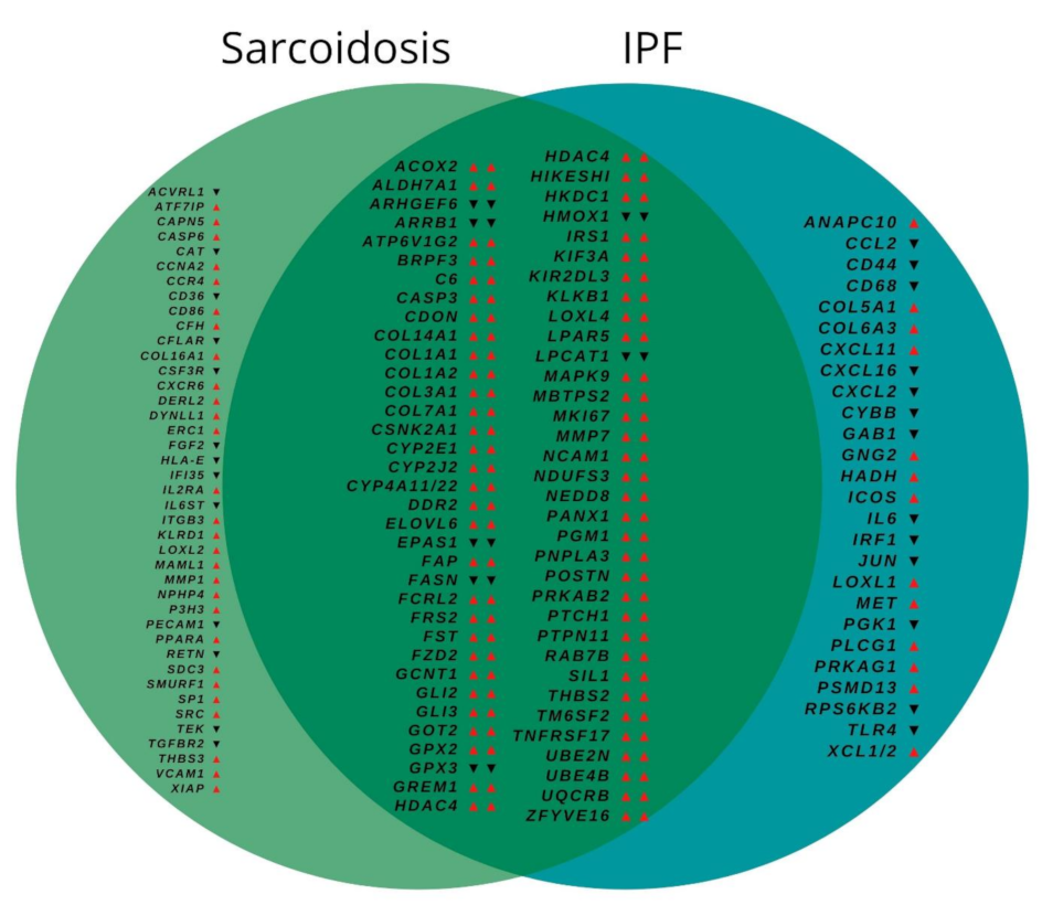
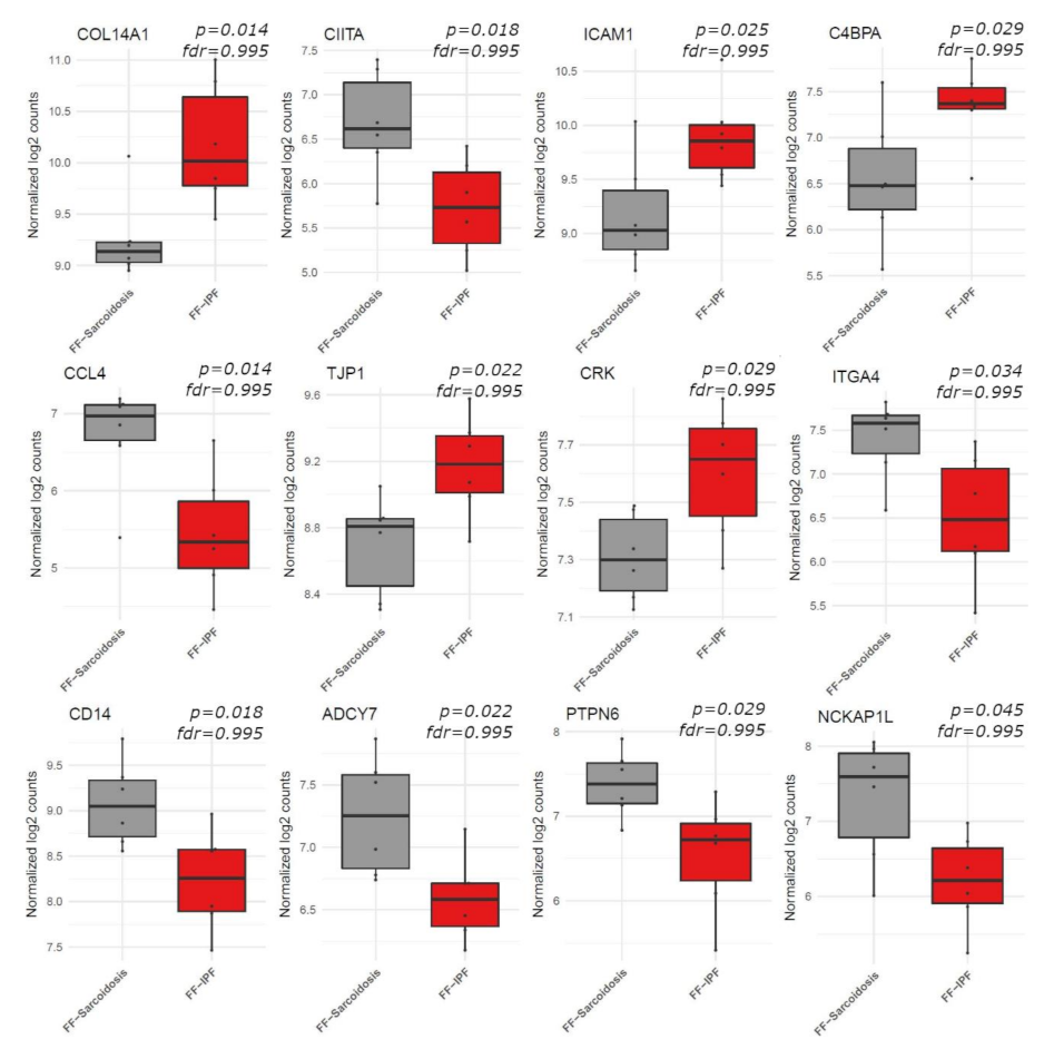
3.2. Gene Expression

The mRNA expression analysis revealed a significantly altered expression signature for 375 out of 760 genes compared to controls as depicted in Figure S1. Of these, 264 showed similar regulation in FF from both disease entities. After Holm Bonferroni correction, 136 genes still showed a significantly altered expression compared to controls. Of these, 69 showed a similar expression in both groups, while 41 showed an altered expression solely in the sarcoidosis group and 26 solely in the IPF group, as depicted in Figure 2. While directly comparing FF from sarcoidosis and IPF, there were no differentially regulated genes. The 12 genes which showed the strongest, although not significant difference between both entities are depicted in Figure 3.
Based on the Holm Bonferroni corrected results, 67 of these 136 genes showed an altered expression compared to controls solely in sarcoidosis or in IPF. However, with regard to the fdr-values, only 2 of all these genes remained differentially expressed as depicted in Figure S1, i.e., decreased expression of calcium transport protein 1 (CAT1) and increased expression of SMAD specific E3 ubiquitin protein ligase 1 (SMURF1), both in the sarcoidosis group. Raw data are deposited in Tables S3 and S4.

3.3. Biological Pathway Analysis

In comparison of FF from both entities with healthy control lungs, a number of biological pathways showed increased activity in FF from both entities as shown in Table 1. In FF from sarcoidosis, these were only tendencies. While comparing FF from sarcoidosis and IPF with each other, IPF showed a distinct up-regulation of a number of fibrosis-related pathways and a down-regulation of certain inflammatory pathways.

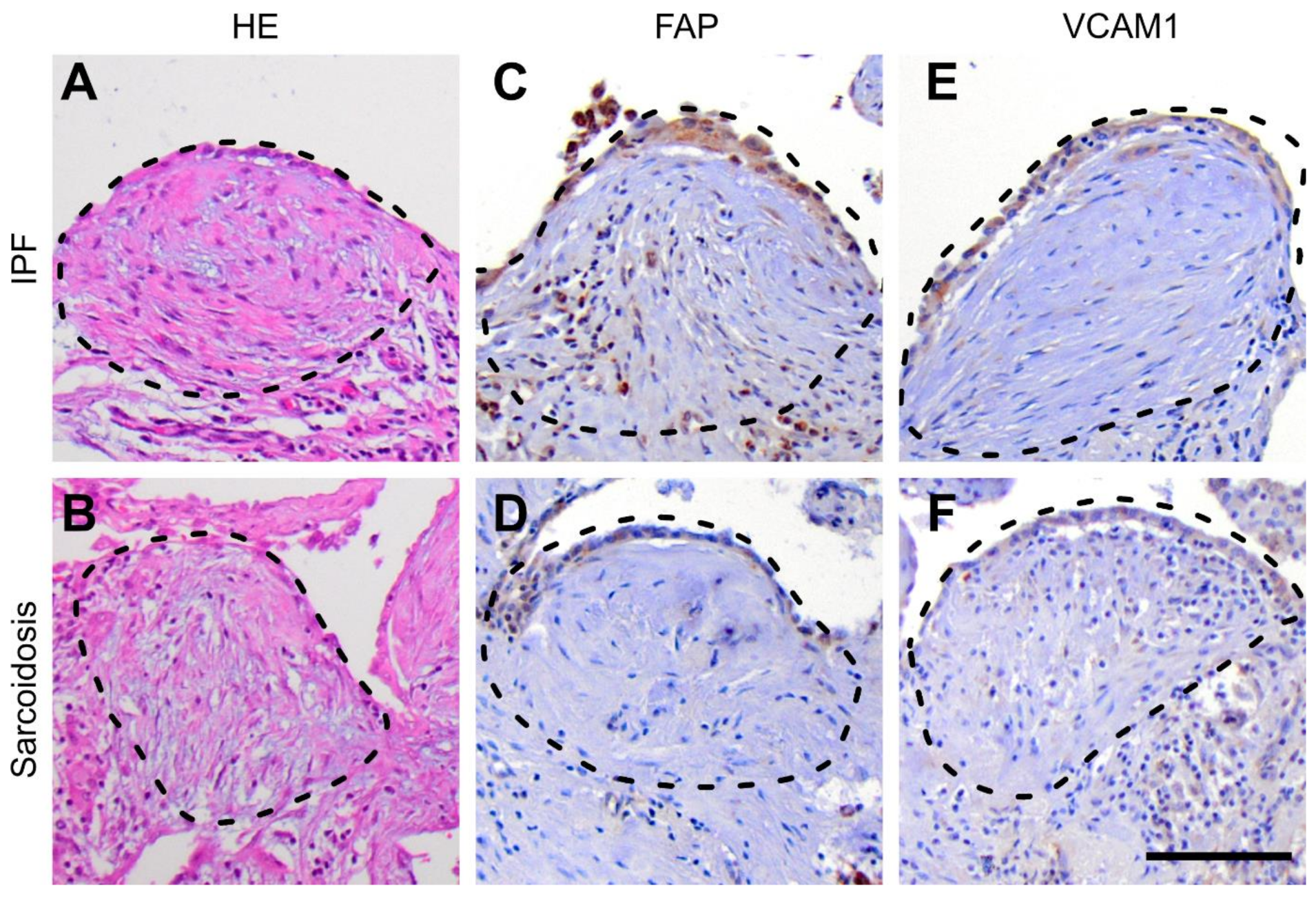
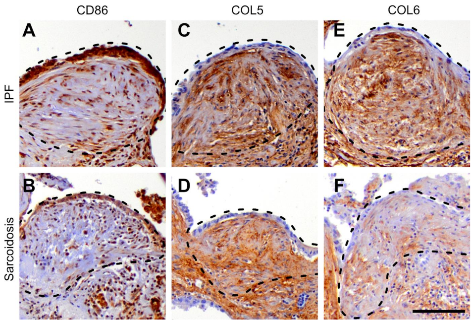
3.4. Protein Expression

Based on the disease specific mRNA expression signatures, we selected fibroblast activation protein alpha (FAP), vascular cell adhesion molecule 1 (VCAM1), cluster of differentiation 86 (CD86) as well as collagen types 5 (COL5) and 6 (COL6) as appropriate markers for immunostaining. In addition, we used marker of proliferation Ki-67 (Ki-67) as a proliferation marker. As depicted in Figure 4, FAP and VCAM1 showed a similar expression pattern in both entities. As shown in Figure 5, CD86 and COL5 showed a similar expression in both IPF and sarcoidosis lungs while COL6 was enhanced in IPF lungs. Ki-67 showed no enhancement in fibroblastic foci from both disease entities. These results are in line with the mRNA expression profile.

4. Discussion

IPF and sarcoidosis represent chronic pulmonary diseases with various degrees of progressive pulmonary fibrosis. In end-stage disease of both entities, bilateral lung transplantation remains the only viable treatment option. Although histopathological findings differ between IPF and sarcoidosis, FF can be present in both entities. However, usually FF are larger and more prevalent in IPF, than in sarcoidosis [6] or other fibrotic lung diseases [4]. To date, it is still unclear which molecular mechanisms contribute to their evolution and in particular if these mechanisms – and FF, for that matter - are the same in different entities. In this work, compartment-specific analysis of FF was performed in end-stage organs from IPF and sarcoidosis patients who underwent lung transplantation.
One fundamental finding of our study was the striking similarity of differentially regulated genes when directly comparing IPF and sarcoidosis lungs, distinctly suggesting a common nature of FF, independent from the underlying disease. Even when comparing sarcoidosis and IPF lungs with healthy controls, 69 of the 136 genes with an altered expression profile showed a similar expression in both entities and only 2 genes showed a contrary-regulated expression. With regard to the significantly regulated biological pathways, regulatory patterns were basically the same in both entities, while particularly the fibrogenesis-related pathways were pronounced in IPF lungs. The fact that particularly inflammation-related pathways such as interferon type I/II, tumor necrosis factor production, toll-like receptor, chemokine, and T-cell receptor signaling were less pronounced in tissue from FF compared to healthy controls, might be a consequence of the compartment-specific tissue analysis and the comparison to mRNA from whole lung slices consisting of many other cell-types such as myocytes, endothelial and immune cells.
At first glance, there was a number of single genes with an altered expression signature between sarcoidosis and IPF lungs, as compared to controls, respectively. Here we focused on selected genes with an established relationship to fibrogenesis, sarcoidosis, and/or IPF.
In a former study by Guillotin et al., myofibroblasts within fibroblastic foci from IPF lungs had been isolated using laser microdissection and specifically analyzed [14]. Among others, clusters of genes associated with cell-cycle and cell adhesion were identified to be involved in the fibrogenesis in IPF. In our study, there was also an increased expression of several genes encoding for cell adhesion molecules or the cell cycle in both groups, e.g., VCAM1 and integrin subunit beta 3 (ITGB3) [15] in sarcoidosis, neural cell adhesion molecule 1 (NCAM1) in both entities as well as anaphase promoting complex subunit 10 (ANAPC10) and proteasome 26S subunit, non-ATPase 13 (PSMD13) in IPF lungs. Hence, cell cycle and cell adhesion molecules appear to play an important role in the development of FF in both entities, supporting the hypothesis of a common nature of FF in variant disease entities.
Lysyl oxidase-like (LOXL) protein coding genes belong to a family of ECM crosslinking enzymes and particularly LOXL1 and LOXL2 are known to be strongly involved in the process of pulmonary fibrogenesis [16]. Increased LOXL1 and LOXL2 levels both on the mRNA and on the protein level have been described in IPF [17] and LOXL1 protein deficiency has been shown to prevent pulmonary fibrosis induced by transforming growth factor beta 1 (TGF-B1) overexpression in knockout mice by blocking fibrillar collagen organization and subsequent tissue stiffening [18]. In addition, LOXL1 and LOXL2 have been suggested as potential therapeutic targets in IPF [17]. In our study, LOXL1 and LOXL4 showed increased expression in the IPF group, as well as LOXL2 and LOXL4 in the sarcoidosis group. Thus, although not identical, there are distinct similarities with regard to the expression of LOXL proteins in FF from sarcoidosis and IPF lungs.
Matrix metalloproteinases (MMP) represent a family of proteins involved in the turnover of extracellular matrix in physiological and pathological processes, such as embryonic organogenesis and fibrotic tissue remodeling. Of these, MMP7 is known to play a pivotal role in the pathogenesis of IPF [19,20]. In addition, increased MMP1 expression was related to IPF associated with non-small cell lung cancer in a recent work [21]. However, data on MMP1 and MMP7 as potential serum biomarkers for IPF and/or sarcoidosis are controversial. Some groups suggested both markers as IPF-specific predictors [22,23] while another group reported higher serum and bronchoalveolar lavage fluid concentrations of MMP1 in sarcoidosis compared with higher concentrations of MMP7 in IPF in each fluid [24]. One study also demonstrated an increased mRNA expression of MMP7 in FF from UIP patients within a compartment-specific analysis [25] and one further group found increased plasma concentrations of MMP7 in sarcoidosis patients compared to healthy controls [26]. In the present study, MMP7 showed an increased expression in FF not only from IPF but also from sarcoidosis lungs. Additionally, MMP1 showed increased expression in sarcoidosis. These results demonstrate again the molecular conformity of FF from both entities.
Basically, tyrosine kinases can be divided into receptor and non-receptor tyrosine kinases depending on their location in cells. Receptor tyrosine kinases (RTK) have been shown to contribute substantially to the fibrogenesis in IPF [27,28]. On that basis, the multi-kinase inhibitor Nintedanib has been approved for the treatment of IPF [29,30]. RTK targeted by Nintendanib are platelet-derived growth factor receptor (PDGFR), fibroblast growth factor receptor (FGR) and vascular endothelial growth factor receptor (VEGFR) [31].
The RTK MET proto-oncogene (MET) serves as a key regulator of invasive growth. In relation to fibrogenesis in IPF, MET regulates the proliferation and survival of fibroblasts as well as their migration across the ECM, making it a potential molecular target for antifibrotic therapies [27]. Phospholipase C Gamma 1 (PLCG1) mediates the production of two second messenger molecules involved in the regulation of intracellular signaling cascades [32]. Of note, PLCG1 becomes activated in response to ligand-mediated activation of several RTK, including platelet-derived growth factor receptor (PDGFR) and fibroblast growth factor receptor (FGFR) [33]. In our study, there was an increased expression of MET and PLCG1 in FF from IPF lungs within the IPF group, reinforcing the important role of RTK for the fibrogenesis in IPF.
SRC proto-oncogenes (SRC) subsume a number of non-receptor tyrosine kinases pivotal for several signal transduction and regulatory processes, as well as for cell proliferation and differentiation [34]. Phosphorylation and activation of these kinases is promoted among others by PDGFR [35]. SRC are involved in various molecular pathways such as epithelial to mesenchymal transition and myofibroblast differentiation, particularly in response to inflammatory stimuli. In addition, in vitro and in vivo experiments in which inhibitory effects on pulmonary fibrogenesis were observed using specific SRC kinase inhibitors [36,37] corroborate the crucial role of SRC for the development of variant fibrotic lung diseases. In line with these previous studies, SRC showed a higher expression in sarcoidosis lungs in our study.
Taken together, these results indicate the molecular similarity of FF in sarcoidosis and IPF suggesting that the pathogenic mechanisms behind FF are independent from the underlying disease: a FF is a FF, regardless of the underlying disease. This in turn encourages the use of antifibrotic agents approved for IPF also in progressive fibrosing sarcoidosis. Moreover, in the light of the appearance of FF in other fibrotic lung disease entities, the impact of their presence on diagnosis should be re-evaluated in current guidelines. To draw a comparison, in a former work we could show that bronchiolitis obliterans (BO) and alveolar fibroelastosis (AFE) are based on the same molecular processes independent from the respective disease entities [38]. Interestingly, by analogy here we see again a divergence of histomorphological and clinical presentations emphasizing the importance of multidisciplinary boards for interstitial lung diseases. Limitations of this study are the small sample size and the monocentric design. Future studies are needed to explore the portability to other fibrotic lung disorders.

Supplementary Materials

The following are available online at https://www.mdpi.com/article/10.3390/cells11040664/s1; Figure S1: Venn diagram of differentially regulated genes, Table S1: Clinical information of the study group, Table S2: Target antibodies used for immunohistochemistry; Table S3: Raw data of the mRNA expression analysis, Table S4: p-values and fdr-values of the mRNA expression analysis.

Author Contributions

Conceptualization, J.C.K., L.N., M.P.K., and D.D.J.; Data curation, H.S.; Formal analysis, J.C.K. and H.S.; Funding acquisition, M.P.K. and D.D.J.; Investigation, J.C.K., L.N., amd M.P.K.; Methodology, J.C.K., L.N., C.B., A.D.S., M.P.K., and D.D.J.; Project administration, D.D.J.; Resources, F.I., A.H., and M.P.K.; Software, H.S.; Supervision, M.M.H. and D.D.J.; Visualization, J.C.K., H.S., J.B.H. and P.B.; Writing–original draft, J.C.K., L.N., M.P.K., and D.D.J.; Writing–review and editing, H.S., J.B.H., C.B., A.D.S., F.I., A.H., J.G., T.W., P.B., F.L. and M.M.H. All authors have read and agreed to the published version of the manuscript.

Funding

This research as well as the APC were funded by the German Research Foundation; Clinical Research Group 311 (DFG; KFO 311; 286251789). DDJ is supported by the European Research Council (ERC); European Consolidator Grant, XHaLe (ref. no. 771883) as well as by collaborative research agreement with Boehringer Ingelheim (contract no. 459989). JCK is supported by PRACTIS – Clinician Scientist Program of Hannover Medical School, funded by the German Research Foundation (DFG, ME 3696/3-1).

Institutional Review Board Statement

This study was approved by the local ethics committee at Hannover Medical School (ethics vote no. 2702-2015).

Informed Consent Statement

Informed consent was obtained from all subjects involved in the study.

Data Availability Statement

Data is contained within the supplementary materials. The data presented in this study are available in Tables S3 and S4.

Acknowledgments

The authors thank Annette Müller-Brechlin, Christina Petzold-Mügge, Regina Engelhardt, Nicole Krönke, Edwin Lennart Busch, and Lennart Brandt for their excellent technical support as well as Nikki Chapman for editing the manuscript.

Conflicts of Interest

J.C.K., L.N., H.S., J.B.H., C.B., A.D.S., F.I., A.H., J.G., T.W., P.B., F.L., M.P.K., and D.D.J. have nothing to disclose. M.M.H. has received fees for lectures and/or consultations from Acceleron, Actelion, Bayer, GSK, Janssen, MSD and Pfizer. The funders had no role in the design of the study; in the collection, analyses, or interpretation of data; in the writing of the manuscript, or in the decision to publish the results.

References

  1. King, T.E., Jr.; Pardo, A.; Selman, M. Idiopathic pulmonary fibrosis. Lancet 2011, 378, 1949–1961. [Google Scholar] [CrossRef]
  2. Raghu, G.; Collard, H.R.; Egan, J.J.; Martinez, F.J.; Behr, J.; Brown, K.K.; Colby, T.V.; Cordier, J.-F.; Flaherty, K.R.; Lasky, J.A.; et al. An Official ATS/ERS/JRS/ALAT Statement: Idiopathic Pulmonary Fibrosis: Evidence-based Guidelines for Diagnosis and Management. Am. J. Respir. Crit. Care Med. 2011, 183, 788–824. [Google Scholar] [CrossRef] [PubMed]
  3. Lynch, D.A.; Sverzellati, N.; Travis, W.D.; Brown, K.K.; Colby, T.V.; Galvin, J.R.; Goldin, J.G.; Hansell, D.M.; Inoue, Y.; Johkoh, T.; et al. Diagnostic criteria for idiopathic pulmonary fibrosis: A Fleischner Society White Paper. Lancet Respir. Med. 2018, 6, 138–153. [Google Scholar] [CrossRef]
  4. Harada, T.; Watanabe, K.; Nabeshima, K.; Hamasaki, M.; Iwasaki, H. Prognostic significance of fibroblastic foci in usual interstitial pneumonia and non-specific interstitial pneumonia. Respirology 2013, 18, 278–283. [Google Scholar] [CrossRef] [PubMed]
  5. Raghu, G.; Remy-Jardin, M.; Myers, J.L.; Richeldi, L.; Ryerson, C.J.; Lederer, D.J.; Behr, J.; Cottin, V.; Danoff, S.K.; Morell, F.; et al. Diagnosis of Idiopathic Pulmonary Fibrosis. An Official ATS/ERS/JRS/ALAT Clinical Practice Guideline. Am. J. Respir. Crit. Care Med. 2018, 198, e44–e68. [Google Scholar] [CrossRef] [PubMed]
  6. Zhang, C.; Chan, K.M.; Schmidt, L.A.; Myers, J.L. Histopathology of Explanted Lungs From Patients With a Diagnosis of Pulmonary Sarcoidosis. Chest 2016, 149, 499–507. [Google Scholar] [CrossRef] [PubMed] [Green Version]
  7. Flaherty, K.R.; Martinez, F.J.; Travis, W.; Lynch, I.J.P. Nonspecific Interstitial Pneumonia (NSIP). Semin. Respir. Crit. Care Med. 2001, 22, 423–434. [Google Scholar] [CrossRef]
  8. Churg, A.; Bilawich, A.; Wright, J.L. Pathology of Chronic Hypersensitivity Pneumonitis What Is It? What Are the Diagnostic Criteria? Why Do We Care? Arch. Pathol. Lab. Med. 2017, 142, 109–119. [Google Scholar] [CrossRef] [Green Version]
  9. Baughman, R.P.; Culver, D.A.; Judson, M.A. A Concise Review of Pulmonary Sarcoidosis. Am. J. Respir. Crit. Care Med. 2011, 183, 573–581. [Google Scholar] [CrossRef] [Green Version]
  10. Rossi, A.; Cavazza, A.; Colby, T.V. Pathology of Sarcoidosis. Clin. Rev. Allergy Immunol. 2015, 49, 36–44. [Google Scholar] [CrossRef]
  11. Spagnolo, P.; Rossi, G.; Trisolini, R.; Sverzellati, N.; Baughman, R.P.; Wells, A.U. Pulmonary sarcoidosis. Lancet Respir. Med. 2018, 6, 389–402. [Google Scholar] [CrossRef]
  12. Länger, F.; Stark, H.; Braubach, P.; Ackermann, M.; Hussein, K.; Teiken, K.; Maegel, L.; Kuehnel, M.; Jonigk, D. Schädigungsmuster interstitieller Lungenerkrankungen. Der Pathol. 2018, 39, 262–271. [Google Scholar] [CrossRef] [PubMed]
  13. Katzenstein, A.L.; Myers, J.L. Idiopathic Pulmonary Fibrosis Clinical Relevance of Pathologic Classification Clinical Features of the Idiopathic Interstitial Pneumonias. Am. J. Respir. Crit. Care Med. 1998, 157, 1301–1315. [Google Scholar] [CrossRef]
  14. Guillotin, D.; Taylor, A.R.; Platé, M.; Mercer, P.F.; Edwards, L.M.; Haggart, R.; Miele, G.; McAnulty, R.J.; Maher, T.M.; E Hynds, R.; et al. Transcriptome analysis of IPF fibroblastic foci identifies key pathways involved in fibrogenesis. Thorax 2020, 76, 73–82. [Google Scholar] [CrossRef] [PubMed]
  15. Hynes, R.O. Integrins: Bidirectional, Allosteric Signaling Machines. Cell 2002, 110, 673–687. [Google Scholar] [CrossRef] [Green Version]
  16. Chen, L.; Li, S.; Li, W. LOX/LOXL in pulmonary fibrosis: Potential therapeutic targets. J. Drug Target. 2018, 27, 790–796. [Google Scholar] [CrossRef]
  17. Tjin, G.; White, E.S.; Faiz, A.; Sicard, D.; Tschumperlin, D.J.; Mahar, A.; Kable, E.P.W.; Burgess, J.K. Correction: Lysyl oxidases regulate fibrillar collagen remodelling in idiopathic pulmonary fibrosis. Dis. Models Mech. 2017, 10, 1301–1312. [Google Scholar] [CrossRef] [Green Version]
  18. Bellaye, P.-S.; Shimbori, C.; Upagupta, C.; Sato, S.; Shi, W.; Gauldie, J.; Ask, K.; Kolb, M. Lysyl Oxidase–Like 1 Protein Deficiency Protects Mice from Adenoviral Transforming Growth Factor-β1–induced Pulmonary Fibrosis. Am. J. Respir. Cell Mol. Biol. 2018, 58, 461–470. [Google Scholar] [CrossRef]
  19. Zuo, F.; Kaminski, N.; Eugui, E.; Allard, J.; Yakhini, Z.; Ben-Dor, A.; Lollini, L.; Morris, D.; Kim, Y.; DeLustro, B.; et al. Gene expression analysis reveals matrilysin as a key regulator of pulmonary fibrosis in mice and humans. Proc. Natl. Acad. Sci. USA 2002, 99, 6292–6297. [Google Scholar] [CrossRef] [Green Version]
  20. Vuorinen, K.; Myllärniemi, M.; Lammi, L.; Piirilä, P.; Rytilä, P.; Salmenkivi, K.; Kinnula, V.L. Elevated matrilysin levels in bronchoalveolar lavage fluid do not distinguish idiopathic pulmonary fibrosis from other interstitial lung diseases. APMIS 2007, 115, 969–975. [Google Scholar] [CrossRef]
  21. Leng, D.; Yi, J.; Xiang, M.; Zhao, H.; Zhang, Y. Identification of common signatures in idiopathic pulmonary fibrosis and lung cancer using gene expression modeling. BMC Cancer 2020, 20, 1–15. [Google Scholar] [CrossRef] [PubMed]
  22. Rosas, I.O.; Richards, T.J.; Konishi, K.; Zhang, Y.; Gibson, K.; Lokshin, A.E.; Lindell, K.O.; Cisneros, J.; MacDonald, S.D.; Pardo, A.; et al. MMP1 and MMP7 as Potential Peripheral Blood Biomarkers in Idiopathic Pulmonary Fibrosis. PLoS Med. 2008, 5, e93. [Google Scholar] [CrossRef] [PubMed] [Green Version]
  23. Morais, A.; Beltrão, M.; Sokhatska, O.; Costa, D.; Melo, N.; Mota, P.; Marques, A.; Delgado, L. Serum metalloproteinases 1 and 7 in the diagnosis of idiopathic pulmonary fibrosis and other interstitial pneumonias. Respir. Med. 2015, 109, 1063–1068. [Google Scholar] [CrossRef] [PubMed] [Green Version]
  24. Zhou, L.-F.; Jiang, L.; Li, Z.-H.; Kang, J. Change of matrix metalloproteinase-1 and matrix metalloproteinase-7 in serum and bronchoalveolar lavage fluid of patients with idiopathic pulmonary fibrosis and sarcoidosis. Chin. J. Tuberc. Respir. Dis. 2010, 33. [Google Scholar]
  25. Kelly, M.M.; Leigh, R.; Gilpin, S.E.; Cheng, E.; Martin, G.E.M.; Radford, K.; Cox, G.; Gauldie, J. Cell-specific Gene Expression in Patients with Usual Interstitial Pneumonia. Am. J. Respir. Crit. Care Med. 2006, 174, 557–565. [Google Scholar] [CrossRef] [PubMed] [Green Version]
  26. Isshiki, T.; Matsuyama, H.; Yamaguchi, T.; Morita, T.; Ono, J.; Nunomura, S.; Izuhara, K.; Sakamoto, S.; Homma, S.; Kishi, K. Plasma matrix metalloproteinase 7, CC-chemokine ligand 18, and periostin as markers for pulmonary sarcoidosis. Respir. Investig. 2020, 58, 479–487. [Google Scholar] [CrossRef] [PubMed]
  27. Stella, G.M.; Gentile, A.; Balderacchi, A.; Meloni, F.; Milan, M.; Benvenuti, S. Ockham’s razor for the MET-driven invasive growth linking idiopathic pulmonary fibrosis and cancer. J. Transl. Med. 2016, 14, 1–12. [Google Scholar] [CrossRef] [Green Version]
  28. Grimminger, F.; Günther, A.; Vancheri, C. The role of tyrosine kinases in the pathogenesis of idiopathic pulmonary fibrosis. Eur. Respir. J. 2015, 45, 1426–1433. [Google Scholar] [CrossRef] [Green Version]
  29. Richeldi, L.; Du Bois, R.M.; Raghu, G.; Azuma, A.; Brown, K.K.; Costabel, U.; Cottin, V.; Flaherty, K.R.; Hansell, D.M.; Inoue, Y.; et al. Efficacy and Safety of Nintedanib in Idiopathic Pulmonary Fibrosis. N. Engl. J. Med. 2014, 370, 2071–2082. [Google Scholar] [CrossRef] [Green Version]
  30. Wollin, L.; Distler, J.H.; Redente, E.F.; Riches, D.W.H.; Stowasser, S.; Schlenker-Herceg, R.; Maher, T.; Kolb, M. Potential of nintedanib in treatment of progressive fibrosing interstitial lung diseases. Eur. Respir. J. 2019, 54, 1900161. [Google Scholar] [CrossRef]
  31. Wollin, L.; Wex, E.; Pautsch, A.; Schnapp, G.; Hostettler, K.; Stowasser, S.; Kolb, M. Mode of action of nintedanib in the treatment of idiopathic pulmonary fibrosis. Eur. Respir. J. 2015, 45, 1434–1445. [Google Scholar] [CrossRef] [PubMed]
  32. Lattanzio, R.; Piantelli, M.; Falasca, M. Role of phospholipase C in cell invasion and metastasis. Adv. Biol. Regul. 2013, 53, 309–318. [Google Scholar] [CrossRef] [PubMed]
  33. Wang, Y.; Tomar, A.; George, S.P.; Khurana, S. Obligatory role for phospholipase C-γ1in villin-induced epithelial cell migration. Am. J. Physiol. Physiol. 2007, 292, C1775–C1786. [Google Scholar] [CrossRef] [PubMed] [Green Version]
  34. Li, H.; Zhao, C.; Tian, Y.; Lu, J.; Zhang, G.; Liang, S.; Chen, D.; Liu, X.; Kuang, W.; Zhu, M. Src family kinases and pulmonary fibrosis: A review. Biomed. Pharmacother. 2020, 127, 110183. [Google Scholar] [CrossRef] [PubMed]
  35. Mishra, R.; Zhu, L.; Eckert, R.L.; Simonson, M.S. TGF-β-regulated collagen type I accumulation: Role of Src-based signals. Am. J. Physiol. Physiol. 2007, 292, C1361–C1369. [Google Scholar] [CrossRef] [Green Version]
  36. Beyer, C.; Distler, J.H. Tyrosine kinase signaling in fibrotic disorders. Biochim. Biophys. Acta (BBA) Mol. Basis Dis. 2012, 1832, 897–904. [Google Scholar] [CrossRef] [Green Version]
  37. Hu, M.; Che, P.; Han, X.; Cai, G.-Q.; Liu, G.; Antony, V.; Luckhardt, T.; Siegal, G.P.; Zhou, Y.; Liu, R.-M.; et al. Therapeutic Targeting of Src Kinase in Myofibroblast Differentiation and Pulmonary Fibrosis. J. Pharmacol. Exp. Ther. 2014, 351, 87–95. [Google Scholar] [CrossRef] [Green Version]
  38. Jonigk, D.; Rath, B.; Borchert, P.; Braubach, P.; Maegel, L.; Izykowski, N.; Warnecke, G.; Sommer, W.; Kreipe, H.; Blach, R.; et al. Comparative analysis of morphological and molecular motifs in bronchiolitis obliterans and alveolar fibroelastosis after lung and stem cell transplantation. J. Pathol. Clin. Res. 2016, 3, 17–28. [Google Scholar] [CrossRef]
Figure 1. Radiological and histopathological features of sarcoidosis and IPF. (A) Exemplary axial high resolution computed tomography (CT) slide showing typical radiological predominantly subpleural distributed features of idiopathic pulmonary fibrosis (IPF) including septal thickening, mixed ground glass opacities, reticular densities (arrowheads), and areas of subpleural honeycombing (arrows); (B) typical histopathological features of IPF including disturbed pulmonary architecture (i.e., aberrant alveoli with hyperplastic bronchiolar epithelial lining, extensive myogenic metaplasia, and multiple fibroblastic foci (FF) magnified in (C); (D) exemplary axial high resolution CT slide showing typical hallmarks of fibrotic pulmonary sarcoidosis including reticular and cystic changes, traction bronchiectasis (arrowheads), and subpleural bullous changes (arrows); (E) histopathological hallmarks of sarcoidosis including focal organizing pneumonia and interstitial pneumonitis as well as the in (F) magnified multiple granuloma (Gra) with central giant cells and surrounding epithelioid cells and FF; HE, hematoxylin-eosin stained; scale bar equals 500 µm.
Cells 11 00664 g001
Figure 2. Venn diagram of differentially regulated genes. Arrows indicate significantly increased (arrow up) or significantly decreased (arrow down) expression of the respective genes compared to controls in fibroblastic foci from both disease entities compared to healthy control lungs. Holm Bonferroni corrected p-values < 0.05 were considered as statistically significant. In the overlapping area (middle), left and right arrow indicate the expression in sarcoidosis and IPF, respectively. IPF, idiopathic pulmonary fibrosis.
Cells 11 00664 g002
Figure 3. Gene expression—sarcoidosis vs. IPF. This figure shows the top 12 regulated genes between sarcoidosis and idiopathic pulmonary fibrosis (IPF) in box-plots. FF, fibroblastic foci; IPF, idiopathic pulmonary fibrosis; fdr, false discovery rate.
Cells 11 00664 g003
Figure 4. Immunohistochemical staining of fibroblastic foci. Histological morphology of fibroblastic foci (FF), hematoxylin-eosin (HE) stained, in (A) idiopathic pulmonary fibrosis (IPF) and (B) sarcoidosis showing characteristic myxoid arranged fibroblasts and myofibroblasts, a background of immature extracellular matrix, protrusion into the alveolar space, and a covering epithelial layer of hypertrophied type II pneumocytes; fibroblast activation protein alpha (FAP) showed a coherent staining pattern in FF from sarcoidosis (C) and IPF (D) lungs with a cytoplasmic enhancement of the covering epithelial layer and a low intensity cytoplasmic staining of scattered myofibroblasts (less pronounced in sarcoidosis); (E,F) vascular cell adhesion molecule 1 (VCAM1) showed a specific cytoplasmic enhancement in respiratory epithelial cells and less intensive also in myofibroblasts / connective tissue components (slightly pronounced in sarcoidosis; scale-bar equals 100 µm.
Cells 11 00664 g004
Figure 5. Immunohistochemical staining of fibroblastic foci. (A,B) Cluster of differentiation (CD86) showed a specific cytoplasmic enhancement in respiratory epithelial cells and also in myofibroblasts similar in both entities; (C,D) collagen type 5 (COL5) showed a similar staining of fibroblastic foci from both entities predominantly in myofibroblasts and the surrounding connective tissue; (E,F) Collagen type 6 (COL6) was also enhanced in myofibroblasts and connective tissue structures but markedly stronger in idiopathic pulmonary fibrosis (IPF) lungs than in sarcoidosis; scale bar equals 100 µm.
Cells 11 00664 g005
Table 1. Changes in the expression of genes involved in several biological pathways.
Signaling PathwayFF-S vs. ControlsFF-IPF vs. ControlsFF-IPF vs. FF-S
RegulationfdrRegulationfdrRegulationfdr
Cell adhesion 1Up0.782Up0.437Up0.018
ECM structure 1Up0.165Up0.063Up0.018
ECM receptor interaction 1Up0.623Up0.437Up0.018
ECM organization 2Up0.83Up0.503Up0.005
Organization of collagen fibrils within ECM 2Up0.261Up0.036Up0.095
Innate immune system 1Down0.165Down0.011Down0.0001
Type I interferon signaling 1Down0.165Down0.031Down0.002
Type II interferon signaling 1Down0.343Down0.206Down0.009
MHC class I antigen presentation 1Down0.13Down0.036Down0.002
Activation of TNF production 2Down0.916Down0.038Down0.005
Activation of ERK1/ERK2 cascade 2Down0.14Down0.036Down0.884
NLR signaling 2Down0.276Down0.036Down0.241
TLR signaling 1Down0.926Down0.202Down0.001
T-cell receptor signaling 1Down0.926Down0.96Down0.013
Chemokine signaling 1Down0.957Down0.769Down0.018
Macrophages 1Down0.787Down0.091Down0.015
1 based on gene-pathway associations supplied by Nanostring; 2 based on the GeneOntology database; significant fdr values are written in bold letters; FF-IPF/FF-S, fibroblastic foci in IPF/sarcoidosis; IPF, idiopathic pulmonary fibrosis; fdr, false discovery rate; MHC, major histocompatibility complex; NLR, Nod-like receptor; HIF1A, hypoxia inducible factor; ERK1/2, extracellular signal-related kinase 1/2; ECM, extracellular matrix; TNF, tumor necrosis factor; TLR, toll-like receptor.
Publisher’s Note: MDPI stays neutral with regard to jurisdictional claims in published maps and institutional affiliations.

