Article

Characterization of Aminoglycoside Resistance and Virulence Genes among Enterococcus spp. Isolated from a Hospital in China

1
Center of Laboratory Medicine, Zhejiang Provincial People's Hospital, 158 Shangtang Road, Hangzhou 310014, China
2
Medical College, Shihezi University, 280 Beisan Road, Shihezi 832000, China
3
Second Clinical Medical College, Zhejiang Chinese Medical University, 548 Binwen Road, Hangzhou 310053, China
4
Wenzhou Hospital of Traditional Chinese Medicine, 27 Dashimen Road Xinhe Street, Wenzhou 325000, China
5
Medical College, Taizhou University, 1139 Shifu Road, Taizhou 318000, China
*
Author to whom correspondence should be addressed.
These authors contributed equally to this work.
Int. J. Environ. Res. Public Health 2015, 12(3), 3014-3025; https://doi.org/10.3390/ijerph120303014
Submission received: 22 December 2014 / Revised: 4 March 2015 / Accepted: 5 March 2015 / Published: 11 March 2015

Abstract

:
This study investigated the aminoglycoside resistance phenotypes and genotypes, as well as the prevalence of virulence genes, in Enterococcus species isolated from clinical patients in China. A total of 160 enterococcal isolates from various clinical samples collected from September 2013 to July 2014 were identified to the species level using the VITEK-2 COMPACT system. The antimicrobial susceptibilities of the identified Enterococcus strains were determined by the Kirby-Bauer (K-B) disc diffusion method. PCR-based assays were used to detect the aminoglycoside resistance and virulence genes in all enterococcal isolates. Of 160 Enterococcus isolates, 105 were identified as E. faecium, 35 as E. faecalis, and 20 isolates were classified as “other” Enterococcus species. High-level aminoglycoside resistance (HLAR) for gentamicin, streptomycin, and both antibiotics was identified in 58.8, 50, and 34.4% of strains, respectively. The most common virulence gene (50.6% of isolates) was efaA, followed by asa1 (28.8%). The most prevalent aminoglycoside resistance genes were aac(6')-Ie-aph(2''), aph(2')-Id, aph(3')-IIIa, and ant(6')-Ia, present in 49.4%, 1.3%, 48.8% and 31.3% of strains, respectively. Overall, E. faecium and E. faecalis were most frequently associated with hospital-acquired enterococcal infections in Zhejiang Province. All aminoglycoside resistance genes, except aph(2'')-Id, were significantly more prevalent in HLAR strains than amongst high level aminoglycoside susceptible (HLAS) strains, while there was no significant difference between HLAR and HLAS strains in regard to the prevalence of virulence genes, apart from esp, therefore, measures should be taken to manage infections caused by multi-drug resistant Enterococcus species.

1. Introduction

Enterococcus species are opportunistic pathogens that are part of the normal gut microbiota of both humans and animals, and can survive in a diverse range of harsh conditions [1,2]. The first Enterococcus species to be identified was isolated from an endocarditis patient in the 1900s. Since then, enterococci have been recognized as an important cause of nosocomial infections [3]. Recently, there has been an increase in invasive therapy because of the frequent inappropriate use of antimicrobial agents. In addition, high-level aminoglycoside-resistant (HLAR) enterococci and vancomycin-resistant enterococci (VRE) have caused significant problems for clinical anti-infective therapy. VRE strains are becoming more widespread in North America, Europe, and Asia, although prevalence rates vary greatly among different geographical areas. Six genotypes (vanA, vanB, vanC, vanD, vanE, and vanG) have been described, of which, vanA (Tn1546) and vanB (Tn1549/Tn5382) are the most common [4]. HLAR strains are becoming more prevalent, mostly because of aminoglycoside modifying enzymes (AMEs) that are encoded within mobile genetic elements, which are widespread amongst enterococci [5]. Among the AMEs, the most prevalent gene is aac(6')-Ie-aph(2''), which encodes a bifunctional enzyme, AAC(6')-APH(2''), that confers resistance to a broad spectrum of aminoglycosides [6]. Other resistance genes, encoding the AMEs 2'-O-phosphotransferase (APH(2')), 3'-O-phosphotransferase (APH(3')), 3'-O-adenyltransferase (ANT(3')), 4-O-adenyltransferase (ANT(4')), and 6'-O-adenyltransferase (ANT(6')) are also found on these mobile genetic elements. Enterococci also produce many virulence factors, including collagen-binding protein (ace), aggregation substance (asa1), cytolysin (cylA), endocarditis antigen (efaA), enterococcal surface protein (esp), gelatinase (gelE), and hyaluronidase (hyl) [7,8,9].
Generally, Enterococcus species are not recognized as dominant pathogens, and usually only cause infections in patients who have a severe underlying disease or are immunocompromised [2]. Penicillin, ampicillin, and aminoglycosides are the first-line drugs for the treatment of enterococcal infection [3]. However, enterococci rapidly acquire antibiotic resistance determinants, as shown by the increasing number of infections caused by HLAR Enterococcus species. The aim of present study was to determine the prevalence of HLAR strains, examine their antibiotic susceptibility phenotypes, screen for the presence of genes coding for AMEs (including aac(6')-Ie-aph(2''), aph(2'')-Ib, aph(2'')-Id, ant(3'')-I, ant(4')-Ia, ant(6')-Ia, and aph(3')-IIIa) and other virulence factors (including ace, asa1, cylA, efaA, esp, gelE, and hyl), and examine the relationships between AME- and virulence-encoding genes of enterococci isolated from clinical patients in China.

2. Materials and Methods

2.1. Bacterial Isolation and Identification

This study was performed between September 2013 and July 2014 at Zhejiang Province People’s Hospital, Hangzhou, China, a 2000-bed tertiary hospital. Rectal swab or stool specimens were collected from all patients admitted to the 40-bed ICU, and all VRE strains isolated from other clinical samples collected from June 2011 to January 2014 were also examined. Duplicate isolates were discarded. Each enterococcal isolate was first identified using the VITEK-2 COMPACT fully automated microbiological system (bioMérieux, Inc., Durham, NC, USA). Genotyping was carried out by screening for the presence of aminoglycoside resistance and other virulence genes using a multiplex polymerase chain reaction (PCR) method.

2.2. Susceptibility Testing

The antibacterial agents tested were gentamicin (120 μg), streptomycin (300 μg), erythromycin (15 μg), vancomycin (30 μg), teicoplanin (30 μg), ciprofloxacin (5 μg), levofloxacin (5 μg), chloramphenicol (30 μg), quinupristin-dalfopristin (15 μg), penicillin (10 U), ampicillin (10 μg), linezolid (30 μg), tetracycline (30 μg), and rifampin (5 μg). Susceptibility phenotypes were determined using the disc diffusion method according to the CLSI guidelines. E. faecalis ATCC29212 was used as a reference strain.

2.3. Amplification of Virulence and Resistance Genes

Template DNA was extracted using an EZ-10 spin column bacterial genomic DNA miniprep kit (Bio Basic Inc., Markham, ON, Canada). Genes encoding AMEs, including aac(6')-Ie-aph(2''), aph(2'')-Ib, aph(2'')-Id, aph(3')-IIIa, ant(3'')-I, ant(4')-Ia, and ant(6')-Ia, along with the virulence genes ace, asa1, cylA, efaA, esp, gelE, and hyl, were examined by PCR. Detection was performed in a final volume of 20 μL, containing 400 nM of each primer (primer sequences are provided in Table 1), 10 µL of premix Taq polymerase (Takara, Otsu, Japan) containing MgCl2, dNTPs, and reaction buffer, 1 µL of DNA template, and dd H2O to 20 µL. The PCR conditions consisted of a pre-denaturation step at 94 °C for 4 min, followed by 35 cycles of 94 °C for 40 s, 55 °C for 40 s, and 72 °C for 45 s. A final extension step was performed at 72 °C for 5 min.
Table 1. PCR primers used in the amplification of virulence and resistance genes.
GeneDescriptionSequence(5 '-3')Amplicon Size (bp)Ref.
aceCollagen-binding proteinGGAATGACCGAGAACGATGGC616[8]
GCTTGATGTTGGCCTGCTTCCG
asa1Aggregation substanceCACGCTATTACGAACTATGA375[10]
TAAGAAAGAACATCACCACGA
ylACytolysinACTCGGGGATTGATAGGC688[8]
GCTGCTAAAGCTGCGCTT
efaAEndocarditis antigenCGTGAGAAAGAAATGGAGGA499[7]
CTACTAACACGTCACGAATG
espEnterococcal surface protein AGATTTCATCTTTGATTCTTGG510[10]
AATTGATTCTTTAGCATCTGG
gelEGelatinaseTATGACAATGCTTTTTGGGAT213[10]
AGATGCACCCGAAATAATATA
hylHyaluronidaseACAGAAGAGCTGCAGGAAATG276[10]
GACTGACGTCCAAGTTTCCAA
aac(6')-Ie-aph(2")-IaAAC(6')-APH(2'')AGGAATTTATCGAAAATGGTAGAAAAG369[6]
CACAATCGACTAAAGAGTACCAATC
aph(3')-IIIaAPH(3')GGCTAAAATGAGAATATCACCGG523[6]
CTTTAAAAAATCATACAGCTCGCG
ant(4')-IaANT(4')CAAACTGCTAAATCGGTAGAAGCC294[6]
GGAAAGTTGACCAGACATTACGAACT
aph(2")-IcAPH(2')CCACAATGATAATGACTCAGTTCCC444[11]
CCACAGCTTCCGATAGCAAGAG
aph(2")-IbAPH(2')CTTGGACGCTGAGATATATGAGCAC867[12]
GTTTGTAGCAATTCAGAAACACCCTT
aph(2")-IdAPH(2')GGTGGTTTTTACAGGAATGCCATC642[11]
CCCTCTTCATACCAATCCATATAACC
ant(3")-IIIANT(3')CACGCTATTACGAACTATGA284[11]
TAAGAAAGAACATCACCACGA
ant(6')-IaANT(6')ACTCGGGGATTGATAGGC597[11]
GCTGCTAAAGCTGCGCTT
The amplified PCR products were analyzed on 1% (w/v) agarose gels. DNA bands were visualized by staining with ethidium bromide and photographed under UV illumination.

2.4. Statistical Analyses

VRE data was managed using the WHONET version 5.6 software (The WHONET Team, Boston, MA, USA). SPSS software version 17.0 (IBM, Inc., Armonk, NY, USA) was used for statistical analyses. Differences in antimicrobial sensitivity profiles and the prevalence of resistance and virulence genes between HLAR and HLAS Enterococcus species were compared using the Chi-square test, with a p-value < 0.05 indicating statistical significance.

3. Results

3.1. Antimicrobial Susceptibility

Of the 160 Enterococcus isolates, 105 were identified as E. faecium, 35 as E. faecalis, and 20 as “otherEnterococcus species. The antimicrobial susceptibility profiles of all of the isolates are shown in Table 2. HLAR for gentamicin, streptomycin, and both antibiotics was identified in 58.8%, 50%, and 34.4% of isolates, respectively. In the present study, resistance to either gentamicin or streptomycin (or both) was regarded as indicating HLAR. In total, 119 (74.4%) of the isolates were classified as HLAR. In contrast, very little resistance to linezolid or teicoplanin was detected amongst the 160 isolates. Amongst the HLAR strains, comparison of the two species showed that rates of resistance were significantly higher in E. faecium than in E. faecalis for all antibiotics examined, except ERY, LZD, and CHL. Among the HLAS strains, a significant difference was again observed for all antibiotics, except TET, CHL, TEC, and VAN. Comparison of HLAR vs. HLAS E. faecium isolates revealed that rates of antibiotic resistance were only significantly different for TEC and VAN, while a significant difference was only observed for erythromycin between the HLAR and HLAS E. faecalis isolates.

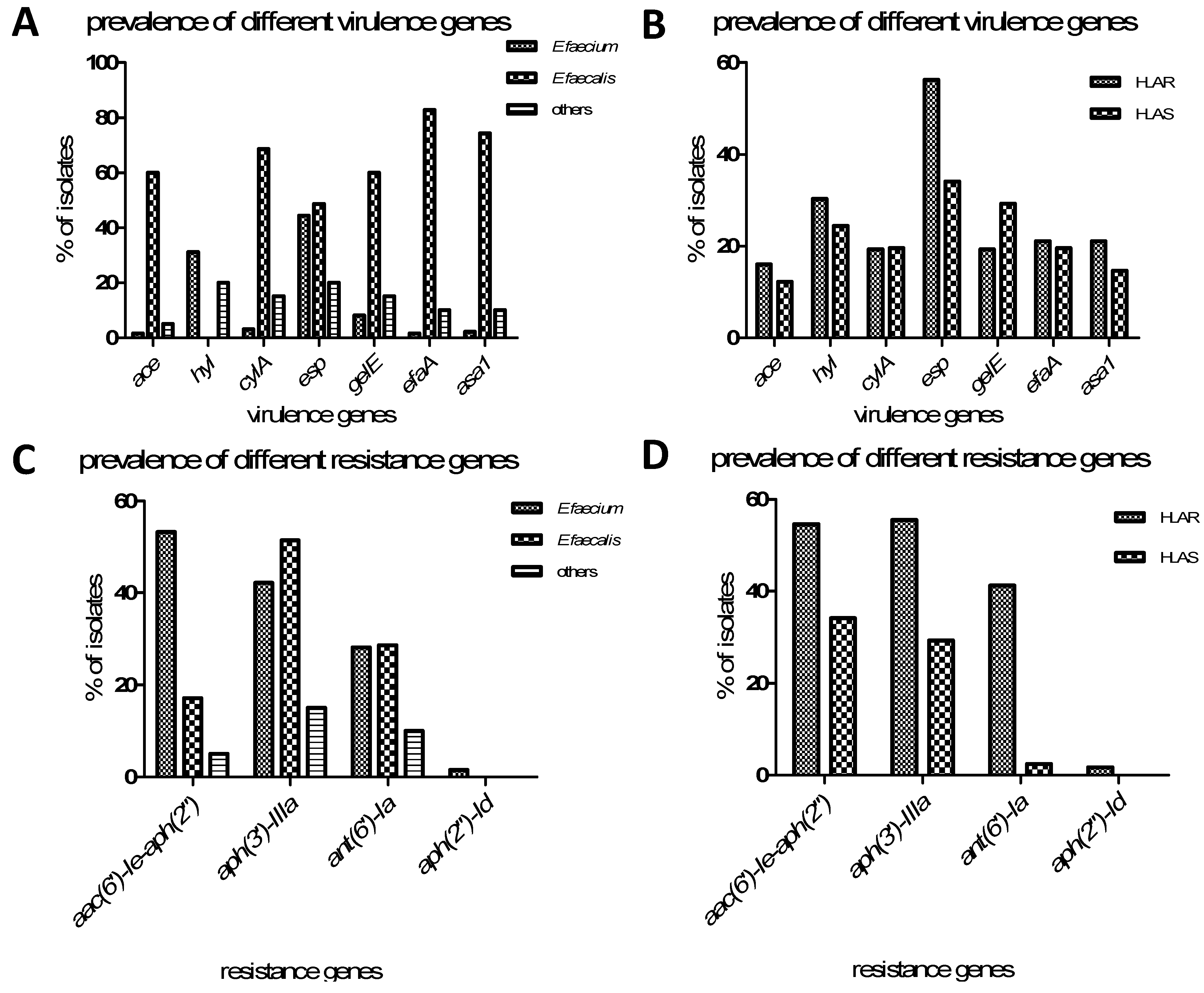
3.2. Distribution of Virulence Genes

The prevalence of the ace, asa1, cylA, efaA, esp, gelE, and hyl virulence genes amongst the 160 Enterococcus species was 15%, 28.8%, 19.4%, 50.6%, 21.9%, 20.6%, and 19.6%, respectively. In bivariate analysis, the overall rate of virulence gene carriage amongst the E. faecalis group was significantly higher (n = 35) than in the E. faecium group (n = 105) or the “other” group (n = 20) (except hyl and esp). ace was significantly (p < 0.001) more prevalent amongst the E. faecalis strains (n = 35, 60%) than the E. faecium strains (n = 105, 1.5%) or the “other” strains (n = 20, 5%), as were cylA (p < 0.001), gelE (p < 0.001), efaA (p < 0.001), and asa1 (p < 0.001). There was no significant difference between E. faecalis and E. faecium with regard to esp (E. faecalis vs. E. faecium, p = 0.377), although esp was significantly more prevalent in both species than in the “otherEnterococcus spp. group (E. faecalis vs.other” strains, p = 0.036; E. faecium vs.other” strains, p = 0.002).
Table 2. Antibiotic susceptibility profiles of the tested Enterococcus species.
Anti-Microbial AgentHLAR (119)HLAS (41)P1P2P3P4
E.faecium (82)E.faecalis (27)Others (10)E.faecium (23)E.faecalis (8)Others (10)
RISRISRISRISRISRIS
AMP *80 (97.6)0 (0)2 (2.4)1 (3.7)0 (0)26 (96.3)2 (20)0 (0)8 (80)22 (95.7)0 (0)1 (4.3)0 (0)0 (0)8 (100)3 (30)0 (0)7 (70)0.000.001.001.00
G *80 (97.6)0 (0)2 (2.4)1 (3.7)0 (0)26 (96.3)2 (20)0 (0)8 (80)22 (95.7)0 (0)1 (4.3)0 (0)0 (0)8 (100)1 (10)0 (0)9 (90)0.000.001.001.00
LEF *80 (97.6)0 (0)2 (2.4)15 (55.6)0 (0)12 (44.4)7 (70)0 (0)3 (30)23 (100)0 (0)0 (0)2 (25)0 (0)6 (75)3 (30)0 (0)7 (70)0.000.001.000.26
ERY *80 (97.6)0 (0)2 (2.4)23 (85.2)3 (11.1)1 (3.7)9 (90)0 (0)1 (10)18 (78.3)4 (17.4)1 (4.3)2 (25)3 (37.5)3 (37.5)8 (80)1 (10)1 (10)1.000.071.000.04
CIP *80 (97.6)0 (0)2 (2.4)15 (55.6)4 (14.8)8 (29.6)8 (80)1 (10)1 (10)23 (100)0 (0)0 (0)1 (12.5)2 (25)5 (62.5)3 (30)2 (20)5 (50)0.000.001.000.20
LZD *3 (3.7)0 (0)79 (96.3)0 (0)0 (0)27 (100)0 (0)0 (0)10 (100)0 (0)0 (0)23 (100)0 (0)0 (0)8 (100)0 (0)0 (0)10 (100)0.57--0.82--
Q/D *8 (9.8)6 (7.3)68 (82.9)------7 (70)2 (20)1 (10)3 (13.0)2 (8.7)18 (78.3)------5 (50)2 (20)3 (30)----0.84--
TET *36 (43.9)1 (1.2)45 (54.9)20 (74.1)1 (3.7)6 (22.2)10 (100)0 (0)0 (0)12 (52.2)0 (0)11 (47.8)4 (50)0 (0)4 (50)8 (80)0 (0)2 (20)0.001.000.550.27
RIF *64 (78.1)7 (8.5)11 (13.4)8 (29.6)5 (18.5)14 (51.9)1 (10)0 (0)9 (90)16 (69.6)5 (21.7)2 (8.7)1 (12.5)1 (12.5)6 (75)3 (30)0 (0)7 (70)0.000.000.800.45
CHL *4 (4.9)3 (3.7)75 (91.5)2 (7.4)0 (0)25 (92.6)0 (0)1 (10)9 (90)1 (4.3)1 (4.3)21 (91.4)1 (12.5)1 (12.5)6 (75)0 (0)0 (0)10 (100)1.000.571.000.46
TEC *15 (18.3)18 (22.0)49 (59.7)0 (0)1 (3.7)26 (96.3)0 (0)0 (0)10 (100)1 (4.3)1 (4.3)21 (91.4)1 (12.5)0 (0)7 (87.5)0 (0)0 (0)10 (100)0.001.000.000.94
VAN *43 (52.4)0 (0)39 (47.6)2 (7.4)1 (3.7)24 (88.9)0 (0)0 (0)10 (100)2 (8.7)0 (0)21 (91.3)1 (12.5)0 (0)7 (87.5)0 (0)0 (0)10 (100)0.001.000.001.00
* AMP (ampicillin); G (penicillins); LEF (levofloxacin); ERY (erythromycin); CIP (ciprofloxacin); LZD (linezolid); Q/D (quinupristin-dalfopristin); TET (tetracycline); RIF (rifampin); CHL (chloramphenicol); TEC (teicoplanin); VAN (vancomycin). P1 In HLAR strains compares E.faecium with E.faecalis. P2 In HLAS compare E.faecium with E.faecalis. P3 Compare HLAR E.faecium with HLAS E.faecium. P4 Compare HLAR E.faecalis with HLAS E.faecalis.
hyl was significantly more prevalent in E. faecium and “other” strains than in E. faecalis (E. faecalis vs. E. faecium, p < 0.001; E. faecalis vs. other strains, p = 0.027), but there was no significant difference between E. faecium and “other” strains (p = 0.089).

3.3. Distribution of Aminoglycoside Resistance Genes

All the Enterococcus species were tested for the presence of aminoglycoside resistance genes. aac(6')-Ie-aph(2''), aph(2')-Id, aph(3')-IIIa, and ant(6')-Ia were found in 49.4%, 1.3%, 48.8%, and 31.3% of isolates, respectively. The distribution of aminoglycoside resistance genes according to phenotype is presented in Table 3. Neither ant(3'')-I nor aph(2')-Ib were detected among the HLAR isolates. The most prevalent resistance gene was aph(3')-IIIa (n = 66 isolates), followed by aac(6')-Ie-aph(2'') (n = 65). Overall, 10 isolates contained aph(3')-IIIa, 22 had aac(6')-Ie-aph(2''), seven carried both aac(6')-Ie-aph(2'') and aph(3')-IIIa, 32 harbored aph(3')-IIIa, ant(6')-Ia, and aac(6')-Ie-aph(2''), and 15 had aph(3')-IIIa and ant(6')-Ia. Only two isolates had both aac(6')-Ie-aph(2'') and ant(6')-Ia, while two others had aac(6')-Ie-aph(2''), aph(2'')-Id, and aph(3')-IIIa. However, among the HLAS isolates, the most prevalent resistance gene was aac(6')-Ie-aph(2'') (n = 14), followed by aph(3')-IIIa (n = 12). ant(6')-Ia was only was detected in one isolate. Chi-square tests showed that several resistance genes were significantly more prevalent in HLAR strains than HLAS strains, including aac(6')-Ie-aph(2'') (p = 0.024), aph(3')-IIIa (p = 0.004), and ant(6')-Ia (p < 0.001). In bivariate analysis, aac(6')-Ie-aph(2'') was significantly more prevalent in E. faecium than in E. faecalis and “other” strains (p < 0.001 in both cases). aph(3')-IIIa and ant(6')-Ia were significantly more prevalent in E. faecalis than in “other” strains (p = 0.001 and p = 0.024, respectively).

3.4. Correlation between Aminoglycoside Resistance Genes and Virulence Genes

Of the 82 esp-positive isolates, the majority (n = 66, 80.5%) were resistant to aminoglycosides (Table 3). However, only 52 of the 78 (66.7%) esp-negative isolates were resistant to high-level aminoglycosides. Bivariate analysis confirmed that this difference between the esp-positive and negative isolates was significant (p = 0.047). Of the 119 HLAR isolates, 67 (56.3%) were esp-positive; however, only 14 of the 41 (34.1%) HLAS isolates were esp-positive (p = 0.014) (Figure 1).

4. Discussion

In recent years, infections caused by multidrug-resistant Gram-positive organisms have become a significant cause of morbidity and mortality [13,14]. Among these Gram-positive organisms, Enterococcus species are the second most common nosocomial bloodstream pathogens isolated in the United States [15,16]. Enterococci have been considered nosocomial pathogens since the early 1970s, and high-level resistance to aminoglycosides has become a serious problem in most healthcare facilities [17]. Schouten, et al. [18] reported that high level resistance to gentamicin has been detected in all investigated European countries, with prevalence ranging from 1% to 48% (mean, 22.6% ± 12.3%) in 1997. Moreover, there were no geographic relationships among the studied countries. However, in the present study, HLAR was detected in 119 (74.4%) of the isolates, which is significantly higher than previously reported.
Table 3. Frequency of different virulence genes in Enterococcus species.
No.of IsolatesPhenotypeGenotypeNo.of Virulence Genes
aac(6')-Ie-aph(2'')aph(2'')-Idaph(3')-IIIaant(6')-IaacehylcylAespgelEefaAasa1
22R*+ 49112543
10R + 2257344
7R+ + 1405111
2R+ +0001000
15R ++7476577
2R+++ 0200000
32R+ ++113128423
29R 4298577
total119 19362367232525
9S *+ 0305200
6S + 1232121
5S+ + 0220211
1S ++1001011
20S 3336743
total41 5108141286
Notes: + = positive. * R (resistance); S (susceptible).
It appears that since the 1970s, the prevalence of HLAR species has rapidly increased. In the current study, a significantly higher prevalence of resistance to ampicillin, vancomycin, rifampicin, penicillin, teicoplanin, ciprofloxacin, and levofloxacin was detected in E. faecium than in E. faecalis (p < 0.05), while a greater prevalence of resistance to chloramphenicol, minocycline, and tetracycline was found in E. faecalis than in E. faecium (p < 0.05).
This study also investigated the prevalence of aminoglycoside resistance genes among the 160 Enterococcus isolates. In total, 119/160 (74.4%) isolates showed a HLAR phenotype. A previous study by Padmasini et al. found that all HLAR E. faecalis and E. faecium isolates included in their study carried aac(6')-Ie-aph(2'')-Ia [17,19]. However, in our study, only 65 (54.6%) of the 119 HLAR strains identified by the Kirby-Bauer method carried aac(6')-Ie-aph(2'')-Ia. Interestingly, 29/119 strains (24.4%) did not carry any of the examined aminoglycoside resistance genes, which was lower than has been reported previously [6,17]. While previous studies found that aac(6')-Ie-aph(2'')-Ia was the most common aminoglycoside resistance gene, in this study, the most prevalent gene was aph(3')-IIIa, which was detected in 55.5% (66/119) of isolates. In a previous studies Padmasini et al, 20.2% (39/178) of strains carried both aac(6')-Ie-aph(2'')-Ia and aph(3')-IIIa, and Emaneini, et al. [6] reported that 35.9% of Enterococcus isolates examined in their study contained both genes. In our study, aac(6')-Ie-aph(2'')-Ia and aph(3')-IIIa coexisted in 28.8% of the Enterococcus species isolates, which was similar to previous results.
Previous studies by Creti, et al. [8] and Billström, et al. [10] showed that the virulence genes ace, asa1, cylA, efaA, esp, gelE, and hyl are present at varying levels in E. faecalis isolates. In the current study, esp was the most frequently identified virulence gene (50.6% of isolates), followed by hyl (28.8%). These findings differed from the results of Zou et al., who showed that gelE was the most prevalent virulence gene, and that hyl and cylA were not detected, while Vankerckhoven, et al. [20] detected esp in 65% of isolates, and gelE and cylA were not detected. Comparison of virulence genes amongst HLAR and HLAS strains in the current study showed that only esp was significantly more prevalent in HLAR isolates than in HLAS isolates. Other than hyl and esp, all tested virulence genes, including ace, cylA, efaA, gelE, and asa1, were significantly more prevalent in E. faecalis compared with E. faecium. These differences indicate that virulence genes are present at different levels between human and animal isolates [9], and that with the passage of time, Enterococcus species have acquired an increasing number of virulence genes.
Figure 1. Frequency of different virulence and resistance genes in Enterococcus species.
Ijerph 12 03014 g001

5. Conclusions

In summary, enterococci have become a significant cause of hospital-acquired infections. Therefore, measures should be taken to manage infection caused by multidrug resistant Enterococcus species. More and more focus is being placed on mechanisms by many health care facilities to prevent such infections [16,21]. There are some programs to control such infections, including better stewardship of antimicrobial agents and better awareness of the source for pathogen transmission is hospital environment, and health care societies should be widely endorsed [16,22].

Acknowledgments

This study was supported by the research grants from the Zhejiang Province Natural Science Fund (Y2100445), and the National Natural Science Fund (81101291).

Author Contributions

Huoyang Lv designed and supported the study; Wanxiang Li and Jing Li conducted the study, collected the data, and performed analysis of the data; Wanxiang Li and Jing Li prepared the manuscript; Quhao Wei, Mengquan Chen, Renji Ye, Qingfeng Hu and Xiaowei Lin provided advice and edited the manuscript. All authors read and approved the final version of the manuscript.

Conflicts of Interest

The authors declare no conflict of interest.

References

  1. Soheili, S.; Ghafourian, S.; Sekawi, Z.; Neela, V.; Sadeghifard, N.; Ramli, R.; Hamat, R.A. Wide distribution of virulence genes among Enterococcus faecium and Enterococcus faecalis clinical isolates. Sci. World J. 2014, 2014. [Google Scholar] [CrossRef]
  2. Garcia-Migura, L.; Liebana, E.; Jensen, L.B. Transposon characterization of vancomycin-resistant Enterococcus faecium (VREF) and dissemination of resistance associated with transferable plasmids. J. Antimicrob. Chemother. 2007, 60, 263–268. [Google Scholar]
  3. Tsai, H.Y.; Liao, C.H.; Chen, Y.H.; Lu, P.L.; Huang, C.H.; Lu, C.T.; Chuang, Y.C.; Tsao, S.M.; Chen, Y.S.; Liu, Y.C.; et al. Trends in susceptibility of vancomycin-resistant Enterococcus faecium to tigecycline, daptomycin, and linezolid and molecular epidemiology of the isolates: Results from the tigecycline in vitro surveillance in Taiwan (TIST) study, 2006 to 2010. Antimicrob. Agents Chemother. 2012, 56, 3402–3405. [Google Scholar]
  4. Valdezate, S.; Labayru, C.; Navarro, A.; Mantecón, M.A.; Ortega, M.; Coque, T.M.; García, M.; Saéz-Nieto, J.A. Large clonal outbreak of multidrug-resistant CC17 ST17 Enterococcus faecium containing Tn5382 in a Spanish hospital. J. Antimicrob. Chemother. 2009, 63, 17–20. [Google Scholar]
  5. Shaw, K.J.; Rather, P.N.; Hare, R.S.; Miller, G.H. Molecular genetics of aminoglycoside resistance genes and familial relationships of the aminoglycoside-modifying enzymes. Microbiol. Rev. 1993, 57, 138–163. [Google Scholar]
  6. Emaneini, M.; Aligholi, M.; Aminshahi, M. Characterization of glycopeptides, aminoglycosides and macrolide resistance among Enterococcus faecalis and Enterococcus faecium isolates from hospitals in Tehran. Pol. J. Microbiol. 2008, 57, 173–178. [Google Scholar]
  7. Duprè, I.; Zanetti, S.; Schito, A.M.; Fadda, G.; Sechi, L.A. Incidence of virulence determinants in clinical Enterococcus faecium and Enterococcus faecalis isolates collected in Sardinia (Italy). J. Med. Microbiol. 2003, 52, 491–498. [Google Scholar]
  8. Creti, R.; Imperi, M.; Bertuccini, L.; Fabretti, F.; Orefici, G.; Di Rosa, R.; Baldassarri, L. Survey for virulence determinants among Enterococcus faecalis isolated from different sources. J. Med. Microbiol. 2004, 53, 13–20. [Google Scholar]
  9. Zou, L.K.; Wang, H.N.; Zeng, B.; Li, J.N.; Li, X.T.; Zhang, A.Y.; Zhou, Y.S.; Yang, X.; Xu, C.W.; Xia, Q.Q. Erythromycin resistance and virulence genes in Enterococcus faecalis from swine in China. New Microbiol. 2011, 34, 73–80. [Google Scholar]
  10. Billström, H.; Lund, B.; Sullivan, A.; Nord, C.E. Virulence and antimicrobial resistance in clinical Enterococcus faecium. Int. J. Antimicrob. Agents 2008, 32, 374–377. [Google Scholar]
  11. Vakulenko, S.B.; Donabedian, S.M.; Voskresenskiy, A.M.; Zervos, M.J.; Lerner, S.A.; Chow, J.W. Multiplex PCR for detection of aminoglycoside resistance genes in enterococci. Antimicrob. Agents Chemother. 2003, 47, 1423–1426. [Google Scholar]
  12. Kao, S.J.; You, I.; Clewell, D.B.; Donabedian, S.M.; Zervos, M.J.; Petrin, J.; Shaw, K.J.; Chow, J.W. Detection of the high-level aminoglycoside resistance gene aph(2d)-Ib in Enterococcus faecium. Antimicrob. Agents Chemother. 2000, 44, 2876–2879. [Google Scholar]
  13. Hayakawa, K.; Martin, E.T.; Gudur, U.M.; Marchaim, D.; Dalle, D.; Alshabani, K.; Muppavarapu, K.S.; Jaydev, F.; Bathina, P.; Sundaragiri, P.R.; et al. Impact of different antimicrobial therapies on clinical and fiscal outcomes of patients with bacteremia due to vancomycin-resistant enterococci. Antimicrob. Agents Chemother. 2014, 58, 3968–3975. [Google Scholar]
  14. Guzek, A.; Tomaszewski, D.; Rybicki, Z.; Truszczyński, A.; Barański, M.; Korzeniewski, K. Comparison of in vitro efficacy of ertapenem, imipenem and meropenem in the infections caused by the Enterobacteriaceae strains family. Anaesthesiol. Intensive Ther. 2013, 45, 67–72. [Google Scholar]
  15. Casapao, A.M.; Kullar, R.; Davis, S.L.; Levine, D.P.; Zhao, J.J.; Potoski, B.A.; Goff, D.A.; Crank, C.W.; Segreti, J.; Sakoulas, G.; Cosgrove, S.E.; Rybak, M.J. Multicenter study of high-dose daptomycin for treatment of enterococcal infections. Antimicrob Agents Chemother. 2013, 57, 4190–4196. [Google Scholar]
  16. Stiefel, U.; Nerandzic, M.M.; Pultz, M.J.; Donskey, C.J. Gastrointestinal colonization with a cephalosporinase-producing bacteroides species preserves colonization resistance against vancomycin-resistant enterococcus and Clostridium difficile in cephalosporin-treated mice. Antimicrob. Agents Chemother. 2014, 58, 4535–4542. [Google Scholar]
  17. Padmasini, E.; Padmaraj, R.; Ramesh, S.S. High level aminoglycoside resistance and distribution of aminoglycoside resistant genes among clinical isolates of Enterococcus species in Chennai, India. Sci. World J. 2014, 2014. [Google Scholar] [CrossRef]
  18. Schouten, M.A.; Voss, A.; Hoogkamp-Korstanje, J.A. Antimicrobial susceptibility patterns of enterococci causing infections in Europe. The European VRE Study Group. Antimicrob. Agents Chemother. 1999, 43, 2542–2546. [Google Scholar]
  19. Zarrilli, R.; Tripodi, M.F.; Di Popolo, A.; Fortunato, R.; Bagattini, M.; Crispino, M.; Florio, A.; Triassi, M.; Utili, R. Molecular epidemiology of high-level aminoglycoside-resistant enterococci isolated from patients in a university hospital in southern Italy. J. Antimicrob. Chemother. 2005, 56, 827–835. [Google Scholar]
  20. Vankerckhoven, V.; van Autgaerden, T.; Vael, C.; Lammens, C.; Chapelle, S.; Rossi, R.; Jabes, D.; Goossens, H. Development of a multiplex PCR for the detection of asa1, gelE, cylA, esp, and hyl genes in enterococci and survey for virulence determinants among European hospital isolates of Enterococcus faecium. J. Clin. Microbiol. 2004, 42, 4473–4479. [Google Scholar]
  21. Furtula, V.; Jackson, C.R.; Farrell, E.G.; Barrett, J.B.; Hiott, L.M.; Chambers, P.A. Antimicrobial resistance in Enterococcus spp. isolated from environmental samples in an area of intensive poultry production. Int. J. Environ. Res. Public Health. 2013, 10, 1020–1036. [Google Scholar]
  22. Jia, W.; Li, G.; Wang, W. Prevalence and antimicrobial resistance of Enterococcus species: A hospital-based study in China. Int. J. Environ. Res. Public Health 2014, 11, 3424–3442. [Google Scholar]

