The Association between Molar-Incisor Hypomineralization and Dental Caries with Socioeconomic Status as an Explanatory Variable in a Group of Finnish Children

Abstract
:1. Introduction
2. Study Design
2.1. Study Population
2.2. Clinical Examination
2.3. Questionnaire
- Family income: The question about annual family gross income had six classes, from the lowest, less than 13,500€, to the highest, more than 50,400€. For further analysis, to yield a more informative distribution of the sample, the lowest three classes were combined, resulting in a total of four categories.
- Education of the mother: The question about mother’s education had four classes: primary and secondary school, vocational school or equivalent, high school or higher vocational school and college/university graduate. The first level also included former forms of the 9-year-long Finnish basic education.
2.4. Statistical Analysis
2.5. Study Ethics
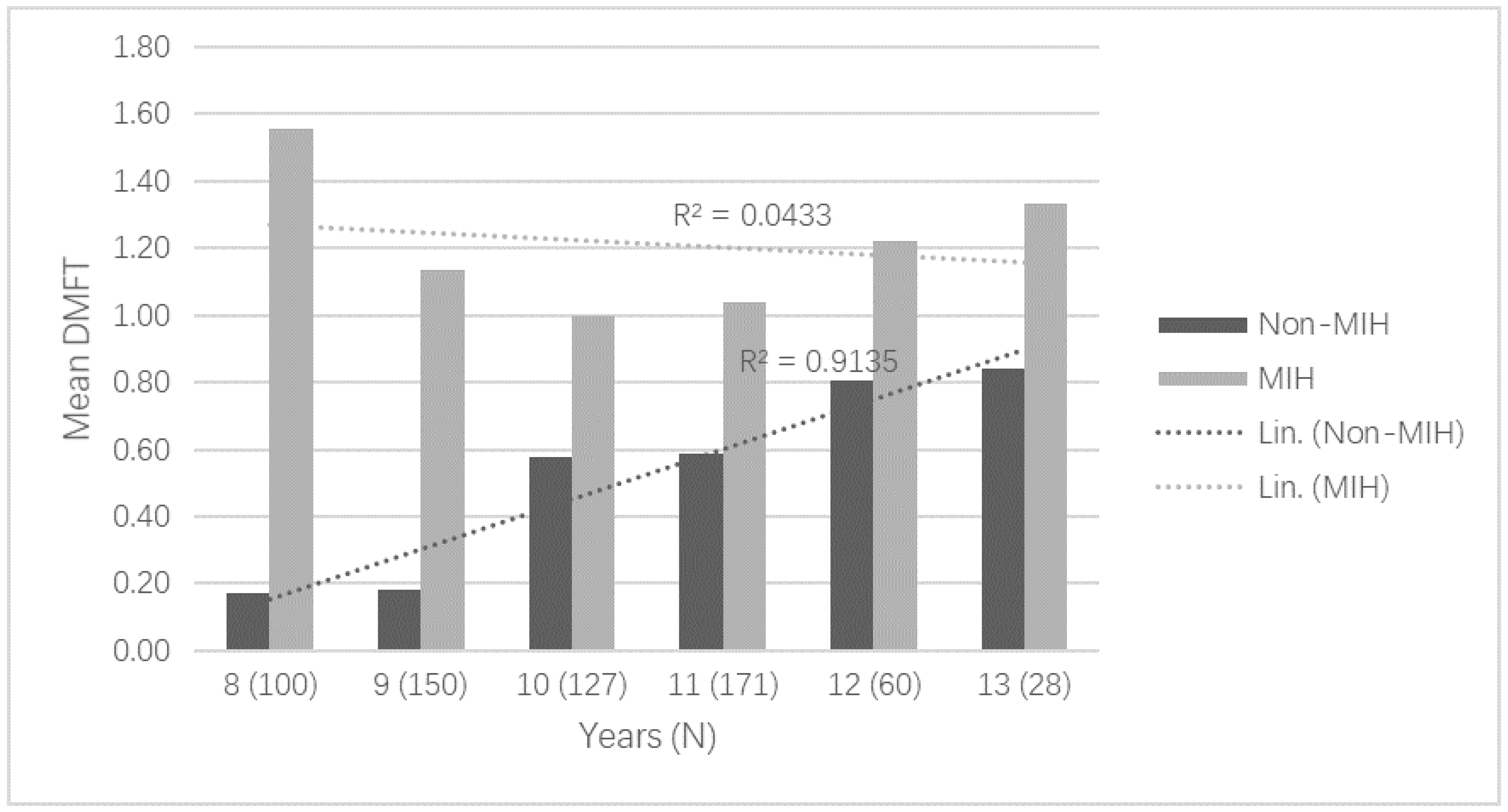
3. Results
4. Discussion
5. Conclusions
Author Contributions
Funding
Acknowledgments
Conflicts of Interest
References
- Schwendicke, F.; Dorfer, C.E.; Schlattmann, P.; Foster, P.L.; Thomson, W.M.; Paris, S. Socioeconomic inequality and caries: A systematic review and meta-analysis. J. Dent. Res. 2015, 94, 10–18. [Google Scholar] [CrossRef] [PubMed]
- Vargas-Ferreira, F.; Salas, M.M.S.; Nascimento, G.G.; Tarquinio, S.B.C.; Faggion, C.M.; Peres, M.A.; Thomson, W.M.; Demarco, F.F. Association between developmental defects of enamel and dental caries: A systematic review and meta-analysis. J. Dent. 2015, 43, 619–628. [Google Scholar] [CrossRef] [PubMed]
- Weerheijm, K.L.; Jalevik, B.; Alaluusua, S. Molar-incisor hypomineralisation. Caries Res. 2001, 35, 390–391. [Google Scholar] [CrossRef] [PubMed]
- Jalevik, B.; Norén, J.G. Enamel hypomineralization of permanent first molars: A morphological study and survey of possible aetiological factors. Int. J. Paediatr. Dent. 2000, 10, 278–289. [Google Scholar] [CrossRef] [PubMed]
- Nystrom, M.E.; Ranta, H.M.; Peltola, J.S.; Kataja, J.M. Timing of developmental stages in permanent mandibular teeth of Finns from birth to age 25. Acta Odontol. Scand. 2007, 65, 36–43. [Google Scholar] [CrossRef] [PubMed]
- Reid, D.J.; Dean, M.C. Variation in modern human enamel formation times. J. Hum. Evol. 2006, 50, 329–346. [Google Scholar] [CrossRef] [PubMed]
- Rodd, H.D.; Morgan, C.R.; Day, P.F.; Boissonade, F.M. Pulpal expression of TRPV1 in molar incisor hypomineralisation. Eur. Arch. Paediatr. Dent. 2007, 8, 184–188. [Google Scholar] [CrossRef] [PubMed]
- Fagrell, T.G.; Dietz, W.; Jalevik, B.; Noren, J.G. Chemical, mechanical and morphological properties of hypomineralized enamel of permanent first molars. Acta Odontol. Scand. 2010, 68, 215–222. [Google Scholar] [CrossRef] [PubMed]
- Kotsanos, N.; Kaklamanos, E.G.; Arapostathis, K. Treatment management of first permanent molars in children with Molar-Incisor Hypomineralisation. Eur. J. Paediatr. Dent. 2005, 6, 179–184. [Google Scholar] [PubMed]
- Lygidakis, N.A.; Wong, F.; Jalevik, B.; Vierrou, A.M.; Alaluusua, S.; Espelid, I. Best Clinical Practice Guidance for clinicians dealing with children presenting with Molar-Incisor-Hypomineralisation (MIH): An EAPD Policy Document. Eur. Arch. Paediatr. Dent. 2010, 11, 75–81. [Google Scholar] [CrossRef] [PubMed]
- Elfrink, M.E.; Ghanim, A.; Manton, D.J.; Weerheijm, K.L. Standardised studies on Molar Incisor Hypomineralisation (MIH) and Hypomineralised Second Primary Molars (HSPM): A need. Eur. Arch. Paediatr. Dent. 2015, 16, 247–255. [Google Scholar] [CrossRef] [PubMed]
- Schwendicke, F.; Elhennawy, K.; Reda, S.; Bekes, K.; Manton, D.J.; Krois, J. Global burden of molar incisor hypomineralization. J. Dent. 2018, 68, 10–18. [Google Scholar] [CrossRef] [PubMed]
- Brogardh-Roth, S.; Matsson, L.; Klingberg, G. Molar-incisor hypomineralization and oral hygiene in 10- to-12-yr-old Swedish children born preterm. Eur. J. Oral Sci. 2011, 119, 33–39. [Google Scholar] [CrossRef] [PubMed]
- Tourino, L.F.; Correa-Faria, P.; Ferreira, R.C.; Bendo, C.B.; Zarzar, P.M.; Vale, M.P. Association between molar incisor hypomineralization in schoolchildren and both prenatal and postnatal factors: A population-based study. PLoS ONE 2016, 11, e0156332. [Google Scholar] [CrossRef] [PubMed]
- Wuollet, E.; Laisi, S.; Salmela, E.; Ess, A.; Alaluusua, S. Molar-incisor hypomineralization and the association with childhood illnesses and antibiotics in a group of Finnish children. Acta Odontol. Scand. 2016, 74, 416–422. [Google Scholar] [CrossRef] [PubMed]
- Babajko, S.; Jedeon, K.; Houari, S.; Loiodice, S.; Berdal, A. Disruption of steroid axis, a new paradigm for molar incisor hypomineralization (MIH). Front. Physiol. 2017, 8, 343. [Google Scholar] [CrossRef] [PubMed]
- Jeremias, F.; Koruyucu, M.; Kuchler, E.C.; Bayram, M.; Tuna, E.B.; Deeley, K.; Pierri, R.A.; Souza, J.F.; Fragelli, C.M.; Paschoal, M.A.; et al. Genes expressed in dental enamel development are associated with molar-incisor hypomineralization. Arch. Oral Biol. 2013, 58, 1434–1442. [Google Scholar] [CrossRef] [PubMed] [Green Version]
- Jeremias, F.; Pierri, R.A.; Souza, J.F.; Fragelli, C.M.; Restrepo, M.; Finoti, L.S.; Bussaneli, D.G.; Cordeiro, R.C.; Secolin, R.; Maurer-Morelli, C.V.; et al. Family-Based Genetic Association for Molar-Incisor Hypomineralization. Caries Res. 2016, 50, 310–318. [Google Scholar] [CrossRef] [PubMed]
- Kuhnisch, J.; Thiering, E.; Heitmuller, D.; Tiesler, C.M.; Grallert, H.; Heinrich-Weltzien, R.; Hickel, R.; Heinrich, J. GINI-10 Plus Study Group; LISA-10 plus study group. Genome-wide association study (GWAS) for molar-incisor hypomineralization (MIH). Clin. Oral Investig. 2014, 18, 677–682. [Google Scholar] [CrossRef] [PubMed]
- Americano, G.C.; Jacobsen, P.E.; Soviero, V.M.; Haubek, D. A systematic review on the association between molar incisor hypomineralization and dental caries. Int. J. Paediatr. Dent. 2017, 27, 11–21. [Google Scholar] [CrossRef] [PubMed]
- Weerheijm, K.L.; Duggal, M.; Mejare, I.; Papagiannoulis, L.; Koch, G.; Martens, L.C.; Hallonsten, A.L. Judgement criteria for molar incisor hypomineralisation (MIH) in epidemiologic studies: A summary of the European meeting on MIH held in Athens, 2003. Eur. J. Paediatr. Dent. 2003, 4, 110–113. [Google Scholar] [PubMed]
- FDI Working Group. A review of the developmental defects of enamel index (DDE Index). Commission on oral health, research & epidemiology. Report of an FDI Working Group. Int. Dent. J. 1992, 42, 411–426. [Google Scholar]
- Sucking, G.W.; Brown, R.H.; Herbison, G.P. The prevalence of developmental defects of enamel in 696 nine-year-old New Zealand children participating in a health and development study. Community Dent. Health 1985, 2, 303–313. [Google Scholar]
- Calderara, P.C.; Gerthoux, P.M.; Mocarelli, P.; Lukinmaa, P.L.; Tramacere, P.L.; Alaluusua, S. The prevalence of molar incisor hypomineralisation (MIH) in a group of Italian school children. Eur. J. Paediatr. Dent. 2005, 6, 79–83. [Google Scholar] [PubMed]
- Jalevik, B.; Klingberg, G.; Barregard, L.; Noren, J.G. The prevalence of demarcated opacities in permanent first molars in a group of Swedish children. Acta Odontol. Scand. 2001, 59, 255–260. [Google Scholar] [CrossRef] [PubMed]
- Leppäniemi, A.; Lukinmaa, P.L.; Alaluusua, S. Nonfluoride hypomineralizations in the first molars and their impact on treatment need. Caries Res. 2001, 35, 36–40. [Google Scholar] [PubMed]
- World Health Organization. Oral Health Surveys: Basic Methods, 4th ed.; World Health Organization: Geneva, Switzerland, 1997; pp. 39–44. ISBN 92 4 154493 7. [Google Scholar]
- Grossi, J.A.; Cabral, R.N.; Leal, S.C. Caries experience in children with and without molar-incisor hypomineralisation: A case-control study. Caries Res. 2017, 51, 419–424. [Google Scholar] [CrossRef] [PubMed]
- Balmer, R.; Toumba, J.; Godson, J.; Duggal, M. The prevalence of molar incisor hypomineralisation in Northern England and its relationship to socioeconomic status and water fluoridation. Int. J. Paediatr. Dent. 2012, 22, 250–257. [Google Scholar] [CrossRef] [PubMed]
- Petrou, M.A.; Giraki, M.; Bissar, A.R.; Basner, R.; Wempe, C.; Altarabulsi, M.B.; Schäfer, M.; Schiffner, U.; Beikler, T.; Schulte, A.G.; et al. Prevalence of molar-incisor-hypomineralisation among school children in four German cities. Int. J. Paediatr. Dent. 2014, 24, 434–440. [Google Scholar] [CrossRef] [PubMed]
- Wuollet, E.; Laisi, S.; Salmela, E.; Ess, A.; Alaluusua, S. Background factors of molar-incisor hypomineralization in a group of Finnish children. Acta Odontol. Scand. 2014, 72, 963–969. [Google Scholar] [CrossRef] [PubMed]
- Jeremias, F.; de Souza, J.F.; Silva, C.M.; Cordeiro, R.C.; Zuanon, A.C.; Santos-Pinto, L. Dental caries experience and molar-incisor hypomineralization. Acta Odontol. Scand. 2013, 71, 870–876. [Google Scholar] [CrossRef] [PubMed]
- Da Costa-Silva, C.M.; Jeremias, F.; de Souza, J.F.; Cordeiro, R.C.; Santos-Pinto, L.; Zuanon, A.C. Molar incisor hypomineralization: Prevalence, severity and clinical consequences in Brazilian children. Int. J. Paediatr. Dent. 2010, 20, 426–434. [Google Scholar] [CrossRef] [PubMed]
- Bhaskar, S.; Hedge, S. Molar-incisor hypomineralization: Prevalence, severity and clinical characteristics in 8- to 13-year-old children of Udaipur, India. J. Indian Soc. Pedod. Prev. Dent. 2014, 32, 322–329. [Google Scholar] [CrossRef] [PubMed]
- Heitmuller, D.; Thiering, E.; Hoffmann, U.; Heinrich, J.; Manton, D.; Kuhnisch, J.; Neumann, C.; Bauer, C.P.; Heinrich-Weltzien, R.; Hickel, R.; et al. Is there a positive relationship between molar incisor hypomineralisations and the presence of dental caries? Int. J. Paediatr. Dent. 2013, 23, 116–124. [Google Scholar] [CrossRef] [PubMed]
- Xie, Z.H.; Mahoney, E.K.; Kilpatrick, N.M.; Swain, M.V.; Hoffman, M. On the structure-property relationship of sound and hypomineralized enamel. Acta Biomater. 2007, 3, 865–872. [Google Scholar] [CrossRef] [PubMed]
- Caufield, P.W.; Li, Y.; Bromage, T.G. Hypoplasia-associated severe early childhood Caries-a proposed definition. J. Dent. Res. 2012, 91, 544–550. [Google Scholar] [CrossRef] [PubMed]
- Fagrell, T.G.; Lingstrom, P.; Olsson, S.; Steiniger, F.; Noren, J.G. Bacterial invasion of dentinal tubules beneath apparently intact but hypomineralized enamel in molar teeth with molar incisor hypomineralization. Int. J. Paediatr. Dent. 2008, 18, 333–340. [Google Scholar] [CrossRef] [PubMed]
- Nelson, S.; Albert, J.M.; Geng, C.; Curtan, S.; Lang, K.; Miadich, S.; Heima, M.; Malik, A.; Ferretti, G.; Eggertsson, H.; et al. Increased enamel hypoplasia and very low birthweight infants. J. Dent. Res. 2013, 92, 788–794. [Google Scholar] [CrossRef] [PubMed]
- Hamilton, B.E.; Martin, J.A.; Ventura, S.J. Births: Preliminary data for 2011. Natl. Vital Stat. Rep. 2012, 61, 1–18. [Google Scholar] [PubMed]
- Wells, G.; Shea, B.; O’Connell, D.; Peterson, J.; Welch, V.; Losos, M.; Tugwell, P. The Newcastle-Ottawa Scale (NOS) for Assessing the Quality of Nonrandomized Studies in Meta-Analyses. 2016. Available online: http://www.ohri.ca/programs/clinical_epidemiology/oxford.asp (accessed on 23 September 2017).


| Variable | MIH | Non-MIH | Total | p-Value | |
|---|---|---|---|---|---|
| Children | 115 | 521 | 636 | ||
| Region | Lammi | 32 (27.8) | 156 (29.9) | 188 (29.6) | 0.000 |
| Jalasjärvi | 15 (13.0) * | 172 (33.0) | 187 (29.4) | ||
| Lappeenranta | 68 (59.1) * | 193 (37.0) | 261 (41.0) | ||
| DMFT value | 1.17 ± 1.39 | 0.46 ± 1.12 | 0.59 ± 1.21 | 0.000 a | |
| DMFT in FPMs | 1.03 ± 1.25 | 0.32 ± 0.80 | 0.45 ± 0.94 | 0.000 a | |
| Age, year | 10.5 ± 1.4 | 10.5 ± 1.3 | 10.5 ± 1.4 | 0.848 a | |
| Gender | Girls | 57 (49.6) | 250 (48.0) | 307 (48.3) | 0.094 |
| Boys | 58 (50.4) | 271 (52.0) | 329 (51.7) | ||
| Family income, € | <25,200 | 16 (16.7) * | 117 (27.0) | 133 (25.1) | 0.037 |
| 25,200–33,600 | 15 (15.6) | 90 (20.8) | 105 (19.8) | ||
| 33,600–50,400 | 32 (33.3) | 123 (28.4) | 155 (29.3) | ||
| >50,400 | 33 (34.4) * | 103 (23.8) | 136 (25.7) | ||
| Mother’s education | Prim. & second. school | 20 (17.5) | 74 (14.5) | 94 (15.0) | 0.357 |
| Vocational school | 29 (25.4) | 146 (28.5) | 175 (28.0) | ||
| High school/Higher vocational school | 46 (40.4) | 232 (45.3) | 278 (44.4) | ||
| University | 19 (16.7) | 60 (11.7) | 79 (12.6) | ||
| Variable | Categories | Coefficient | OR (95% CI) | p-Value |
|---|---|---|---|---|
| MIH | MIH | 1.88 | 6.60 (3.83–11.39) ** | 0.000 |
| Age of the child | Years | 0.25 | 1.29 (1.08–1.53) ** | 0.004 |
| Region | Lammi (reference) | 0 | 1 | |
| Jalasjärvi | 1.29 | 3.62 (1.67–7.87) ** | 0.001 | |
| Lappeenranta | 0.88 | 2.41 (1.16–4.99) * | 0.018 | |
| Family income, € | <25,200 (reference) | 0 | 1 | |
| 25,200–33,600 | −0.50 | 0.61 (0.30–1.25) | 0.177 | |
| 33,600–50,400 | −0.93 | 0.39 (0.20–0.78) ** | 0.008 | |
| >50,400 | −0.47 | 0.63 (0.30–1.32) | 0.217 | |
| Mother’s education | Prim. & second. school (reference) | 0 | 1 | |
| Vocational school | 0.58 | 1.79 (0.79–4.07) | 0.166 | |
| High school/Higher vocational school | −0.0.3 | 0.97 (0.42–2.22) | 0.944 | |
| University | 0.06 | 1.06 (0.38–2.96) | 0.909 |
© 2018 by the authors. Licensee MDPI, Basel, Switzerland. This article is an open access article distributed under the terms and conditions of the Creative Commons Attribution (CC BY) license (http://creativecommons.org/licenses/by/4.0/).
Share and Cite
Wuollet, E.; Laisi, S.; Alaluusua, S.; Waltimo-Sirén, J. The Association between Molar-Incisor Hypomineralization and Dental Caries with Socioeconomic Status as an Explanatory Variable in a Group of Finnish Children. Int. J. Environ. Res. Public Health 2018, 15, 1324. https://doi.org/10.3390/ijerph15071324
Wuollet E, Laisi S, Alaluusua S, Waltimo-Sirén J. The Association between Molar-Incisor Hypomineralization and Dental Caries with Socioeconomic Status as an Explanatory Variable in a Group of Finnish Children. International Journal of Environmental Research and Public Health. 2018; 15(7):1324. https://doi.org/10.3390/ijerph15071324
Chicago/Turabian StyleWuollet, Emma, Sakari Laisi, Satu Alaluusua, and Janna Waltimo-Sirén. 2018. "The Association between Molar-Incisor Hypomineralization and Dental Caries with Socioeconomic Status as an Explanatory Variable in a Group of Finnish Children" International Journal of Environmental Research and Public Health 15, no. 7: 1324. https://doi.org/10.3390/ijerph15071324