Article

Novel SFRP2 DNA Methylation Profile Following Neoadjuvant Therapy in Colorectal Cancer Patients with Different Grades of BMI

by
Amanda Cabrera-Mulero
1,2,†,
Ana B. Crujeiras
2,3,†,
Andrea G. Izquierdo
2,3,
Esperanza Torres
4,
Duncan Ayers
5,6,
Felipe F. Casanueva
2,3,
Francisco J. Tinahones
1,2,
Sonsoles Morcillo
1,2,7,* and
Manuel Macias-Gonzalez
1,2
1
Department of Endocrinology and Nutrition, Virgen de la Victoria University Hospital, University of Malaga (IBIMA), 29010 Málaga, Spain
2
CIBEROBN (CIBER in Physiopathology of Obesity and Nutrition CB06/03/0018), “Instituto de Salud Carlos III”, 28029 Madrid, Spain
3
Epigenomics in Endocrinology and Nutrition Group, Instituto de Investigación Sanitaria (IDIS), Complejo Hospitalario Universitario de Santiago (CHUS/SERGAS), 15706 Santiago de Compostela, Spain
4
Unidad de Gestión Clínica de Oncología Intercentros Hospital Universitario Virgen de la Victoria, 29010 Málaga, Spain
5
Centre for Molecular Medicine and Biobanking, University of Malta, 2080 Msida MSD, Malta
6
Faculty of Biology, Medicine and Health, The University of Manchester, Manchester M1 7DN, UK
7
Laboratorio Investigación Biomédica 1ª Planta, Hospital Universitario Virgen de la Victoria, Campus de Teatinos s/n 29010, 29010 Málaga, Spain
*
Author to whom correspondence should be addressed.
These authors contributed equally to this work.
J. Clin. Med. 2019, 8(7), 1041; https://doi.org/10.3390/jcm8071041
Submission received: 10 June 2019 / Revised: 10 July 2019 / Accepted: 15 July 2019 / Published: 17 July 2019
(This article belongs to the Section Clinical Laboratory Medicine)

Abstract

:
The relationship between body weight and different cancers is now well-recognized and among such cancers, colorectal cancer (CRC) is reported most frequently. Our group recently published findings, through an epigenome-wide association study, suggesting that body mass index (BMI) could act as a relevant risk factor in the CRC. In addition, aberrant SFRP2 methylation is one of the major mechanisms for Wnt signaling activation in CRC. Conversely, neoadjuvant chemo-radiotherapy appears to alter the rectal cancer epigenome. This study was aimed to evaluate the effect of obesity, measured by BMI, on the methylation of SFRP2 in tumor samples of patients with CRC. Non-treated CRC patients and CRC patients treated with pre-operative neoadjuvant therapy from 2011 to 2013 were included and classified by BMI < 25.0 kg/m2 and BMI > 25.0 kg/m2. SFRP2 DNA methylation in tumor samples was measured by pyrosequencing. Our findings suggest a possible interaction between SFRP2 methylation levels and BMI in CRC tumor samples. The correlation of SFRP2 hypomethylation with an elevated BMI was stronger within the non-treated CRC patient group than within the treated CRC patient group. We have successfully demonstrated that the beneficial association of tumor SFRP2 hypomethylation is dependent on patient BMI in non-treated CRC, suggesting a possible tumor suppressor role for SFRP2 in overweight and obese patients. Additional studies of clinical pathologies would be necessary to strengthen these preliminary results.

1. Introduction

One of the biggest challenges facing biomedical science is finding promising diagnostic, prognostic, and predictive biomarkers of non-communicable diseases of multi-factorial origin, such as colorectal cancer (CRC). Based on global cancer statistics, CRC is one of the leading causes of mortality from all cancers and the cancer-related mortality has increased by almost 40% in the last 40 years [1]. Consequently, novel promising biomarkers are required to increase the CRC screening sensitivity and predict more effective responses to individual cancer therapies.
DNA and miRNAs methylation of tumor suppressor genes and oncogenes is a key process in human carcinogenesis [2,3]. Moreover, epigenetic changes of tumor suppressor genes are a recognized factor in Knudson´s model of cancer [4]. According to the literature, epigenetic remodeling of several genes (KRAS, APC, p53, DCC, genes of the Rho family of GTPases, MACC1, Met, MTA1, RASSF1A, SFRP1, etc.) are associated with malignant cellular transformation, progression, invasion, and metastasis in colorectal cancers [5,6,7]. Consequently, aberrant DNA methylation also offers an opportunity for its exploitation as ideal biomarkers for CRC prediction, diagnosis, and prognosis [8]. Furthermore, the identification and validation of several hyper- and hypomethylated genes in CRC can be highly useful for employment as novel biomarkers for tumor development. Previous studies in CRC observed that LINE-1 (long interspersed nucleotide element) hypomethylation was associated with more advanced disease stages [9,10].
One important example of such key molecular players is the secreted frizzled-related protein type 2 (SFRP2), which can be deemed as a potential epigenetic biomarker of CRC. Moreover, SFRP2 (a Wnt signaling inhibitor) is hypermethylated in CRC [11,12,13]. Recently, a number of studies demonstrated that SFRP2 is not only hypermethylated in CRC tissue, but also in a spectrum of biological tissues including blood and stools, suggesting that SFRP2 methylation might be a potential non-invasive biomarker for CRC screening [14,15,16].
Currently, it is well established that excess body weight, defined by the body mass index (BMI), can be considered to be a risk factor for the incidence and progression of CRC [17]. The underlying mechanisms linking obesity to CRC are still a matter of debate. However, BMI-associated DNA methylation profile changes could provide one of the missing links between obesity and CRC [18]. Recently, we have reported that the hyper-methylation of the SFRP2 promoter exhibited a strong correlation with BMI in tumor samples, consequently suggesting adiposity as a prognostic factor in patients with CRC [14].
Presently, neoadjuvant chemo-radiotherapy, followed by radical surgery is considered to be the standard of care for patients with locally advanced rectal cancer [19]. Several authors have described how the patient characteristics, such as BMI and internal distribution of adipose tissue, have the potential to interfere with radiation dosing or delivery, leading to appreciable differences in treatment response and survival outcomes [20]. On the other hand, some studies have reported that DNA methylation could be a crucial factor that determines oncological treatment efficacy of, both, chemotherapy and radiotherapy [21]. However, it has been suggested that oncological treatment can also induce epigenetic changes that could mediate treatment response [22]. Ultimately, novel therapeutic approaches opt for utilizing adjuvant methyltransferase inhibitors, in order to increase treatment efficacy [23].
Considering the correlation between the SFRP2 methylation status and BMI in CRC patients [14], together with the potential effect of neoadjuvant treatment on DNA methylation pattern; we propose that SFRP2 methylation could be associated with specific clinical outcome of CRC patients and this relationship could be affected by BMI or treatment. In the present study, we have specifically focused on the analysis of tumor and non-tumor samples for the methylation status of SFRP2 in an independent prospective cohort study, together with its association with the clinical and pathological features of CRC patients.

2. Materials and Methods

2.1. Study Population

A total of 75 CRC patients diagnosed by biopsy or colonoscopy were included in the present study from Virgen de la Victoria University Hospital (Málaga, Spain), recruited from October 2011 to September 2013. One cohort group consisted of 53 overweight or obese patients (BMI > 25 kg/m2), while 22 were non-obese participants (BMI < 25 kg/m2). All participants underwent surgery with curative intention, by hemicolectomy, lower anterior resection with ileostomy (caused by a carcinoma of the CRC), followed by a total mesocolorectal excision. A formalin-fixed paraffin-embedded (FFPE) (10 sections of 14 μm) tumor area and an adjacent tumor-free area were obtained from all CRC patients.
Participants included in the study had primary CRC whose medical records/pathological examinations were complete. The exclusion criteria were patients with inflammatory bowel disease (Crohn´s disease or ulcerative colitis) and patients who had evidence of hereditary non-polyposis colorectal cancer or familial adenomatous polyposis. All participants were anonymized and gave their written informed consent. The study was performed in accordance with the Declaration of Helsinki and was approved by the Ethics Committees of Virgen de la Victoria University Hospital (registration number 0311/PI7) (Málaga, Spain).
The clinical–pathological parameters of the cohort groups were confirmed by reviewing the patient medical records and pathology files. The pathological diagnosis, tumor histology, and staging were performed according to the Classification of the “World Health Organization Classification of Tumours of the Digestive System” (2016) [24]. Rectal cancer patients were treated according to local protocols, after a routine workout, including a turaco abdominal scan and a pelvis MRI. Those classified as T3–4 or N 1–3 were proposed for neoadjuvant chemoradiation treatment with pelvic radiotherapy 50Gy (2 Gy/fraction) and concomitant administration of fluoropyrimidine-based chemotherapy, followed by total mesorectal excision in 6–8 weeks. Patients were sub-classified as treated (those that were designated for neoadjuvant therapy), while those who did not receive any pre-operative treatment were classified as non-treated.

2.2. Biochemical Determination

Fasting blood samples were obtained from the antecubital vein and placed in vacutainer tubes (BD Vacutainer™, Franklin Lakes, NJ, USA). Serum glucose, total cholesterol, triglycerides, and HDL cholesterol (HDL-c) were measured in a Dimension auto analyzer (Dade Behring Inc., Deerfield, IL, USA) through enzymatic methods (Randox Laboratories Ltd., Crumlin, UK). The LDL cholesterol (LDL-c) was calculated using the Friedewald equation [25]. Insulin was quantified by radioimmunoassay (BioSource International, Camarillo, CA, USA). Corrected calcium was assessed using a complex metric method from Boehringer Hitachi 717. Alkaline phosphatase was calculated using ELISA (LifeSpan Biosciences Inc., Madrid, Spain). Insulin growth factor type 1 (IGF-1) was determined using Human IGF1 ELISA Kit (Abcam, Madrid, Spain). The homeostasis model assessment of insulin resistance (HOMA-IR) was calculated using the following equation: HOMA-IR = fasting insulin (IU/mL) x fasting glucose (mmol/L)/22.5 [26].

2.3. Human Colorectal CarcinomaCell Lines and SFRP2 Expression Assay by RT-qPCR

HTC116 cell line (Homo Sapiens colon colorectal carcinoma, ATCC, Manassas, VA, USA), LoVo cell line (Homo Sapiens colorectal adenocarcinoma derived from metastatic site—left supraclavicular region, ATCC, Manassas, VA, USA) and Caco-2 cell line (Homo Sapiens colon colorectal adenocarcinoma, ATCC, Manassas, VA, USA) were used to study the gene expression of SFRP2. Cells were cultured in Dulbecco’s modified Eagle’s medium containing 10% fetal bovine serum, penicillin, and streptomycin at 37 °C and 5% CO2. Cell lines were treated with the DNA demethylation agent 5-aza-2′-deoxycitydine (AZA) (A3656, Sigma Aldrich, Madrid, Spain) at 5μM for 72 hours.
Consequently, RNA from cell lines was isolated using QiampRNA Mini Kit (Qiagen GmbH, Hilden, Germany), according to the manufacturer’s instructions. The purity and concentration of the RNA was determined by the 260/280 absorbance ratio using the Nanodrop 2000 platform (Thermo scientific, USA). For cDNA synthesis, a fixed amount of 1 µg of the total RNA was reverse transcribed using random hexamers as primers and Moloney Murine Leukemia Virus reverse transcriptase (Roche Diagnostic, Rotkreuz, Switzerland). Gene expression was assessed by real-time PCR, using an Applied Biosystems 7500 Fast Real-Time PCR System (Applied Biosystems, Darmstadt, Germany) with TaqMan technology. The reaction was performed following the manufacturer's protocol of TaqMan technologies Premix Ex Taq™ (Probe qPCR) (Takara, Madrid, Spain), in a final volume of 12.5 µL. The cycle program consisted of an initial denaturing of 20 s at 95 °C, then 45 cycles of a 3 s denaturing phase at 95 °C, followed by a 30 s annealing period. The commercially available and revalidated TaqMan primer/probe sets used in our samples were SFRP2 and PPIA TaqMan Probes (IDT technologies, Madrid, Spain). A threshold cycle (Ct value) was obtained for each amplification curve and a ΔCt value was first calculated by subtracting the Ct value for Cyclophilin A cDNA from the Ct value for each sample and transcript. Fold changes compared with the endogenous control were then determined by calculating 2−ΔCt, and relative quantification results were obtained.

2.4. DNA Extraction, Bisulfite Treatment, and Pyrosequencing

Total genomic DNA from 75 CRC paraffin samples (formalin-fixed paraffin embedded (FFPE) (10 sections of 14 μm) was isolated using the QIAmp DNA FFPE Tissue Kit (Qiagen GmbH, Hilden, Germany), with a xylene wash, to remove the paraffin. DNA concentration and quality was determined using NanoDrop 2000 (Thermo scientific, USA). DNA methylation analyses were performed using bisulfite-treated DNA, followed by a highly quantitative analysis, based on PCR-based pyrosequencing. The bisulfite conversion was conducted with 2 μg of genomic DNA isolated from each sample, using Epitect Bisulfite conversion, according to the manufacturer´s instructions (Qiagen GmbH, Hilden, Germany). Consequently, the SFRP2 promoter and LINE-1 amplification steps were performed in a total volume of 25 μL, with a starting primer concentration of 10 μM (SFRP2: Primer 5′3′ forward and reverse respectively: TTTGATTTTTTTAYGGTATTGGGGAGTA and ATAAAACCCRAAACCTACCC. LINE-1: Primer 5′3′ forward and reverse respectively: TAGGGAGTGTTAGATAGTGG and AACTCCCTAACCCCTTAC). The reverse primer was biotinylated, in order to purify the final PCR product, using sepharose beads. Finally, 20 μL of the PCR products were pyro sequenced using the PyroMarkTMQ96 ID Pyrosequencing System, using a 0.4 μM sequencing primer. The primer sequences used in this analysis were designed using Qiagen’s PyroMark Assay Design 2.0 software (Qiagen, Hilden, Germany).
The methylation level was expressed as the percentage of methylated cytosine over the sum of methylated and unmethylated cytosines. Non-CpG cytosine residues were used as built-in controls to verify bisulfite conversion. The values are expressed as the mean for all the sites and individually for seven CpGs at the SFRP2 gene promoter and for six CpGs at the LINE-1 sequence [27,28]. We also included unmethylated and methylated DNA as controls in each run (New England Biolabs, Ipswich, MA, USA).

2.5. Statistical Analysis

The results were expressed as the mean ± standard deviation (or percentage) where appropriate. Student’s t test was used for comparing anthropometric and biochemical data between non-obese and overweight/obese groups. Paired t-test was performed to compare means between tumor and tumor-free area. Student’s t test and Mann–Whitney U-test were used for comparisons of LINE-1 and SFRP2 methylation between study groups. Spearman’s correlation analyses were performed to study the correlations between LINE-1 and SFRP2 methylation. A regression linear model was used to perform multivariate analysis. All analyses were performed using the R statistical software, version 2.8.1 (Department of Statistics, University of Auckland, Auckland, NZ; http://www.rproject.org/).

3. Results

3.1. Clinical, Anthropometrical, and Biochemical Characteristics of the CRC Patients

There were no differences in anthropometric and biochemical variables between subjects with BMI < 25 or BMI > 25, except for the BMI itself (as expected) and the insulin levels (Table 1).

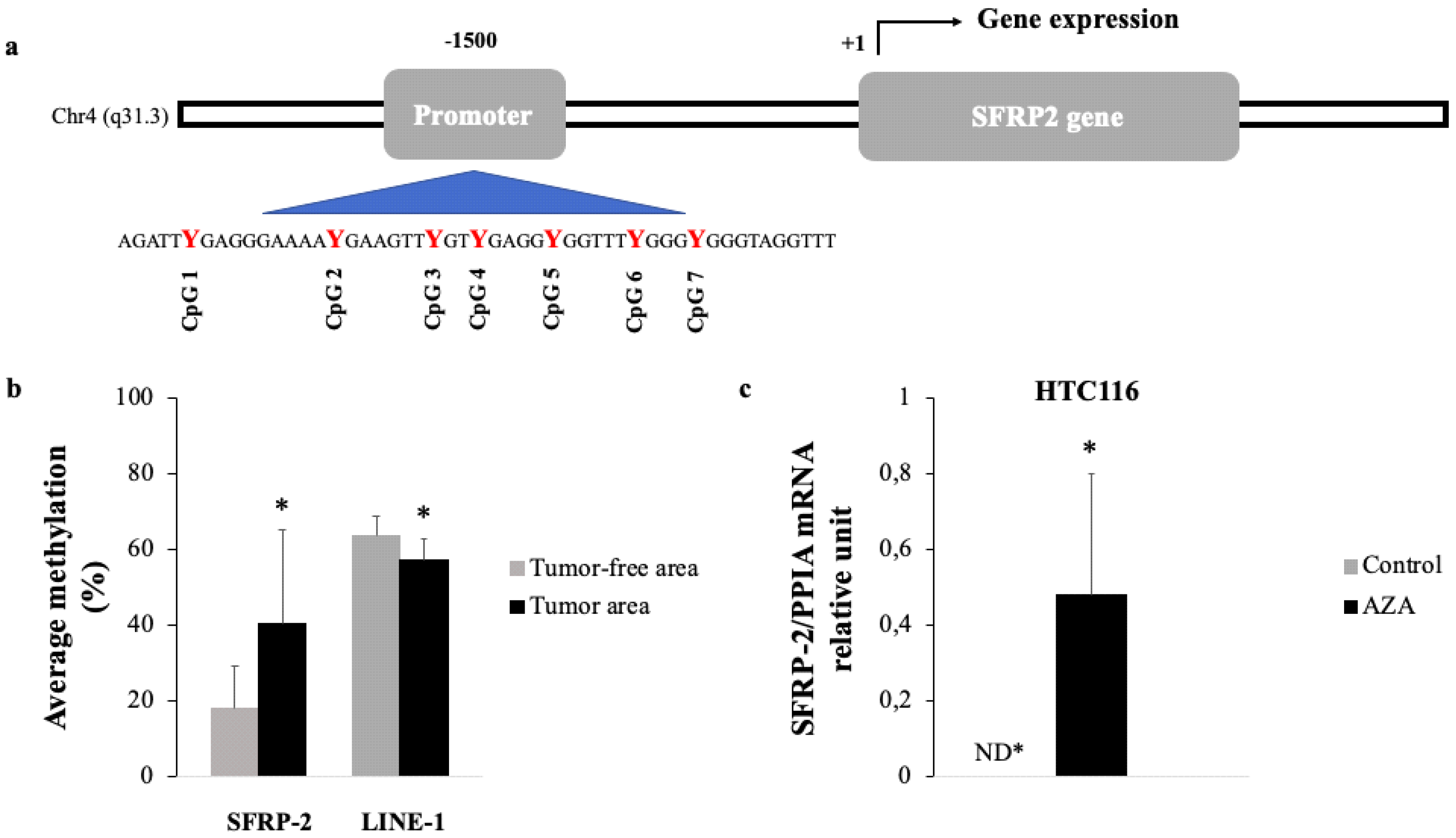
3.2. Methylation Status of SFRP2 in CRC Tissue

First of all, we initiated our analysis based on previous data of the SFRP2 promoter performed and published by our group and obtained from the Infinium Human Methylation 450 BeadChip array [14].The study sequence of the SFRP2 promoter used in our analysis showed seven CpG sites in the position −1500, relative to the expected transcription start site (+1),which was consequently utilized to determine the methylation status of the SFRP2 promoter (Figure 1a).
We aimed to evaluate the SFRP2 methylation status in both CRC tissue and adjacent tumor-free tissue from 75 CRC patients, by pyrosequencing after bisulfite treatment.
The results revealed hyper-methylation of the SFRP2 promoter, since SFRP2 methylation in tumor tissue was significantly higher (40.30%)in comparison to the levels obtained for adjacent tumor-free tissue areas (17.86%). Conversely, LINE-1 methylation was significantly decreased in the tumor area (57.38%), when compared to the tumor-free area (63.50%) (Figure 1b).
In addition, to test whether the hyper-methylation of SFRP2 promoter is functionally associated with a down-regulated expression of SFRP2 at the transcriptomic level, we performed in vitro demethylation assays, using different colorectal carcinoma cell lines.
Accordingly, we employed RT-qPCR assays to evaluate whether SFRP2 mRNA is differentially expressed, before and after treatment, with a methylation inhibitor. First, we treated cell lines for 72 h with 5 μM of AZA, a methylation inhibitor. Consequently, we quantified the mRNA expression of SFRP2 through RT-qPCR. The HTC116 control showed undetectable mRNA expression of SFRP2, while the HCT116 treated with AZA resulted in a significant and clear increase of mRNA SFRP2 expression, as shown in Figure 1c. However, despite measuring the SFRP2 gene expression in three different human colorectal carcinoma cell lines (HCT116, LoVo, and Caco-2), we only observed expression recovery of SFRP2 in HCT116, after the AZA treatment. The lack of results from the other cell lines could be explained by their inherent biological heterogeneity from these cell lines.

3.3. Baseline Characteristics of Colorectal Cancer Patients with Regards to their BMI Category

Clinical, pathological, and tumor molecular features according to the BMI in proximal colon cancer (cecum, ascending colon, and transverse colon), distal colon cancer (descending colon, and sigmoid colon), and rectal colon cancer are summarized in Table 2.
We observed that there were no differences between the two groups for any of the oncological variables studied.

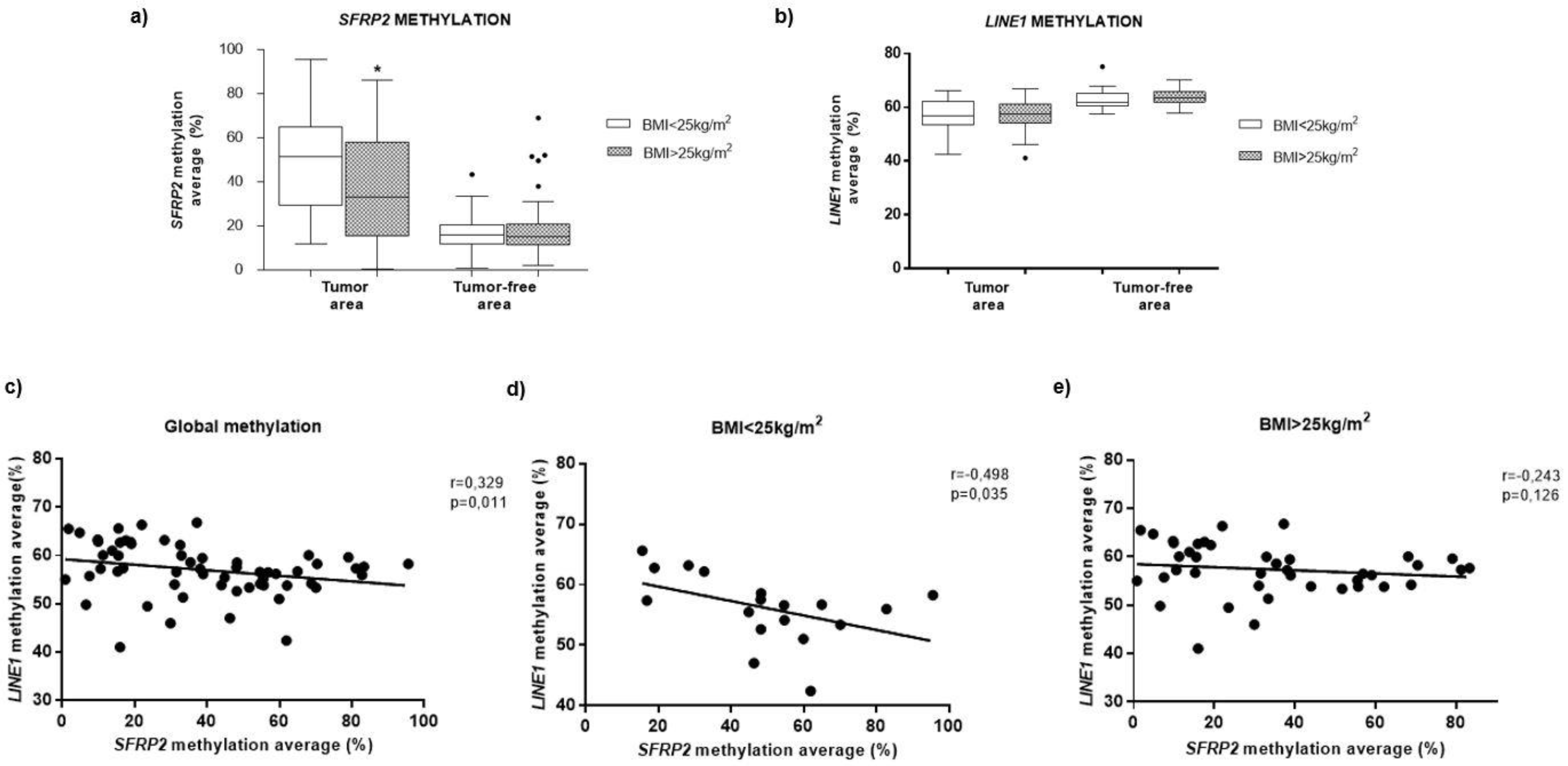
3.4. Methylation of SFRP2 in CRC Tissue is Associated with BMI

In order to investigate whether adiposity affects DNA methylation levels, the methylation pattern of SFRP2 and LINE-1 was compared in tumor tissue and tumor-free area, in both BMI groups. Our results showed that the SFRP2 methylation was significantly lower in overweight/obese individuals (34.06%), compared to the non-obese individuals (51.24%) (Figure 2a), while LINE-1 methylation did not show any significant difference in BMI (56.68% for non-obese subjects and for overweight/obese individuals 57.69%) (Figure 2b). We also observed a significant negative correlation between global LINE-1 and SFRP2 methylation in CRC tumor tissue (r = −0.329, p = 0.011) (Figure 2c). However, when the subjects were divided according to the BMI, this association only remained significant in subjects with BMI < 25 (Figure 2d,e).

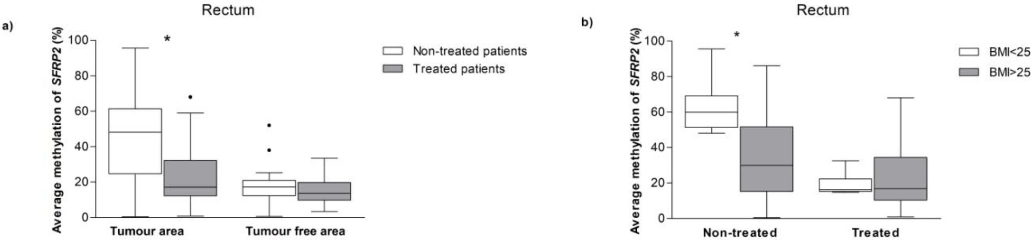
3.5. SFRP2Methylation in CRC Patients with and without Neoadjuvant Treatment

The specific location of the tumor is a factor to be considered, as they could present biological and clinical differences. We analyzed the methylation SFRP2 status according to the tumor location. First of all, we observed that SFRP2 methylation was significantly higher on the right-sided CRC (57.33%), as compared to the left-sided CRC (36.60%). However, in this classification, most patient tumor samples were derived from the rectum.
Primary rectal cancer requires specific surgical treatment (total mesorectal excision, preceded by neoadjuvant radiotherapy or chemoradiotherapy); so we aimed to evaluate whether the neoadjuvant therapy could be affecting the methylation levels. Patients who received treatment displayed lower SFRP2 methylation levels than those without treatment, within the tumor tissue, though not in the tumor-free area (Figure 3a). Finally, we assessed the SFRP2 methylation status according to the BMI, in the treated and the non-treated patients. In the group of patients who received neoadjuvant therapy, there was no statistical difference in the SFRP2 methylation levels regarding BMI. However, within the non-treated patients, those with BMI > 25 showed significantly lower SFRP2 methylation levels, when compared to the BMI < 25 group (Figure 3b), albeit only in the tumor samples.
At last, to evaluate whether this association between SFRP2 methylation and therapy could be due to other potential confounder variables, we performed a multivariable analysis, including age, BMI and presence of diabetes mellitus. We proved that neoadjuvant treatment was the main variable explaining the variability of SFRP2 methylation levels (Table 3).

4. Discussion

Aberrant DNA methylation in gene promoters has been strongly associated with key dysregulations in the oncogenesis for most human tumor models, including colorectal cancer [29]. Presently, there is a large list of established genes subjected to abnormal DNA methylation that are consequently implicated in pivotal physiological processes, including tumor suppression, which can ultimately prove to be of essential utility as novel, epigenetics-based tumor biomarkers [30].
In this study, we evaluated the methylation status of SFRP2, which functionally acts as an inhibitor of Wnt signaling in CRC, together with BMI influences on such a methylation status. CRC is most commonly initiated by accumulation of β catenin in the Wnt signaling pathway, leading to the activation of the Wnt target genes [31].
However, contradicting results were also published in the scientific literature, stating that the modification of the prognostic impact of obesity by a positive status for nuclear β catenin was associated with a significantly improved survival rate for colorectal cancer [32].
The results of our study demonstrated that decreased SFRP2 methylation in CRC was associated with an increased BMI. It has been established that adipose tissue is associated with local inflammation due to increased production of cytokines and pro-inflammatory factors through the macrophages present in adipose tissue [33]. This inflamed state was able to create a chronic low-grade inflammation that can activate the Wnt/β catenin pathway, where SFRP2 plays an important regulatory role in the carcinogenesis process of CRC [34]. In essence, the functional role of SFRP2 in CRC and its potential influence through an increased BMI, could explain, at least partially, the process through which obesity and Wnt/β catenin might exert their effect on carcinogenesis through independent molecular pathways [35].
The SFRP2 methylation status was found to be associated with a decreased gene expression of SFRP2 [36] and, furthermore, was found to be involved in carcinogenesis through down-regulation of tumor suppressor genes. Indeed, SFRP2 can be detected in many biological samples, such as serum and stools, rendering it a potential non-invasive biomarker for many carcinogenic processes [37]. In this study we discovered that SFRP2 was hypermethylated in CRC tumor tissue, when compared to adjacent tumor-free tissue areas. In addition, the hypermethylated SFRP2 in vitro (as found in the HTC116 CRC tumoral cell line) were presumably associated with suppression of gene expression. Following treatment with AZA as a demethylating agent, the expression of SFRP2 was significantly increased and restored. This suggests that SFRP2 demethylation has a functional role in the transcriptional control of SFRP2. Our study was not focused on exploring the potential mechanisms involved in this decrease of DNA methylation pattern, although a possible explanation could be due to a de-regulation of DNA methyltransferase 1 (DNMT1). In most tumors, an altered regulation of this enzyme was described, which is involved in maintaining methylation patterns [38]. Studies have shown that DNMT inhibition promotes gene expression of SFRP2, suggesting DNMTs can modulate SFRP2 methylation status [39].
Genome-wide DNA hypo-methylation of LINE-1 is associated with a worse prognosis in early-stage colorectal cancer, though the majority of the performed studies did not reveal a relation between LINE-1 hypomethylation and clinical outcome in more advanced disease stages [40]. However, our study results confirmed this association, since we revealed a low methylation rate for LINE-1 in tumor samples, without any association or relationship with the clinical–pathological parameters of colorectal tumors.
Previous studies have reported that the location of tumors could influence treatment choice, whereby a large percentage of survival patients were specifically patients who presented primary CRC tumors on the left-side of the abdomen [41]. Consequently, we focused our study on SFRP2 methylation in rectum tumor samples. Treated patients demonstrated a decrease of SFRP2 methylation, independent of the BMI value. This supports the hypothesis that both SFRP2 methylation and BMI could have a potential role in the development of CRC. Additionally, in the non-treated group, we observed higher SFRP2 methylation levels in patients with low BMI, suggesting a possible tumor suppressor role of SFRP2 in overweight/obese patients.
These results point to a possible interaction between BMI and neoadjuvant treatment. Moreover, recently, described epigenetic changes in DNA have also been underlined as mediators of the networking between obesity and cancer [42,43]. Li et al. reported that obesity led to epigenetic remodeling that resulted in differential gene expression related to metabolism and tumorigenesis. Moreover, this epigenetic signature could be reverted by weight loss reinforcing the role of BMI in the epigenetic mark of genes involved in neoplasia [44].
Limitations of this research include the lack of large cohort groups in order to analyze the effect of therapy with more statistical significance and perform survival studies. We could also not demonstrate the causal relation between methylation and gene expression, since expression data were obtained from human colon cancer cell lines. Finally, due to the challenge involved in obtaining high integrity RNA from tumor paraffin-embedded samples, we could not evaluate the gene expression from different DNA methyltransferases.

5. Conclusions

We demonstrated that the DNA methylation levels of SFRP2 are lower in overweight/obese patients, in comparison with non-obese patients, specifically in tumor areas, supporting the hypothesis that BMI could play a protective role in the development of CRC. We also examined that the patients undergoing neoadjuvant treatment showed similar methylation values of SFRP2, independent of their BMI, suggesting that neoadjuvant treatment might have an effect on the epigenetic regulation of SFRP2, though further studies of clinical pathologies for such patients would be necessary to strengthen these preliminary results.

Author Contributions

Conceptualization: F.F.C., F.J.T., S.M. and M.M.-G.; Formal analysis: A.C.-M. and S.M.; Funding acquisition: M.M.-G.; Investigation: A.C.-M.; Methodology: A.C.-M. and A.B.C.; Project administration: M.M.-G.; Resources: A.B.C., E.T., S.M. and M.M.-G.; Supervision: M.M.-G.; Visualization: A.C.-M., A.G.-I. and S.M.; Writing–original draft: A.C.-M. and A.B.C.; Writing–review & editing: D.A., F.F.C., F.J.T., S.M. and M.M.-G.

Funding

This study was supported by “Centros de Investigación En Red” (CIBER, CB06/03/0018) of the “Instituto de Salud Carlos III” (ISCIII) and a grant from ISCIII (PI8/01399) and it was co-financed by the European Regional Development Fund (FEDER). M.M.G. was the recipient of the Nicolas Monardes Program from the “Servicio Andaluz de Salud, Junta de Andalucía”, Spain (RC-0001-2018 and C-0029-2014). S.M. was the recipient of the Nicolas Monardes Program from the “Servicio Andaluz de Salud, Junta de Andalucía”, Spain (C-0050-2017). A.B.C. was funded by a research contract “Miguel Servet” (CP17/00088) from the ISCIII. A.C.-M. was recipient of an FPU grant from Education Ministry, Madrid, Spain.

Acknowledgments

The authors thank all patients for their important contribution.

Conflicts of Interest

The authors declare that no competing interests exist. They are in agreement to be accountable for all aspects of the work in ensuring that questions related to the accuracy or integrity of any part of the work are appropriately investigated and resolved.

Availability of Data and Materials

Data will be not shared. An explicit statement was not included in the informed consent to allow data sharing. Data were not collected anonymously. Research data are stored with patients’ name, contact information, or other identifiers.

Abbreviations

CRCcolorectal cancer
SFRP2secreted frizzled-related protein 2
LINE-1long Interspersed Element-1
BMIbody mass index
HDL-chigh density lipoprotein cholesterol
LDL-clow density lipoprotein cholesterol
FFPEformalin-fixed paraffin-embedded

References

  1. Siegel, R.L.; Miller, K.D.; Jemal, A.; Siegel, R.L.; Miller, K.D.; Jemal, A. Cancer statistics, 2016. CA Cancer J. Clin. 2016, 66, 7–30. [Google Scholar] [CrossRef] [PubMed] [Green Version]
  2. Lopez-Serra, P.; Esteller, M. {DNA} methylation-associated silencing of tumor-suppressor {microRNAs} in cancer. Oncogene 2012, 31, 1609–1622. [Google Scholar] [CrossRef] [PubMed]
  3. Chimonidou, M.; Strati, A.; Tzitzira, A.; Sotiropoulou, G.; Malamos, N.; Georgoulias, V.; Lianidou, E.S. DNA methylation of tumor suppressor and metastasis suppressor genes in circulating tumor cells. Clin. Chem. 2011, 57, 1169–1177. [Google Scholar] [CrossRef]
  4. Kondo, Y.; Issa, J.P.J. Epigenetic changes in colorectal cancer. Cancer Metastasis Rev. 2004, 23, 29–39. [Google Scholar] [CrossRef]
  5. Sakai, E.; Nakajima, A.; Kaneda, A. Accumulation of aberrant DNA methylation during colorectal cancer development. World J. Gastroenterol. 2014, 20, 978–987. [Google Scholar] [CrossRef]
  6. Kumar, A.; Gosipatala, S.B.; Pandey, A.; Singh, P. Prognostic Relevance of Sfrp1 Gene Promoter Methylation in Colorectal Carcinoma. Asian Pac. J. Cancer Prev. 2019, 20, 1571–1577. [Google Scholar] [CrossRef]
  7. Tokarz, P.; Pawlowska, E.; Bialkowska-Warzecha, J.; Blasiak, J. The significance of DNA methylation profile in metastasis-related genes for the progression of colorectal cancer. Cell. Mol. Biol. 2017, 63, 79–87. [Google Scholar] [CrossRef] [PubMed]
  8. Bishnupuri, K.S.; Mishra, M.K. Epigenetics of colorectal cancer. In Epigenetic Advancements in Cancer; Springer International Publishing: Cham, Switzerland, 2016; ISBN 9783319249513. [Google Scholar]
  9. Portela, A.E.M. Epigenetic modifications and human disease. Nat. Biotechnol. 2010, 10, 1057–1068. [Google Scholar] [CrossRef]
  10. Baba, Y.; Huttenhower, C.; Nosho, K.; Tanaka, N.; Shima, K.; Hazra, A.; Schernhammer, E.S.; Hunter, D.J.; Giovannucci, E.L.; Fuchs, C.S.; et al. Epigenomic diversity of colorectal cancer indicated by LINE-1 methylation in a database of 869 tumors. Mol. Cancer 2010, 9, 125. [Google Scholar] [CrossRef]
  11. Sui, C.; Ma, J.; Chen, Q.; Yang, Y. The variation trends of {SFRP}2 methylation of tissue, feces, and blood detection in colorectal cancer development. Eur. J. Cancer Prev. 2016, 25, 288–298. [Google Scholar] [CrossRef]
  12. Hu, H.; Wang, T.; Pan, R.; Yang, Y.; Li, B.; Zhou, C.; Zhao, J.; Huang, Y.; Duan, S. Hypermethylated Promoters of Secreted Frizzled-Related Protein Genes are Associated with Colorectal Cancer. Pathol. Oncol. Res. 2019, 25, 567–575. [Google Scholar] [CrossRef]
  13. Bagci, B.; Sari, M.; Karadayi, K.; Turan, M.; Ozdemir, O.; Bagci, G. KRAS, BRAF oncogene mutations and tissue specific promoter hypermethylation of tumor suppressor SFRP2, DAPK1, MGMT, HIC1 and p16 genes in colorectal cancer patients. Cancer Biomark. 2016, 17, 133–143. [Google Scholar] [CrossRef]
  14. Crujeiras, A.B.; Morcillo, S.; Diaz-Lagares, A.; Sandoval, J.; Castellano-Castillo, D.; Torres, E.; Hervas, D.; Moran, S.; Esteller, M.; Macias-Gonzalez, M.; et al. Identification of an episignature of human colorectal cancer associated with obesity by genome-wide DNA methylation analysis. Int. J. Obes. 2018, 43, 176–188. [Google Scholar] [CrossRef]
  15. Li, H.; Wang, Z.; Zhao, G.; Ma, Y.; Chen, Y.; Xue, Q.; Zheng, M.; Fei, S. Performance of a MethyLight assay for methylated SFRP2 DNA detection in colorectal cancer tissue and serum. Int. J. Biol. Markers 2019, 34, 54–59. [Google Scholar] [CrossRef]
  16. Yang, Q.; Huang, T.; Ye, G.; Wang, B.; Zhang, X. Methylation of SFRP2 gene as a promising noninvasive biomarker using feces in colorectal cancer diagnosis: A systematic meta-analysis. Sci. Rep. 2016, 6, 1–11. [Google Scholar] [CrossRef]
  17. Bardou, M.; Barkun, A.N.; Martel, M. Obesity and colorectal cancer. Gut 2013, 62, 933–947. [Google Scholar] [CrossRef]
  18. Wahl, S.; Drong, A.; Lehne, B.; Loh, M.; Scott, W.R.; Kunze, S.; Tsai, P.C.; Ried, J.S.; Zhang, W.; Yang, Y.; et al. Epigenome-wide association study of body mass index, and the adverse outcomes of adiposity. Nature 2017, 541, 81–86. [Google Scholar] [CrossRef]
  19. Dayde, D.; Tanaka, I.; Jain, R.; Tai, M.C.; Taguchi, A. Predictive and Prognostic Molecular Biomarkers for Response to Neoadjuvant Chemoradiation in Rectal Cancer. Int. J. Mol. Sci. 2017, 18, 573. [Google Scholar] [CrossRef]
  20. Sun, Y.; Xu, Z.; Lin, H.; Lu, X.; Huang, Y.; Huang, S.; Wang, X.; Chi, P. Impact of body mass index on treatment outcome of neoadjuvant chemoradiotherapy in locally advanced rectal cancer. Eur. J. Surg. Oncol. 2017, 43, 1828–1834. [Google Scholar] [CrossRef]
  21. Zhu, X.; Wang, Y.; Tan, L.; Fu, X. The pivotal role of DNA methylation in the radio-sensitivity of tumor radiotherapy. Cancer Med. 2018, 7, 3812–3819. [Google Scholar] [CrossRef]
  22. Sood, S.; Patel, F.D.; Srinivasan, R.; Dhaliwal, L.K. Chemoradiation therapy induces in vivo changes in gene promoter methylation & gene transcript expression in patients with invasive cervical cancer. Indian J. Med. Res. 2018, 147, 151–157. [Google Scholar]
  23. Stone, L. Radiotherapy induces epigenetic changes. Nat. Rev. Urol. 2016, 13, 241. [Google Scholar] [CrossRef]
  24. Bosnan, F.T.; Carneiro, F.; Hruban, R.H.; Theise, N.D. WHO Classification of Tumors of the Digestive System, 4th ed.; International Agency for Research on Cancer (IARC): Lyon, France, 2010; ISBN 978-92-832-2432-7. [Google Scholar]
  25. Friedewald, W.T.; Levy, R.I.; Fredrickson, D.S. Estimation of the concentration of low-density lipoprotein cholesterol in plasma, without use of the preparative ultracentrifuge. Clin. Chem. 1972, 18, 499–502. [Google Scholar]
  26. Matthews, D.R.; Hosker, J.P.; Rudenski, A.S.; Naylor, B.A.; Treacher, D.F.; Turner, R.C. Homeostasis model assessment: Insulin resistance and beta-cell function from fasting plasma glucose and insulin concentrations in man. Diabetologia 1985, 28, 412–419. [Google Scholar] [CrossRef]
  27. Castellano-Castillo, D.; Morcillo, S.; Crujeiras, A.B.; Sánchez-Alcoholado, L.; Clemente-Postigo, M.; Torres, E.; Tinahones, F.J.; Macias-Gonzalez, M. Association between serum 25-hydroxyvitamin D and global DNA methylation in visceral adipose tissue from colorectal cancer patients. BMC Cancer 2019. [Google Scholar] [CrossRef]
  28. Castellano-Castillo, D.; Morcillo, S.; Clemente-Postigo, M.; Crujeiras, A.B.; Fernandez-García, J.C.; Torres, E.; Tinahones, F.J.; Macias-Gonzalez, M. Adipose tissue inflammation and VDR expression and methylation in colorectal cancer. Clin. Epigenet. 2018. [Google Scholar] [CrossRef]
  29. Ashktorab, H.; Brim, H. DNA Methylation and Colorectal Cancer. Curr. Colorectal Cancer Rep. 2014, 10, 425–430. [Google Scholar] [CrossRef] [Green Version]
  30. Leygo, C.; Williams, M.; Jin, H.C.; Chan, M.W.Y.; Chu, W.K.; Grusch, M.; Cheng, Y.Y. DNA Methylation as a Noninvasive Epigenetic Biomarker for the Detection of Cancer. Dis. Markers 2017. [Google Scholar] [CrossRef]
  31. Bovolenta, P.; Esteve, P.; Ruiz, J.M.; Cisneros, E.; Lopez-Rios, J. Beyond Wnt inhibition: New functions of secreted Frizzled-related proteins in development and disease. J. Cell Sci. 2008, 121, 737–746. [Google Scholar] [CrossRef]
  32. Morikawa, T.; Kuchiba, A.; Yamauchi, M.; Meyerhardt, J.A.; Shima, K.; Nosho, K.; Chan, A.T.; Giovannucci, E.; Fuchs, C.S.; Ogino, S. Association of CTNNB1 (β-Catenin) alterations, body mass index, and physical activity with survival in patients with colorectal cancer. J. Am. Med. Assoc. 2011, 305, 1685–1694. [Google Scholar] [CrossRef]
  33. Bai, Y.; Sun, Q. Macrophage recruitment in obese adipose tissue. Obes. Rev. 2015, 16, 127–136. [Google Scholar] [CrossRef] [Green Version]
  34. Chen, N.; Wang, J. Wnt/β-Catenin Signaling and Obesity. Front. Physiol. 2018, 9, 792. [Google Scholar] [CrossRef]
  35. Gomez-Millan, J.; Queipo, M.I.; Delgado, M.M.; Perez-Villa, L.; Roman, A.; Portilla, D.F.; Torres, E.; Luque, D.V.; Bayo, E.; Medina, J.A. The impact of body mass index and nuclear β-catenin on survival in locally advanced rectal cancer treated with preoperative radiochemotherapy. J. Surg. Oncol. 2017, 115, 301–306. [Google Scholar] [CrossRef]
  36. Veeck, J.; Noetzel, E.; Bektas, N.; Jost, E.; Hartmann, A.; Knüchel, R.; Dahl, E. Promoter hypermethylation of the SFRP2 gene is a high-frequent alteration and tumor-specific epigenetic marker in human breast cancer. Mol. Cancer 2008. [Google Scholar] [CrossRef]
  37. Tang, D.; Liu, J.; Wang, D.; Yu, H.; Li, Y.; Zhang, J. Diagnostic and prognostic value of the methylation status of secreted frizzled-related protein 2 in colorectal cancer. Clin. Investig. Med. 2011, 34, 88–95. [Google Scholar] [CrossRef] [Green Version]
  38. Herman, J.G. Hypermethylation of tumor suppressor genes in cancer. Semin. Cancer Biol. 1999, 9, 359–367. [Google Scholar] [CrossRef]
  39. Park, J.; Morley, T.S.; Kim, M.; Clegg, D.J.; Scherer, P.E. Obesity and cancer—Mechanisms underlying tumour progression and recurrence. Nat. Rev. Endocrinol. 2014, 10, 455–465. [Google Scholar] [CrossRef]
  40. Swets, M.; Zaalberg, A.; Boot, A.; Wezel, V.T.; Frouws, M.A.; Bastiaannet, E.; Gelderblom, H.; Velde, C.J.H.; Kuppen, P.J.K. Tumor LINE-1 Methylation Level in Association with Survival of Patients with Stage II Colon Cancer. Int. J. Mol. Sci. 2016, 18, 36. [Google Scholar] [CrossRef]
  41. Moretto, R.; Cremolini, C.; Rossini, D.; Pietrantonio, F.; Battaglin, F.; Mennitto, A.; Bergamo, F.; Loupakis, F.; Marmorino, F.; Berenato, R.; et al. Location of Primary Tumor and Benefit From Anti-Epidermal Growth Factor Receptor Monoclonal Antibodies in Patients With RAS and BRAF Wild-Type Metastatic Colorectal Cancer. Oncologist. 2016, 21, 988–994. [Google Scholar] [CrossRef]
  42. Berger, N.A.; Scacheri, P.C. Targeting Epigenetics to Prevent Obesity Promoted Cancers. Cancer Prev. Res. (Phila.) 2018, 11, 125–128. [Google Scholar] [CrossRef] [Green Version]
  43. Dong, L.; Ma, L.; Ma, G.H.; Ren, H. Genome-wide Analysis Reveals DNA Methylation Alterations in Obesity Associated with High Risk of Colorectal Cancer. Sci. Rep. 2019, 9, 5100. [Google Scholar] [CrossRef] [PubMed]
  44. Bultman, S.J. A Reversible Epigenetic Link between Obesity and Cancer Risk. Trends Endocrinol. Metab. 2018, 29, 529–531. [Google Scholar] [CrossRef] [PubMed]
Figure 1. Methylation status of SFRP2 in tumoral tissue of patients with CRC. (a) A representative picture of the genomic structure of the human SFRP2 gene on chromosome 4q31 and the CpG island included in this study. Bioinformatic analysis demonstrated seven CpG-rich islands located at the −1500 position, relating to the transcription start site (+1). (b) Methylation levels of the SFRP2 and LINE-1 genes in 75 tumor tissue biopsies of CRC and adjacent tumor-free areas were analyzed by bisulfite treatment and pyrosequencing. Comparison through paired t-test (c) Quantitative RT-PCR was used to determine the expression of SFRP2 in HTC116, after treatment with AZA, during 72 h. The mRNA expression of SFRP2 was normalized to the PPIA expression. The results are displayed as mRNA relative mean expression ± SD. Abbreviations: SFRP2—secreted frizzled-related protein type 2; LINE-1—long interspersed element type 1; PPIA—peptidylprolyl Isomerase A; HTC116—Homo sapiens colon colorectal carcinoma; AZA—5-aza-2′-deoxycytidine; ND—not detected.
Jcm 08 01041 g001
Figure 2. Relationship between BMI and SFRP2 methylation in colorectal cancer. Methylation levels of the SFRP2 (a) and LINE-1 (b) genes, in 75 tumor tissue biopsies of CRC and adjacent tumor-free areas, in BMI < 25 and BMI > 25 kg/m2, analyzed by Mann–Whitney U-test. (c) Correlations between global methylation in SFRP2 and LINE-1 genes in all subjects (d) in individuals with BMI < 25 kg/m2 and (e) those with BMI > 25 kg/m2 subjects. Abbreviations: SFRP2—Secreted frizzled-related protein type 2; LINE-1—Long interspersed element type 1; BMI—body mass index.
Jcm 08 01041 g002
Figure 3. Analysis of abdominal location and therapy of CRC on the SRFP2 methylation. Analysis of location and CRC patients treated on SFRP2 methylation status. Methylation levels of the SFRP2 divided by the treated versus the non-treated patients in areas with tumor and in tumor-free areas (a), and rectum separated by BMI in the treated versus the non-treated patients only in the samples with tumors (b). Mann-Whitney U-test was performed to compare the means. Abbreviations: SFRP2—Secreted frizzled-related protein type 2; BMI—body mass index.
Jcm 08 01041 g003
Table 1. Baseline clinical, anthropometrical, and biochemical variables of the colorectal cancer (CRC) patients, according to the body mass index (BMI). Data are expressed as mean ± standard deviations or proportions.
VariablesBMI < 25 kg/m2
(n = 22)
BMI > 25 kg/m2
(n = 53)
p-Value
Age (years)62.91 ± 11.2166.64 ± 7.91NS
Male/female14/838/15NS
BMI (kg/m2)23.33 ± 1.3229.20 ± 3.43p < 0.05
Waist circumference (cm)89.80 ± 12.01101.39 ± 12.93NS
Glucose (mg/dL)122.57 ± 48.51120.00 ± 40.40NS
Insulin (µUI/mL)4.07 ± 2.307.16 ± 5.63p < 0.05
HOMA-IR1.42 ± 1.382.25 ± 2.17NS
Diabetes Mellitus (%)3029.2NS
Triglycerides (mg/dL)146.71 ± 76.36171.92 ± 82.52NS
Total cholesterol (mg/dL)180.76 ± 39.90169.13 ± 36.60NS
HDL-c (mg/dL)42.86 ± 15.0739.51 ± 13.57NS
LDL-c (mg/dL)109.99 ± 30.94100.72 ± 30.88NS
Corrected calcium (mg/dL)9.09±0.328.94 ± 0.54NS
Alkaline phosphatase (U/L)63.09 ± 26.7672.57 ± 30.65NS
IGF-1 (ng/mL)137.26 ± 48.41128.75 ± 73.92NS
Abbreviations: BMI—Body mass index; NS—Non-significant; HOMA-IR—Homeostasis model assessment of insulin resistance; HDL-c—High density lipoprotein cholesterol; LDL-c—Low density lipoprotein cholesterol; IGF-1—Insulin growth factor type 1.
Table 2. Characteristics in CRC patients, locations, therapy, and stages. Comparisons were performed by chi-square test.
VariablesBMI < 25 kg/m2
(n = 22)
BMI > 25 kg/m2
(n = 53)
p-Value
Location 0.37
Proximal (Cecum, Ascending, Transverse)212
Distal (Descending, Sigmoid)59
Rectum1532
Stage 0.43
I613
II812
III720
IV18
Treatments (previous surgery)
Radiotherapy + Chemotherapy4130.25
Radiotherapy21
Chemotherapy04
Non-treatment1635
Abbreviations: BMI—Body mass index; CRC—colorectal cancer.
Table 3. Multivariate regression analysis of SFRP2 methylation levels according to the neoadjuvant therapy.
R2 = 0.28095% IC
VariablesStandardized βp ValueLower BoundUpper Bound
BMI
(BMI < 25 vs. BMI > 25)
−0.3390.022−32.166−2.700
Neoadjuvant therapy
(treated vs. non treated)
−0.419<0.001−33.657−5.860
Diabetes mellitus
(diabetic vs. no diabetic)
−0.2970.064−34.5351.013
Age (years)0.1880.218−0.2841.201

