Antibacterial, Hydrophilic Effect and Mechanical Properties of Orthodontic Resin Coated with UV-Responsive Photocatalyst

 , , , ,
Abstract
:1. Introduction
2. Materials and Methods
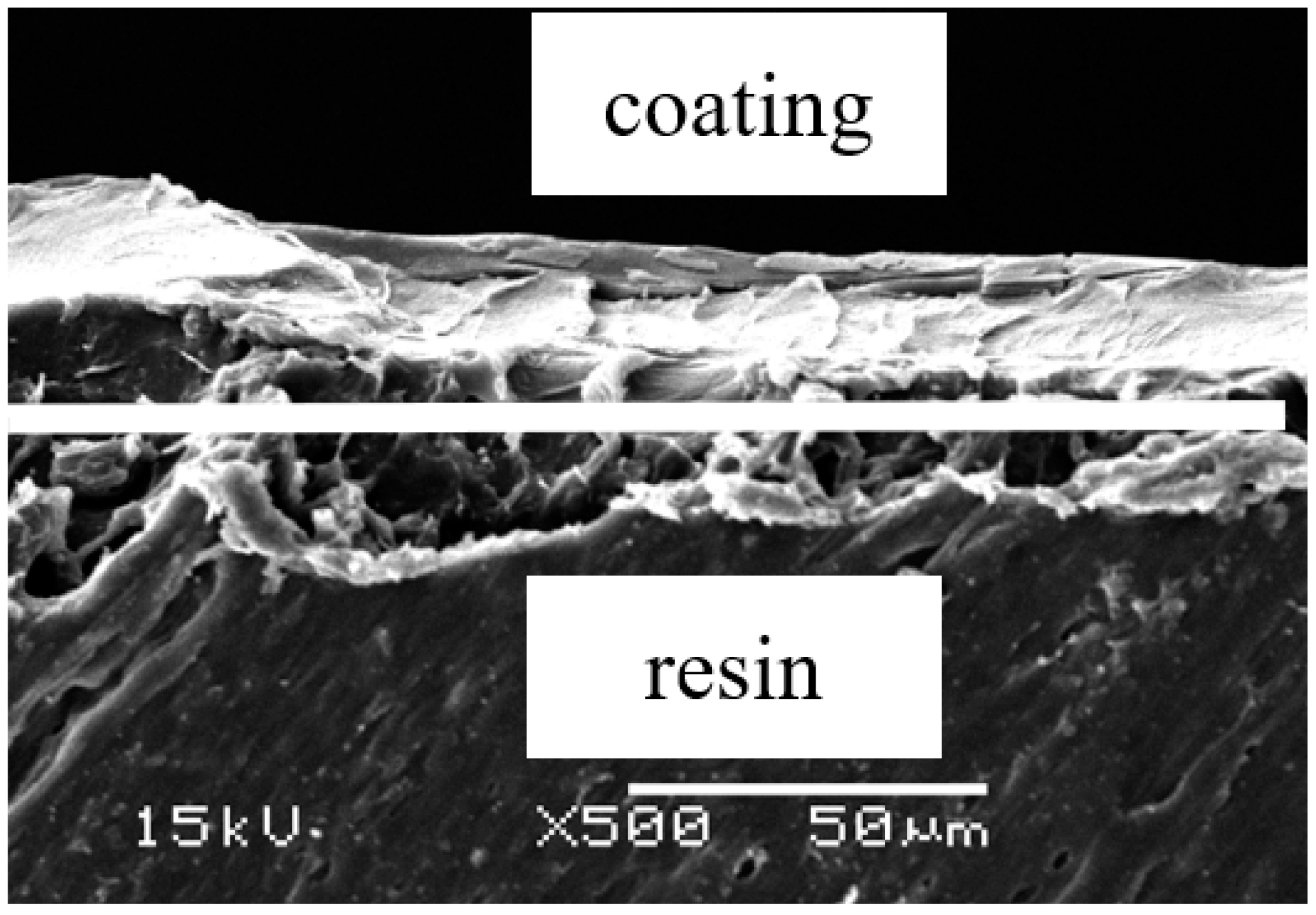
2.1. Sample Preparation
2.2. Bacterial Strains
2.3. Antibacterial Test
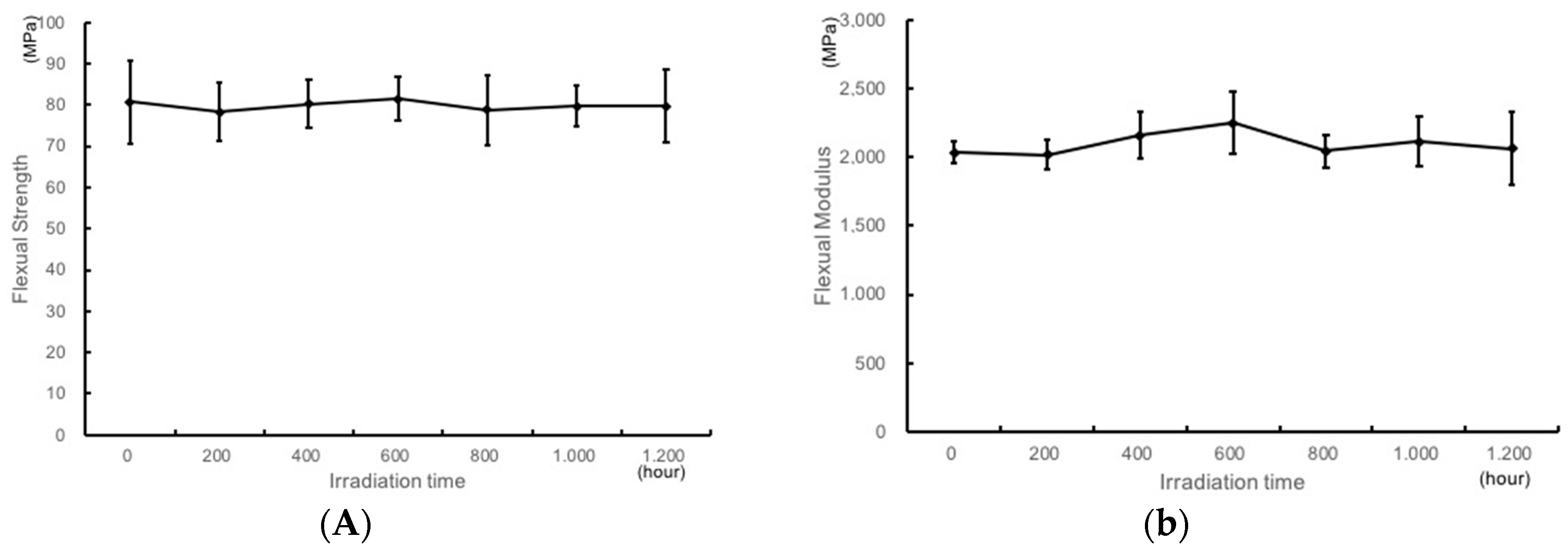
2.4. Bending Test
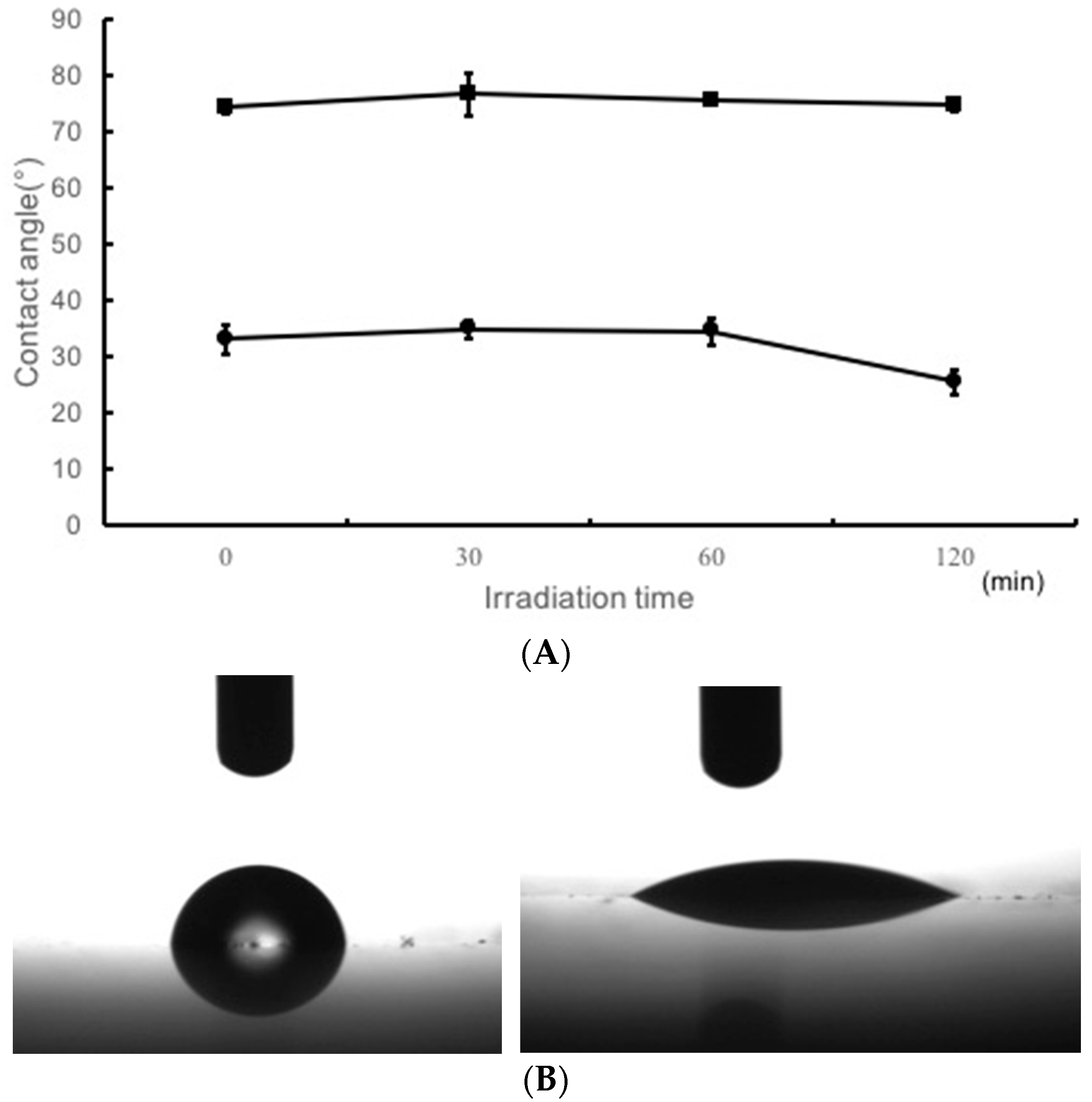
2.5. Water Contact Angle Measurement
2.6. Statistical Analysis
3. Results
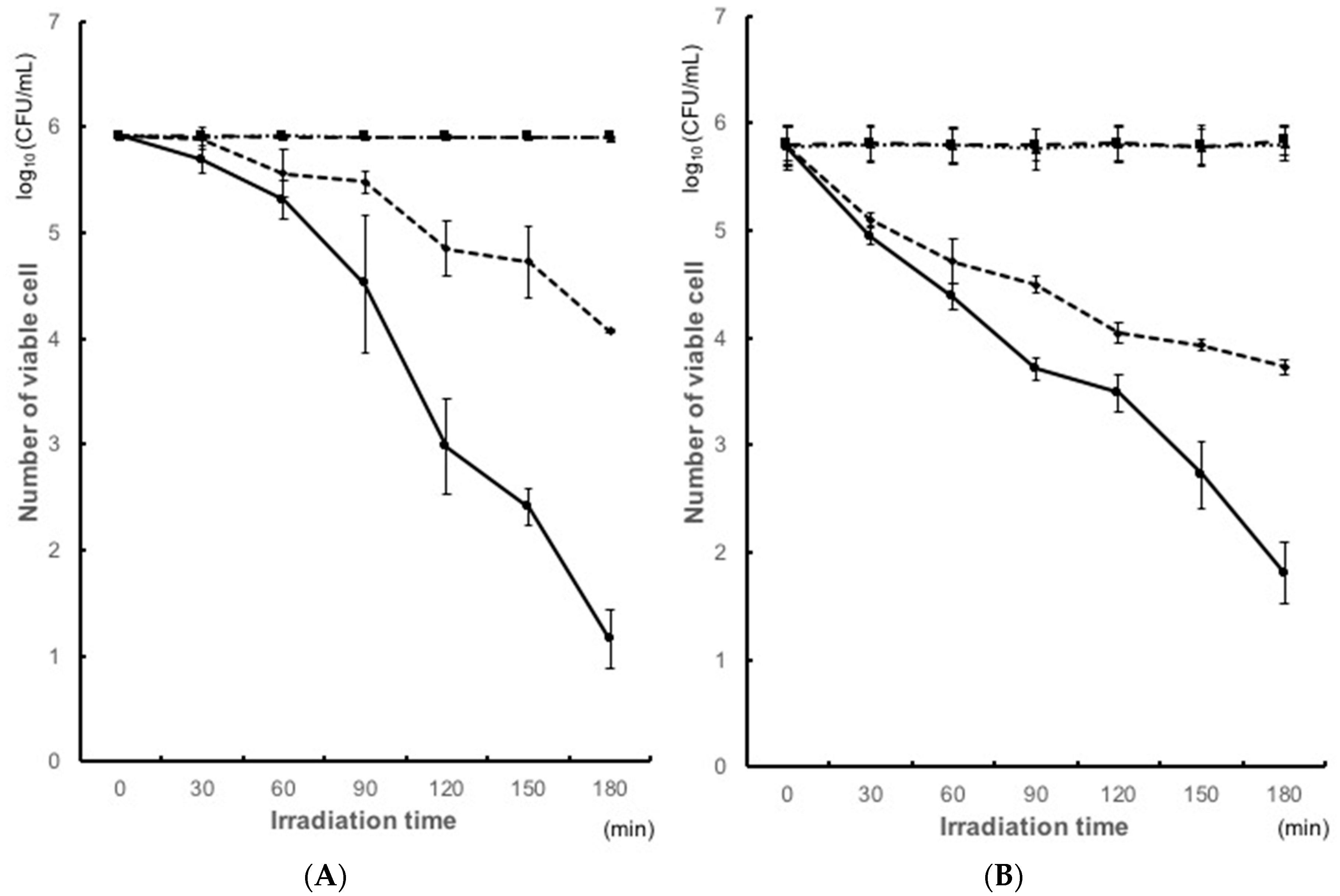
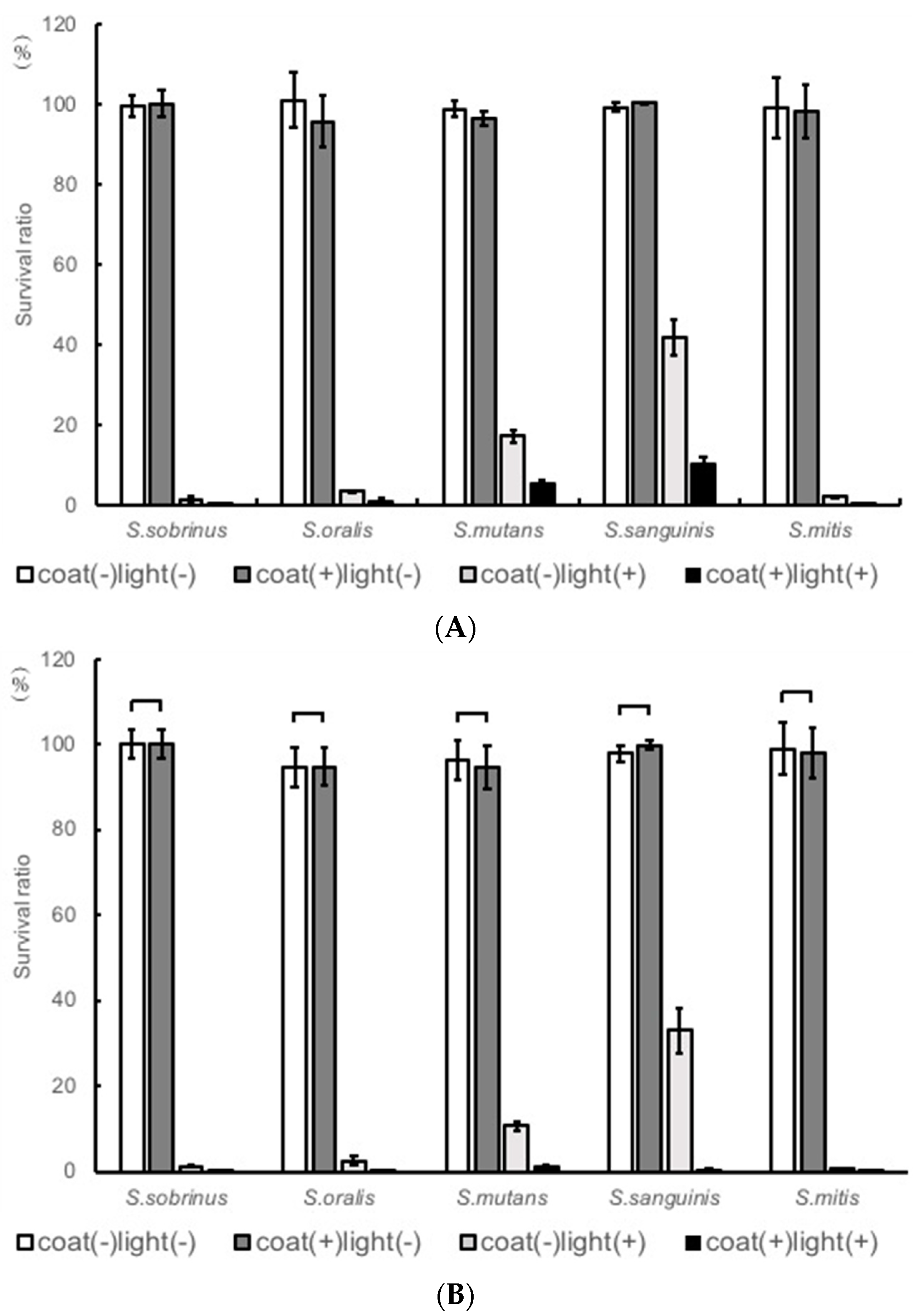
Antibacterial Test
4. Discussion
5. Conclusions
Author Contributions
Funding
Acknowledgments
Conflicts of Interest
References
- Littlewood, S.J.; Millett, D.T.; Doubleday, B.; Bearn, D.R.; Worthington, H.V. Retention procedures for stabilising tooth position after treatment with orthodontic braces. Cochrane Database Syst. Rev. 2006, 51, 94–95. [Google Scholar]
- Batoni, G.; Pardini, M.; Giannotti, A.; Ota, F.; Giuca, M.R.; Gabriele, M.; Campa, M.; Senesi, S. Effect of removable orthodontic appliances on oral colonisation by mutans streptococci in children. Eur. J. Oral Sci. 2001, 109, 388–392. [Google Scholar] [CrossRef] [PubMed]
- Bjerklin, K.; Gärskog, B.; Rönnerman, A. Proximal caries increment in connection with orthodontic treatment with removable appliances. Br. J. Orthod. 1983, 10, 21–24. [Google Scholar] [CrossRef] [PubMed]
- Hibino, K.; Wong, R.W.; Hagg, U.; Samaranayake, L.P. The effects of orthodontic appliances on candida in the human mouth. Int. J. Paediatr. Dent. 2009, 19, 301–308. [Google Scholar] [CrossRef] [PubMed]
- Kiyoko, T.; Kazuhiko, N.; Sonoko, M.; Atsuko, T.; Takashi, O. Clinical and microbiological evaluations of acute periodontitis in areas of teeth applied with orthodontic bands. Pediatr. Dent. J. 2005, 15, 212–218. [Google Scholar]
- Kuroki, K.; Hayashi, T.; Sato, K.; Asai, T.; Okano, M.; Kominami, Y.; Takahashi, Y.; Kawai, T. Effect of self-cured acrylic resin added with an inorganic antibacterial agent on Streptococcus mutans. Dent. Mater. J. 2010, 29, 277–285. [Google Scholar] [CrossRef] [PubMed]
- Madurantakam, P.; Kumar, S. Fixed and removable orthodontic retainers and periodontal health. Evid. Based Dent. 2017, 18, 103–104. [Google Scholar] [CrossRef] [PubMed]
- Ramage, G.; Tomsett, K.; Wickes, B.L.; López-Ribot, J.L.; Redding, S.W. Denture stomatitis: A role for candida biofilms. Oral Surg. Oral Med. Oral Pathol. Oral Radiol. Endodontol. 2004, 98, 53–59. [Google Scholar] [CrossRef]
- Turkoz, C.; Canigur Bavbek, N.; Kale Varlik, S.; Akca, G. Influence of thermoplastic retainers on Streptococcus mutans and Lactobacillus adhesion. Am. J. Orthod. Dentofac. Orthop. 2012, 141, 598–603. [Google Scholar] [CrossRef] [PubMed]
- Zharmagambetova, A.; Tuleutayeva, S.; Akhmetova, S. Microbiological aspects of the orthodontic treatment. Georgian Med. News 2017, 264, 39–43. [Google Scholar]
- Zingler, S.; Pritsch, M. Association between clinical and salivary microbial parameters during treatment with removable orthodontic appliances with or without use of fluoride mouth rinse. Eur. J. Paediatr. Dent. 2016, 17, 181–187. [Google Scholar] [PubMed]
- Shay, K. Denture hygiene: A review and update. J. Contemp. Dent. Pract. 2000, 1, 28–41. [Google Scholar] [PubMed]
- Song, W.S.; Lee, J.K.; Park, S.H.; Um, H.S.; Lee, S.Y.; Chang, B.S. Comparison of periodontitis-associated oral biofilm formation under dynamic and static conditions. J. Periodontal Implant Sci. 2017, 47, 219–230. [Google Scholar] [CrossRef] [PubMed]
- Kolenbrander, P.E.; Andersen, R.N.; Blehert, D.S.; Egland, P.G.; Foster, J.S.; Palmer, R.J. Communication among oral bacteria. Microbiol. Mol. Biol. Rev. 2002, 66, 486–505. [Google Scholar] [CrossRef] [PubMed]
- Teles, F.R.; Teles, R.P.; Sachdeo, A.; Uzel, N.G.; Song, X.Q.; Torresyap, G.; Singh, M.; Papas, A.; Haffajee, A.D.; Socransky, S.S. Comparison of microbial changes in early redeveloping biofilms on natural teeth and dentures. J. Periodontol. 2012, 83, 1139–1148. [Google Scholar] [CrossRef] [PubMed]
- Nyvad, B.; Kilian, M. Microbiology of the early colonization of human enamel and root surfaces in vivo. Scand. J. Dent. Res. 1987, 95, 369–380. [Google Scholar] [CrossRef] [PubMed]
- Verran, J.; Motteram, K.L. The effect of adherent oral streptococci on the subsequent adherence of candida albicans to acrylic in vitro. J. Dent. 1987, 15, 73–76. [Google Scholar] [CrossRef]
- Da Silva, P.M.; Acosta, E.J.; Pinto Lde, R.; Graeff, M.; Spolidorio, D.M.; Almeida, R.S.; Porto, V.C. Microscopical analysis of candida albicans biofilms on heat-polymerised acrylic resin after chlorhexidine gluconate and sodium hypochlorite treatments. Mycoses 2011, 54, e712–e717. [Google Scholar] [CrossRef] [PubMed]
- De Andrade, I.M.; Cruz, P.C.; da Silva, C.H.; de Souza, R.F.; Paranhos Hde, F.; Candido, R.C.; Marin, J.M.; de Souza-Gugelmin, M.C. Effervescent tablets and ultrasonic devices against candida and mutans streptococci in denture biofilm. Gerodontology 2011, 28, 264–270. [Google Scholar] [CrossRef] [PubMed]
- De Freitas Fernandes, F.S.; Pereira-Cenci, T.; da Silva, W.J.; Ricomini Filho, A.P.; Straioto, F.G.; Cury, A.A.D.B. Efficacy of denture cleansers on Candida spp. Biofilm formed on polyamide and polymethyl methacrylate resins. J. Prosthet. Dent. 2011, 105, 51–58. [Google Scholar] [CrossRef]
- De Souza, R.F.; de Freitas Oliveira Paranhos, H.; Lovato da Silva, C.H.; Abu-Naba’a, L.; Fedorowicz, Z.; Gurgan, C.A. Interventions for cleaning dentures in adults. Cochrane Database Syst. Rev. 2009, CD007395. [Google Scholar] [CrossRef] [PubMed]
- Dhamande, M.M.; Pakhan, A.J.; Thombare, R.U.; Ghodpage, S.L. Evaluation of efficacy of commercial denture cleansing agents to reduce the fungal biofilm activity from heat polymerized denture acrylic resin: An in vitro study. Contemp. Clin. Dent. 2012, 3, 168–172. [Google Scholar] [CrossRef] [PubMed]
- Farhadian, N.; Usefi Mashoof, R.; Khanizadeh, S.; Ghaderi, E.; Farhadian, M.; Miresmaeili, A. Streptococcus mutans counts in patients wearing removable retainers with silver nanoparticles vs. those wearing conventional retainers: A randomized clinical trial. Am. J. Orthod. Dentofac. Orthop. 2016, 149, 155–160. [Google Scholar] [CrossRef] [PubMed]
- Monteiro, D.R.; Gorup, L.F.; Takamiya, A.S.; de Camargo, E.R.; Filho, A.C.; Barbosa, D.B. Silver distribution and release from an antimicrobial denture base resin containing silver colloidal nanoparticles. J. Prosthodont. 2012, 21, 7–15. [Google Scholar] [CrossRef] [PubMed]
- Oei, J.D.; Zhao, W.W.; Chu, L.; DeSilva, M.N.; Ghimire, A.; Rawls, H.R.; Whang, K. Antimicrobial acrylic materials with in situ generated silver nanoparticles. J. Biomed. Mater. Res. B Appl. Biomater. 2012, 100, 409–415. [Google Scholar] [CrossRef] [PubMed]
- Regis, R.R.; Zanini, A.P.; Della Vecchia, M.P.; Silva-Lovato, C.H.; Oliveira Paranhos, H.F.; de Souza, R.F. Physical properties of an acrylic resin after incorporation of an antimicrobial monomer. J. Prosthodont. 2011, 20, 372–379. [Google Scholar] [CrossRef] [PubMed]
- Shinonaga, Y.; Arita, K. Antibacterial effect of acrylic dental devices after surface modification by fluorine and silver dual-ion implantation. Acta Biomater. 2012, 8, 1388–1393. [Google Scholar] [CrossRef] [PubMed]
- Sodagar, A.; Kassaee, M.Z.; Akhavan, A.; Javadi, N.; Arab, S.; Kharazifard, M.J. Effect of silver nano particles on flexural strength of acrylic resins. J. Prosthodont. Res. 2012, 56, 120–124. [Google Scholar] [CrossRef] [PubMed]
- Sousa, F.A.; Paradella, T.C.; Koga-Ito, C.Y.; Jorge, A.O. Effect of sodium bicarbonate on candida albicans adherence to thermally activated acrylic resin. Braz. Oral Res. 2009, 23, 381–385. [Google Scholar] [CrossRef] [PubMed]
- Vieira, A.P.; Senna, P.M.; Silva, W.J.; Del Bel Cury, A.A. Long-term efficacy of denture cleansers in preventing Candida spp. Biofilm recolonization on liner surface. Braz. Oral Res. 2010, 24, 342–348. [Google Scholar] [CrossRef] [PubMed]
- Wady, A.F.; Machado, A.L.; Zucolotto, V.; Zamperini, C.A.; Berni, E.; Vergani, C.E. Evaluation of candida albicans adhesion and biofilm formation on a denture base acrylic resin containing silver nanoparticles. J. Appl. Microbiol. 2012, 112, 1163–1172. [Google Scholar] [CrossRef] [PubMed] [Green Version]
- Wolff, M.S.; Larson, C. The cariogenic dental biofilm—Good, bad or just something to control? Braz. Oral. Res. 2009, 23, 31–38. [Google Scholar] [CrossRef] [PubMed]
- Chandra, J.; Mukherjee, P.K.; Leidich, S.D.; Faddoul, F.F.; Hoyer, L.L.; Douglas, L.J.; Ghannoum, M.A. Antifungal resistance of candidal biofilms formed on denture acrylic in vitro. J. Dent. Res. 2001, 80, 903–908. [Google Scholar] [CrossRef] [PubMed]
- Jagger, D.C.; Harrison, A. Denture cleaning the best approach. Br. Dent. J. 1995, 178, 413–417. [Google Scholar] [CrossRef] [PubMed]
- Budtz-Jørgensen, E. Materials and methods for cleaning dentures. J. Prosthet. Dent. 1979, 42, 619–623. [Google Scholar] [CrossRef]
- Fitjer, L.C.; Jonas, I.E.; Kappert, H.F. Corrosion susceptibility of lingual wire extensions in removable appliances. An in vitro study. J. Orofac. Orthop. 2002, 63, 212–226. [Google Scholar] [CrossRef] [PubMed]
- Hong, G.; Murata, H.; Li, Y.; Sadamori, S.; Hamada, T. Influence of denture cleansers on the color stability of three types of denture base acrylic resin. J. Prosthet. Dent. 2009, 10, 205–213. [Google Scholar] [CrossRef]
- Rantala, L.I.; Lastumäki, T.M.; Peltomäki, T.; Vallittu, P.K. Fatigue resistance of removable orthodontic appliancereinforced with glass fibre weave. J. Oral Rehabil. 2003, 30, 501–506. [Google Scholar] [CrossRef] [PubMed]
- Yamada, Y.; Yamada, M.; Ueda, T.; Sakurai, K. Reduction of biofilm formation on titanium surface with ultraviolet-c pre-irradiation. J. Biomater. Appl. 2014, 29, 161–171. [Google Scholar] [CrossRef] [PubMed]
- Tomomi, S.; Susumu, I.; Tsuyoshi, O.; Hiroyuki, K.; Yusuke, M.; Nobuhiro, H.; Yoshiki, N. Evaluation of antibacterial activity of visible light-responsive TiO2-based photocatalyst coating on orthodontic materials against cariogenic bacteria. Asian Pacfic. J. Dent. 2016, 16, 15–22. [Google Scholar]
- Fujishima, A.; Honda, K. Electrochemical photolysis of water at a semiconductor electrode. Nature 1972, 238, 37–38. [Google Scholar] [CrossRef] [PubMed]
- Cho, M.; Chung, H.; Choi, W.; Yoon, J. Linear correlation between inactivation of E. coli and oh radical concentration in TiO2 photocatalytic disinfection. Water Res. 2004, 38, 1069–1077. [Google Scholar] [CrossRef] [PubMed]
- Choi, J.Y.; Chung, C.J.; Oh, K.T.; Choi, Y.J.; Kim, K.H. Photocatalytic antibacterial effect of TiO2 film of tiag on streptococcus mutans. Angle Orthod. 2009, 79, 528–532. [Google Scholar] [CrossRef]
- Choi, J.Y.; Kim, K.H.; Choy, K.C.; Oh, K.T.; Kim, K.N. Photocatalytic antibacterial effect of TiO2 film formed on ti and tiag exposed to lactobacillus acidophilus. J. Biomed. Mater. Res. B Appl. Biomater. 2007, 80, 353–359. [Google Scholar] [CrossRef] [PubMed]
- Ochiai, T.; Fujishima, A. Photoelectrochemical properties of TiO2 photocatalyst and its applications for environmental purification. J. Photochem. Photobiol. C Photochem. Rev. 2012, 13, 247–262. [Google Scholar] [CrossRef]
- Hoffmann, M.R.; Martin, S.T.; Choi, W. Environmental applications of semiconductor photocatalysis. Chem. Rev. 1995, 95, 69–96. [Google Scholar] [CrossRef]
- Hosseinpour, S.; Tang, F.; Wang, F.; Livingstone, R.A.; Schlegel, S.J.; Ohto, T.; Bonn, M.; Nagata, Y.; Backus, E. Chemisorbed and physisorbed water at the TiO2/water interface. J. Phys. Chem. Lett. 2017, 8, 2195–2199. [Google Scholar] [CrossRef] [PubMed]
- Sunada, K.; Watanabe, T.; Hashimoto, K. Studies on photokilling of bacteria on TiO2 thin film. J. Photochem. Photobiol. A Chem. 2003, 156, 227–233. [Google Scholar] [CrossRef]
- Takeuchi, M.; Sakamoto, K.; Martra, G.; Coluccia, S.; Anpo, M. Mechanism of photoinduced superhydrophilicity on the TiO2 photocatalyst surface. J. Phys. Chem. B 2005, 109, 15422–15428. [Google Scholar] [CrossRef] [PubMed]
- Unosson, E.; Tsekoura, E.K.; Engqvist, H.; Welch, K. Synergetic inactivation of staphylococcus epidermidis and streptococcus mutansin a TiO2/H2O2/uv system. Biomatter 2013, 3, e26727. [Google Scholar] [CrossRef] [PubMed]
- ÖZyildiz, F.; Uzel, A.; Hazar, A.S.; GÜDen, M.; ÖLmez, S.; Aras, I.; Karaboz, İ. Photocatalytic antimicrobial effect of TiO2 anatase thin-film–coated orthodontic arch wires on 3 oral pathogens. Turkish J. Biol. 2014, 38, 289–295. [Google Scholar] [CrossRef]
- Uchimaru, M.; Sakai, T.; Moroi, R.; Shiota, S.; Shibata, Y.; Deguchi, M.; Sakai, H.; Yamashita, Y.; Terada, Y. Antimicrobial and antifungal effects of tissue conditioners containing a photocatalyst. Dent. Mater. J. 2011, 30, 691–699. [Google Scholar] [CrossRef] [PubMed]
- Westas, E.; Hayashi, M.; Cecchinato, F.; Wennerberg, A.; Andersson, M.; Jimbo, R.; Davies, J.R. Bactericidal effect of photocatalytically-active nanostructured TiO2 surfaces on biofilms of the early oral colonizer, streptococcus oralis. J. Biomed. Mater. Res. A 2017, 105, 2321–2328. [Google Scholar] [CrossRef] [PubMed]
- Ahn, S.; Han, J.; Lim, B.; Lim, Y. Comparison of ultraviolet light-induced photocatalytic bactericidal effect on modified titanium implant surfaces. Int. J. Oral Maxillofac. Implants 2011, 26, 39–44. [Google Scholar] [PubMed]
- Kayano, S.; Yoshihiko, K.; Kazuhito, H.; Akira, F. Batericidal and detoxification effects of TiO2 film photocatalysts. Environ. Sci. Technol. 1998, 32, 726–728. [Google Scholar]
- Saito, T.; Iwase, T.; Horie, J.; Morioka, T. Mode of photocatalytic bactericidal action of powdered semiconductor TiO2 on mutans streptococci. J. Photochem. Photobiol. B 1992, 14, 369–379. [Google Scholar] [CrossRef]
- Dongari, A.I.; Miyasaki, K.T. Sensitivity of actinobaciiius actinomycetemcomitans and haemophiius apiiropiiiius to oxicjative killing. Oral Microbiol. Immunol. 1991, 6, 363–372. [Google Scholar] [CrossRef] [PubMed]
- Jagger, J. Near-uv radiation effects on microorganisms. Photochem. Photobiol. 1981, 34, 761–768. [Google Scholar] [CrossRef] [PubMed]
- Maness, P.; Smolinski, S.; Blake, D.; Huang, Z.; Wolfrum, E.; Jacoby, W. Bactericidal activity of photocatalytic TiO2 reaction: Toward an understanding of its killing mechanism. Appl. Environ. Microbiol. 1999, 65, 4094–4098. [Google Scholar] [PubMed]
- Matallana-Surget, S.; Meador, J.; Joux, F.; Douki, T. Effect of the gc content of DNA on the distribution of uvb-induced bipyrimidinephotoproducts. Photochem. Photobiol. Sci. 2008, 7, 794–801. [Google Scholar] [CrossRef] [PubMed]
- Arrieta, J.M.; Weinbauer, M.G.; Herndl, G.J. Interspecific variability in sensitivity to uv radiation and subsequent recovery in selected isolates of marine bacteria. Appl. Environ. Microbiol. 2000, 66, 1468–1473. [Google Scholar] [CrossRef] [PubMed]
- Matallana-Surget, S.; Douki, T.; Cavicchioli, R.; Joux, F. Remarkable resistance to uvb of the marine bacterium photobacterium angustum explained by an unexpected role of photolyase. Photoch. Photobio. Sci. 2009, 8, 1313–1320. [Google Scholar] [CrossRef] [PubMed]
- Santos, A.L.; Lopes, S.; Baptista, I.; Henriques, I.; Gomes, N.C.; Almeida, A.; Correia, A.; Cunha, A. Diversity in uv sensitivity and recovery potential among bacterioneuston and bacterioplankton isolates. Lett. Appl. Microbiol. 2011, 52, 360–366. [Google Scholar] [CrossRef] [PubMed]
- Santos, A.L.; Oliveira, V.; Baptista, I.; Henriques, I.; Gomes, N.C.; Almeida, A.; Correia, A.; Cunha, A. Wavelength dependence of biological damage induced by uv radiation on bacteria. Arch. Microbiol. 2013, 195, 63–74. [Google Scholar] [CrossRef] [PubMed]
- Gilbert, P.; Das, J.; Foley, I. Biofilm susceptibility to antimicrobials. Adv. Dent. Res. 1997, 11, 160–167. [Google Scholar] [CrossRef] [PubMed]
- Kado, D.; Sakurai, K.; Sugiyama, T.; Ueda, T. Evaluation of cleanability of a titanium dioxide (TiO2)-coated acrylic resin denture base. Prosthodont. Res. Pract. 2005, 4, 69–76. [Google Scholar] [CrossRef]
- Arai, T.; Ueda, T.; Sugiyama, T.; Sakurai, K. Inhibiting microbial adhesion to denture base acrylic resin by titanium dioxide coating. J. Oral Rehabil. 2009, 36, 902–908. [Google Scholar] [CrossRef] [PubMed]
- Obata, T.; Ueda, T.; Sakurai, K. Inhibition of denture plaque by TiO2 coating on denture base resins in the mouth. J. Prosthet. Dent. 2017, 118, 759–764. [Google Scholar] [CrossRef] [PubMed]
- Tsuji, M.; Ueda, T.; Sawaki, K.; Kawaguchi, M.; Sakurai, K. Biocompatibility of a titanium dioxide-coating method for denture base acrylic resin. Gerodontology 2016, 33, 539–544. [Google Scholar] [CrossRef] [PubMed]
 coat(+)light(+): experiment group containing powdered TiO2 with irradiation.
  coat(−)light(−): control group without both TiO2 and irradiation.
  coat(+)light(−): experiment group in the presence of powdered TiO2 without irradiation.
  coat(−)light(−): experiment group without TiO2, but with irradiation.
coat(+)light(+): experiment group containing powdered TiO2 with irradiation. coat(−)light(−): control group without both TiO2 and irradiation. coat(+)light(−): experiment group in the presence of powdered TiO2 without irradiation. coat(−)light(−): experiment group without TiO2, but with irradiation.
coat(−)light(−): experiment group without TiO2, but with irradiation.
coat(+)light(+): experiment group containing powdered TiO2 with irradiation. coat(−)light(−): control group without both TiO2 and irradiation. coat(+)light(−): experiment group in the presence of powdered TiO2 without irradiation. coat(−)light(−): experiment group without TiO2, but with irradiation.


| S. gordoniiATCC10558 | S. mitis | S. oralisATCC35037 | S. oralis GTC276 | ||||||||||
| Coefficient | 95% CI | p-Value | Coefficient | 95% CI | p-Value | Coefficient | 95% CI | p-Value | Coefficient | 95% CI | p-Value | ||
| Intercept | 6.563 | 6.04–7.08 | 0.999< | 8.267 | 7.57–8.96 | 0.999< | 6.584 | 6.18–6.98 | 0.999< | 8.840 | 8.30–9.37 | 0.999< | |
| time | 30 min | −0.061 | −0.67–0.55 | 0.845 | −0.306 | −1.13–0.52 | 0.462 | −0.372 | −0.84–0.10 | 0.123 | −0.498 | −1.13–0.13 | 0.122 |
| 60 min | −0.238 | −0.85–0.37 | 0.444 | −0.753 | −1.57–0.07 | 0.073 | −0.616 | −1.09–−0.14 | 0.012 | −0.700 | −1.33–−0.06 | 0.031 | |
| 90 min | −0.458 | −1.07–0.15 | 0.143 | −1.246 | −2.07–−0.42 | <0.001 | −0.846 | −1.32–−0.37 | <0.001 | −0.877 | −1.51–−0.24 | <0.001 | |
| 120 min | −1.001 | −1.61–−0.38 | <0.001 | −1.671 | −2.49–−0.84 | <0.001 | −1.003 | −1.47–−0.52 | <0.001 | −1.260 | −1.89–−0.62 | <0.001 | |
| 150 min | −1.175 | −1.79–−0.55 | <0.001 | −1.935 | −2.75–−1.10 | <0.001 | −1.231 | −1.70–−0.75 | <0.001 | −1.576 | −2.21–−0.94 | <0.001 | |
| 180 min | −1.654 | −2.27–−1.03 | <0.001 | −2.432 | −3.25–−1.60 | <0.001 | −1.491 | −1.96–−1.01 | <0.001 | −1.820 | −2.45–−1,18 | <0.001 | |
| coat(+) | −0.001 | −0.46–0.46 | 0.996 | −0.015 | −0.63–0.60 | 0.963 | 0.017 | −0.34–0.37 | 0.925 | −0.008 | −0.48–0.47 | 0.975 | |
| light(+) | −0.695 | −1.16–−0.22 | <0.001 | −1.457 | −2.08–−0.83 | <0.001 | −1.247 | −1.60–−0.88 | <0.001 | −1.338 | −1.81–−0.85 | <0.001 | |
| interaction of light and coat | −1.216 | −1.87–−0.55 | <0.001 | −1.845 | −2.72–−0.96 | <0.001 | −0.727 | −1.23–−0.21 | 0.006 | −1.206 | −1.88–−0.52 | <0.001 | |
| S. mutans | S. sobrinus | S. sanguinis | |||||||||||
| Coefficient | 95% CI | p-Value | Coefficient | 95% CI | p-Value | Coefficient | 95% CI | p-Value | |||||
| Intercept | 5.609 | 5.38–5.83 | 0.999< | 11.066 | 10.68–11.44 | 0.999< | 9.280 | 8.90–9.65 | 0.999< | ||||
| time | 30 min | −0.119 | −0.38–0.14 | 0.376 | −0.585 | −1.03–−0.13 | 0.011 | −0.105 | −0.54–0.33 | 0.638 | |||
| 60 min | −0.272 | −0.53–−0.01 | 0.044 | −0.938 | −1.38–−0.49 | <0.001 | −0.231 | −0.67–0.20 | 0.299 | ||||
| 90 min | −0.347 | −0.61–−0.82 | 0.011 | −1.121 | −1.56–−0.67 | <0.001 | −0.348 | −0.78–0.09 | 0.120 | ||||
| 120 min | −0.512 | −0.77–−0.24 | <0.001 | −1.146 | −1.59–−0.70 | <0.001 | −0.718 | −1.15–−0.27 | 0.002 | ||||
| 150 min | −0.512 | −0.77–−0.24 | <0.001 | −1.404 | −1.85–−0.95 | <0.001 | −0.848 | −1.28–−0.40 | <0.001 | ||||
| 180 min | −0.908 | −1.17–−0.64 | <0.001 | −1.555 | −2.00–−1.10 | <0.001 | −1.239 | −1.67–−0.79 | <0.001 | ||||
| coat(+) | <0.001 | −0.20–0.20 | 0.998 | 0.001 | −0.33–0.33 | 0.995 | <0.001 | −0.33–0.33 | 0.999< | ||||
| light(+) | −0.480 | −0.67–−0.27 | <0.001 | −1.587 | −1.92–−1.25 | <0.001 | −0.514 | −0.84–−0.18 | 0.003 | ||||
| interaction of light and coat | −0.557 | −0.84–−0.27 | <0.001 | −0.680 | −1.15–−0.20 | 0.006 | −0.948 | −1.41–−0.47 | <0.001 | ||||
© 2018 by the authors. Licensee MDPI, Basel, Switzerland. This article is an open access article distributed under the terms and conditions of the Creative Commons Attribution (CC BY) license (http://creativecommons.org/licenses/by/4.0/).
Share and Cite
Kuroiwa, A.; Nomura, Y.; Ochiai, T.; Sudo, T.; Nomoto, R.; Hayakawa, T.; Kanzaki, H.; Nakamura, Y.; Hanada, N. Antibacterial, Hydrophilic Effect and Mechanical Properties of Orthodontic Resin Coated with UV-Responsive Photocatalyst. Materials 2018, 11, 889. https://doi.org/10.3390/ma11060889
Kuroiwa A, Nomura Y, Ochiai T, Sudo T, Nomoto R, Hayakawa T, Kanzaki H, Nakamura Y, Hanada N. Antibacterial, Hydrophilic Effect and Mechanical Properties of Orthodontic Resin Coated with UV-Responsive Photocatalyst. Materials. 2018; 11(6):889. https://doi.org/10.3390/ma11060889
Chicago/Turabian StyleKuroiwa, Akira, Yoshiaki Nomura, Tsuyoshi Ochiai, Tomomi Sudo, Rie Nomoto, Tohru Hayakawa, Hiroyuki Kanzaki, Yoshiki Nakamura, and Nobuhiro Hanada. 2018. "Antibacterial, Hydrophilic Effect and Mechanical Properties of Orthodontic Resin Coated with UV-Responsive Photocatalyst" Materials 11, no. 6: 889. https://doi.org/10.3390/ma11060889