
A Review of Extraction and Analysis of Bioactives in Oat and Barley and Scope for Use of Novel Food Processing Technologies


Abstract
:1. Introduction

| Total Production’000 tons | Area’000 hectares | Average Yield tons/hectare | |
|---|---|---|---|
| Oats | 8083 | 2536 | 3.1 |
| Barley | 67,050 | 12,398 | 5.4 |
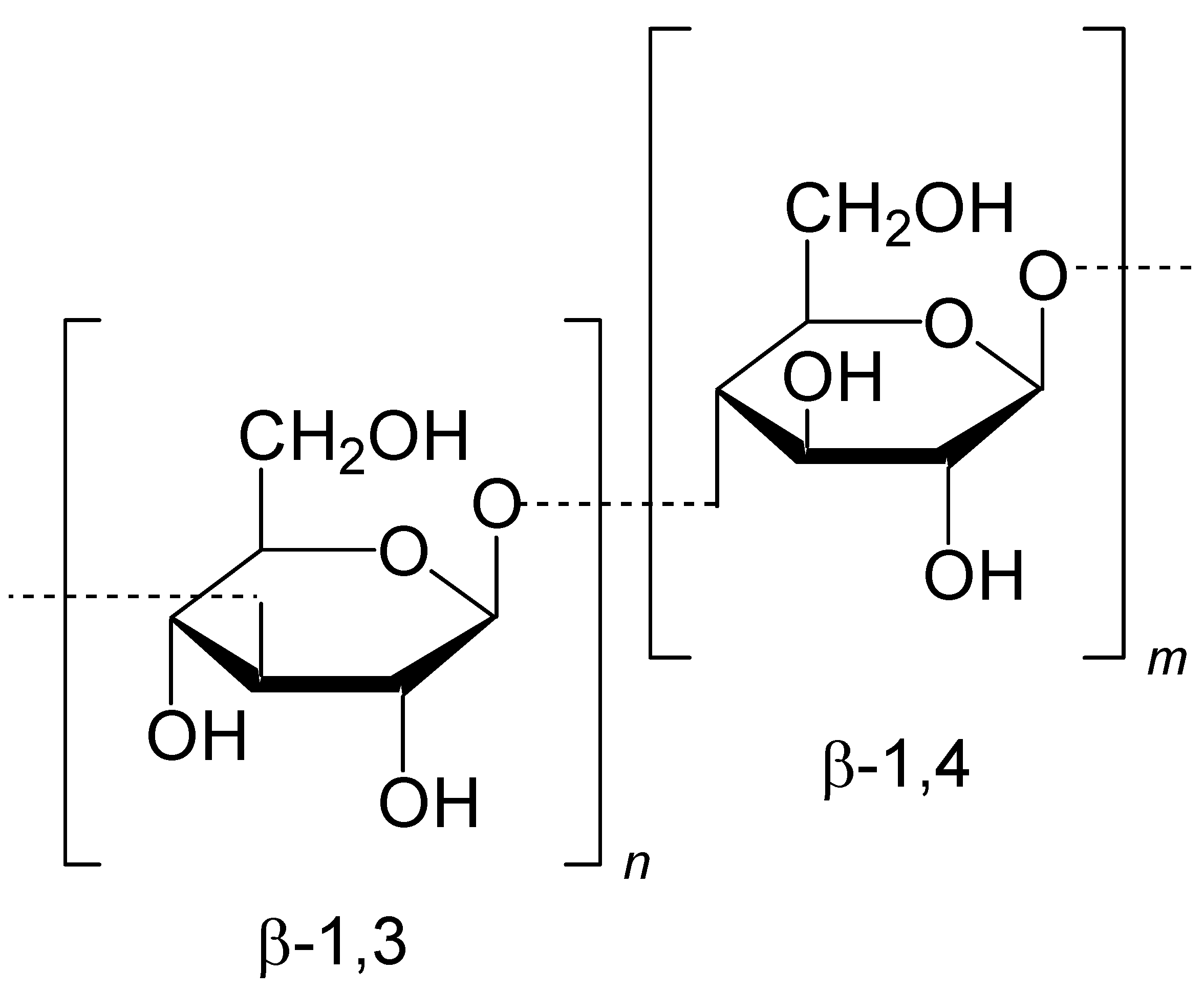
2. β-Glucan

3. Phenolic Compounds

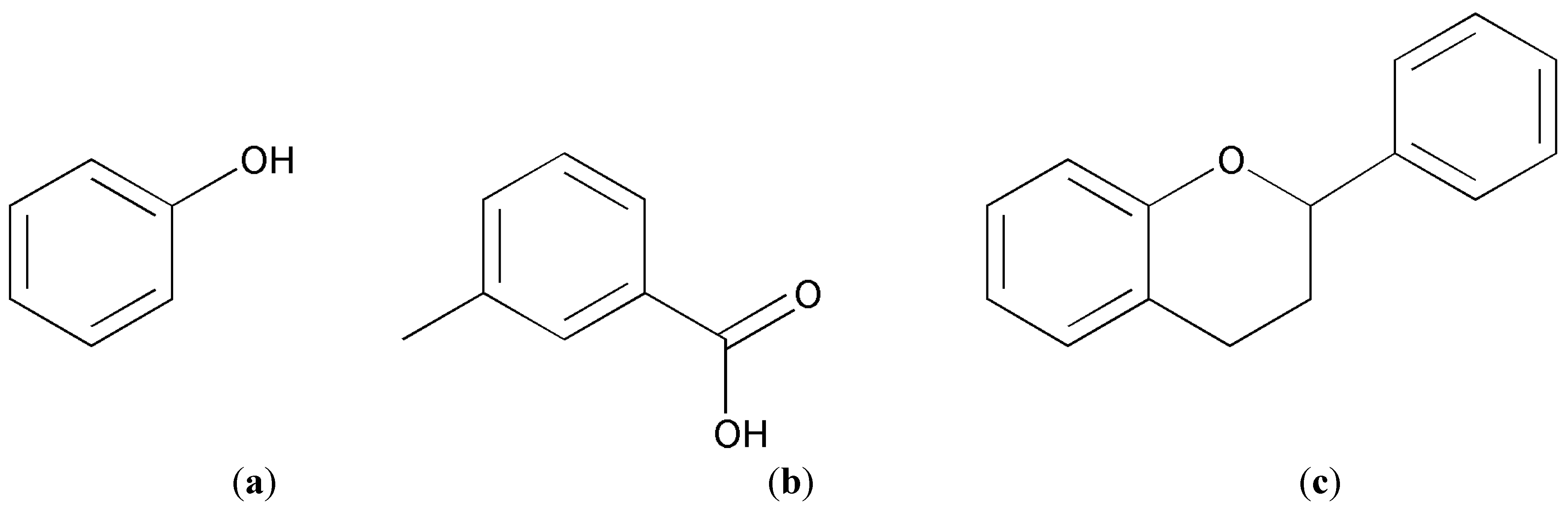
3.1. Phenolic Acids
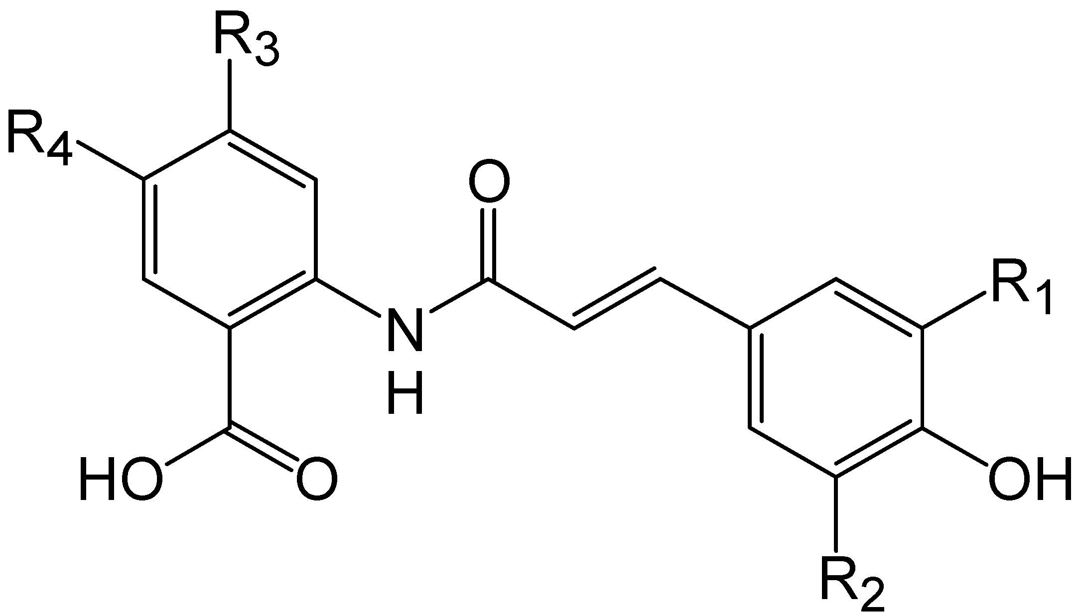
3.2. Avenanthramides

| Avenanthramide | Collin’s Nomenclature | R1 | R2 | R3 | R4 |
|---|---|---|---|---|---|
| 1p | D | H | H | H | H |
| 1c | H | OH | H | H | |
| 1f | E | H | OCH3 | H | H |
| 2p | A | OH | H | H | H |
| 2c | C | OH | OH | H | H |
| 2f | B | OH | OCH3 | H | H |
| 3f | OH | OCH3 | H | OCH3 |
3.3. Flavonoid
4. Lipids
4.1. Phospholipids
4.2. Phytosterols
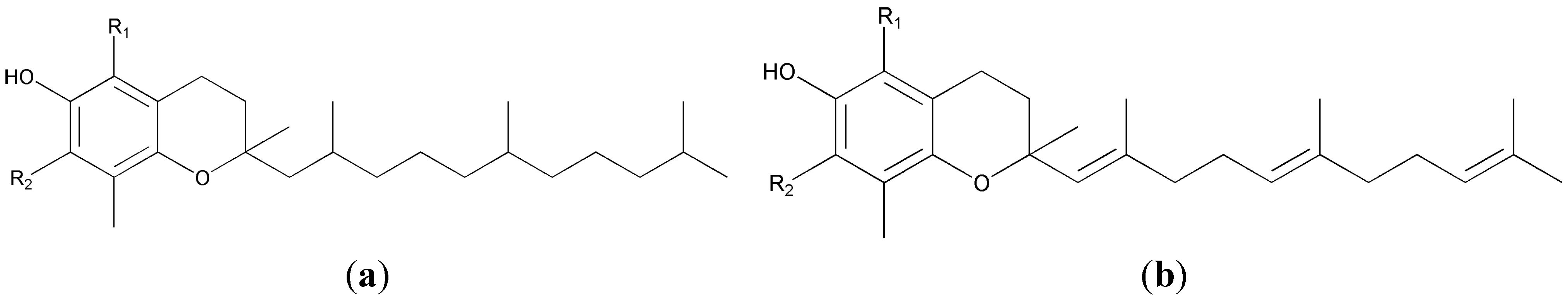
4.3. Tocols

| R1 | R2 | |
|---|---|---|
| α | CH3 | CH3 |
| β | CH3 | H |
| γ | H | CH3 |
| δ | H | H |
5. Proteins and Bioactive Peptides
| Phytochemical | Source | Extraction Solvent/Technique Used | Bioactivity | Commercial Availability |
|---|---|---|---|---|
| β-glucan | Barley | 4% 1 M NaOH [27]; Water at 55 °C and pH 7.0 [32]; Pressurized hot water extraction at 155 °C, 18 min and 50 bar [91] | Lowers cholesterol [92] reducing the chances of coronary and ischemic heart disease. Lowers glycaemic index and postprandial glucose levels [93]. | Barley β-glucan is available as Glucagel®, PromOat®, Glucan 300® |
| Oat | Ethanol reflux followed by extraction with a sodium carbonate solution at pH 10 [26]. | |||
| Phenolic acids (PA) | Barley | Free phenolics—Acetone/water (4/1) [51,94]; ethanol/water (4/1) [51]; pressurized liquid extraction [61]. Bound phenolics—20 h prolonged alkaline hydrolysis [51]; Acid hydrolysis [53]; Sequential acid, α amylase and cellulose hydrolysis [55] | PA act as antioxidants and protect against the destructive activity of free radicals. They reduce the risk of chronic age-related diseases such as cardiovascular diseases, diabetes and ageing, by reducing oxidative stress [95]. PA bound to dietary fiber may prevent cancer of the colon [96]. | Green tea extracts rich in phenolic acids are available as Bulk Powders® and Super Green Tea Diet® |
| Oat | Free phenolics—Methanol [97], Methanol/water (4/1) [52]; Acetone/water (1/1) [95]; 80% ethanol [46,57]; Bound phenolics—Alkaline hydrolysis [52], acid hydrolysis [52,53] | |||
| Avenanthramides (AV) | Oat | 80% methanol [56,98]; 80% ethanol [57,58]. | AV are bioavailable and act as antioxidants by inhibiting LDL oxidation in synergy with Vitamin C [99]. They demonstrate anti-allergic activities. | |
| Flavonoid | Barley | 40% ethanol [100] | Proanthocyanidins have ferrous chelating activity and influence the bioavailability of iron in the body [101]. Tricin shows chemopreventive effects against breast and colon cancer cells [102]. | Citrus flavonoids are available as Citrus Bioflavonoid Caps® and a similar product from soy is available as Phytosoya®, SoyChoice® |
| Oat | Free flavonoids—80% ethanol [103]; Bound flavonoids—Digestion using NaOH, followed by acidification, removal of lipids using hexane and further extraction using ethyl acetate [103]. | |||
| Tocols | Barley | Extraction with methanol, followed by drying and extraction using hexane [79]; Saponification using KOH, ethanol, NaCl and pyrogallol at 70 °C for 25 min. This is followed by cooling on ice bath and extraction using hexane:ethyl acetate (9:1) [76,77]. | Tocols are generically known as Vitamin E and exhibit antioxidant activity. Tocotrienols demonstrate reduction in serum total cholesterol and LDL cholesterol levels in vivo [104]. | |
| Oat | Extraction techniques used are same as those for barley [76,79,81]. | |||
| Proteins | Barley | Alkaline extraction using NaOH and addition of HCl at pH 4.6 for precipitation of protein [105]; A peptide lunasin has been isolated from barley with phosphate-buffered saline supplemented with protease inhibitor cocktail, followed by column and immunoaffinity chromatography [89]. | Hydrolyzed peptides from barley proteins have demonstrated anti-oxidant, antihypertensive and anti-diabetic effects in vitro [105]. The peptide lunasin is a cancer preventive agent and is bioavailable [89]. Hydrolysates from oat proteins demonstrate ACE-I enzyme inhibitory activity in vitro [88]. | Milk derived peptides is available as Calpis® and a sardine fish derived peptide product is available as Valtyron®, (Source—Fish) Valtyron® |
| Oat | Alkaline extraction followed by isoelectric precipitation [88]. |
6. Novel Processing Technologies
7. Conclusions
Acknowledgments
Conflicts of Interest
References
- Trowell, H. Ischemic heart disease and dietary fiber. Am. J. Clin. Nutr. 1972, 25, 926–932. [Google Scholar] [PubMed]
- Anderson, J.W. Whole grains protect against atherosclerotic cardiovascular disease. Proc. Nutr. Soc. 2003, 62, 135–142. [Google Scholar] [CrossRef] [PubMed]
- Chatenoud, L.; Tavani, A.; la Vecchia, C.; Jacobs, D.R.; Negri, E.; Levi, F.; Franceschi, S. Whole grain food intake and cancer risk. Int. J. Cancer 1998, 77, 24–28. [Google Scholar] [CrossRef]
- Venn, B.J.; Mann, J.I. Cereal grains, legumes and diabetes. Eur. J. Clin. Nutr. 2004, 58, 1443–1461. [Google Scholar] [CrossRef] [PubMed]
- Slavin, J.L. Dietary fiber and body weight. Nutrition 2005, 21, 411–418. [Google Scholar] [CrossRef] [PubMed]
- Slavin, J.L.; Martini, M.C.; Jacobs, D.R.; Marquart, L. Plausible mechanisms for the protectiveness of whole grains. Am. J. Clin. Nutr. 1999, 70, 459s–463s. [Google Scholar] [PubMed]
- Slavin, J. Why whole grains are protective: Biological mechanisms. Proc. Nutr. Soc. 2003, 62, 129–134. [Google Scholar] [CrossRef] [PubMed]
- EUROSTAT (2014). Crop Products-Annual Data. Available online: http://ec.europa.eu/eurostat/data/database (accessed on 10 June 2015).
- Baik, B.K.; Ullrich, S.E. Barley for food: Characteristics, improvement, and renewed interest. J. Cereal Sci. 2008, 48, 233–242. [Google Scholar] [CrossRef]
- Biel, W.; Bobko, K.; Maciorowski, R. Chemical composition and nutritive value of husked and naked oats grain. J. Cereal Sci. 2009, 49, 413–418. [Google Scholar] [CrossRef]
- Webster, F.H. Oats. In Cereal Grain Quality; Springer: London, United Kingdom, 1996; pp. 179–203. [Google Scholar]
- Yao, N.; Jannink, J.L.; Alavi, S.; White, P.J. Physical and sensory characteristics of extruded products made from two oat lines with different β-glucan concentrations. Cereal Chem. 2006, 83, 692–699. [Google Scholar] [CrossRef]
- Flander, L.; Salmenkallio-Marttila, M.; Suortti, T.; Autio, K. Optimization of ingredients and baking process for improved wholemeal oat bread quality. LWT-Food Sci. Technol. 2007, 40, 860–870. [Google Scholar] [CrossRef]
- Bhatty, R.S. Nonmalting uses of barley. In Barley: Chemistry and Technology; MacGregor, A.W., Bhatty, R.S., Eds.; Am. Assoc. Cereal Chem. Press: St. Paul, MN, USA, 1993; pp. 355–417. [Google Scholar]
- Newman, R.K.; Newman, C.W. Barley for Food and Health: Science, Technology, and Products; John Wiley & Sons: Hoboken, NJ, USA, 2008; pp. 204–208. [Google Scholar]
- Adil, G. Whole-grain cereal bioactive compounds and their health benefits: A review. J. Food Process. Technol. 2012, 3, 146. [Google Scholar] [CrossRef]
- Garsed, K.; Scott, B.B. Can oats be taken in a gluten-free diet? A systematic review. Scand. J. Gastroenterol. 2007, 42, 171–178. [Google Scholar] [CrossRef] [PubMed]
- Liu, S.; Stampfer, M.J.; Hu, F.B.; Giovannucci, E.; Rimm, E.; Manson, J.E.; Willett, W.C. Whole-grain consumption and risk of coronary heart disease: Results from the Nurses’ Health Study. Am. J. Clin. Nutr. 1999, 70, 412–419. [Google Scholar] [PubMed]
- Munter, J.S.; Hu, F.B.; Spiegelman, D.; Franz, M.; van Dam, R.M. Whole grain, bran, and germ intake and risk of type 2 diabetes: A prospective cohort study and systematic review. PLoS Med. 2007, 4, e261. [Google Scholar] [CrossRef] [PubMed]
- Tighe, P.; Duthie, G.; Vaughan, N.; Brittenden, J.; Simpson, W.G.; Duthie, S.; Thies, F. Effect of increased consumption of whole-grain foods on blood pressure and other cardiovascular risk markers in healthy middle-aged persons: A randomized controlled trial. Am. J. Clin. Nutr. 2010, 92, 733–740. [Google Scholar] [CrossRef] [PubMed]
- Liu, R.H. Potential synergy of phytochemicals in cancer prevention: Mechanism of action. J. Nutr. 2004, 134, 3479S–3485S. [Google Scholar] [PubMed]
- Anderson, J.W.; Gilinsky, N.H.; Deakins, D.A.; Smith, S.F.; O’Neal, D.S.; Dillon, D.W.; Oeltgen, P.R. Lipid responses of hypercholesterolemic men to oat-bran and wheat-bran intake. Am. J. Clin. Nutr. 1991, 54, 678–683. [Google Scholar] [PubMed]
- Åman, P. Cholesterol-lowering effects of barley dietary fiber in humans: Scientific support for a generic health claim. Scand. J. Food Nutr. 2006, 50, 173–176. [Google Scholar] [CrossRef]
- Regand, A.; Chowdhury, Z.; Tosh, S.M.; Wolever, T.M.S.; Wood, P. The molecular weight, solubility and viscosity of oat beta-glucan affect human glycemic response by modifying starch digestibility. Food Chem. 2011, 129, 297–304. [Google Scholar] [CrossRef]
- Wood, P.J.; Siddiqui, I.R.; Paton, D. Extraction of high-viscosity gums from oats. Cereal Chem. 1978, 55, 1038–1049. [Google Scholar]
- Bhatty, R.S. Extraction and enrichment (1 leads to 3), (1 leads to 4)-beta-d-glucan from barley and oat brans. Cereal Chem. 1993, 70, 73–77. [Google Scholar]
- Bhatty, R.S. Laboratory and pilot plant extraction and purification of β-glucans from hull-less barley and oat brans. J. Cereal Sci. 1995, 22, 163–170. [Google Scholar] [CrossRef]
- Burkus, Z.; Temelli, F. Effect of extraction conditions on yield, composition, and viscosity stability of barley β-glucan gum. Cereal Chem. 1998, 75, 805–809. [Google Scholar] [CrossRef]
- Irakli, M.; Biliaderis, C.G.; Izydorczyk, M.S.; Papadoyannis, I.N. Isolation, structural features and rheological properties of water-extractable β-glucans from different Greek barley cultivars. J. Sci. Food Agric. 2004, 84, 1170–1178. [Google Scholar] [CrossRef]
- Temelli, F. Extraction and functional properties of barley β-glucan as affected by temperature and pH. J. Food Sci. 1997, 62, 1194–1201. [Google Scholar] [CrossRef]
- Dawkins, N.L.; Nnanna, I.A. Oat gum and β glucan extraction from oat bran and rolled rats: Temperature and pH effects. J. Food Sci. 1993, 58, 562–566. [Google Scholar] [CrossRef]
- Benito-Román, O.; Alonso, E.; Lucas, S. Optimization of the β-glucan extraction conditions from different waxy barley cultivars. J. Cereal Sci. 2011, 53, 271–276. [Google Scholar] [CrossRef]
- Gangopadhyay, N.; Hossain, M.B.; Rai, D.K.; Brunton, N.P. Optimisation of yield and molecular weight of β-glucan from barley flour using response surface methodology. J. Cereal Sci. 2015, 62, 38–44. [Google Scholar] [CrossRef]
- Morgan, K.R.; Ofman, D.J. Glucagel, a gelling β-glucan from barley. Cereal Chem. 1998, 75, 879–881. [Google Scholar] [CrossRef]
- Clarke, A.E.; Stone, B.A. Enzymic hydrolysis of barley and other beta-glucans by a beta-(1→4)-glucan hydrolase. J. Biochem. 1966, 99, 582–588. [Google Scholar]
- Wood, P.J.; Weisz, J.; Blackwell, B.A. Molecular characterization of cereal β-d-glucans. Structural analysis of oat β-d-glucan and rapid structural evaluation of β-d-glucans from different sources by high-performance liquid chromatography of oligosaccharides released by lichenase. Cereal Chem. 1991, 68, 31–39. [Google Scholar]
- Edney, M.J.; Marchylo, B.A.; MacGregor, A.W. Structure of total barley beta-glucan. J. Inst. Brew. 1991, 97, 39–44. [Google Scholar] [CrossRef]
- Wood, P.J.; Weisz, J.; Blackwell, B.A. Structural studies of (1→3),(1→4)-beta-d-glucans by 13C-nuclear magnetic resonance spectroscopy and by rapid analysis of cellulose-like regions using high-performance anion-exchange chromatography of oligosaccharides released by lichenase. Cereal Chem. 1994, 71, 301–307. [Google Scholar]
- Jiang, G.; Vasanthan, T. MALDI-MS and HPLC quantification of oligosaccharides of lichenase-hydrolyzed water-soluble β-glucan from ten barley varieties. J. Agric. Food Chem. 2000, 48, 3305–3310. [Google Scholar] [CrossRef] [PubMed]
- Johansson, L.; Virkki, L.; Maunu, S.; Lehto, M.; Ekholm, P.; Varo, P. Structural characterization of water soluble β-glucan of oat bran. Carbohydr. Polym. 2000, 42, 143–148. [Google Scholar] [CrossRef]
- Johansson, L.; Tuomainen, P.; Ylinen, M.; Ekholm, P.; Virkki, L. Structural analysis of water-soluble and-insoluble β-glucans of whole-grain oats and barley. Carbohydr. Polym. 2004, 58, 267–274. [Google Scholar] [CrossRef]
- Ryu, J.H.; Lee, S.; You, S.; Shim, J.H.; Yoo, S.H. Effects of barley and oat β-glucan structures on their rheological and thermal characteristics. Carbohydr. Polym. 2012, 89, 1238–1243. [Google Scholar] [CrossRef] [PubMed]
- Visioli, F.; Galli, C. The role of antioxidants in the Mediterranean diet. Lipids 2001, 36, S49–S52. [Google Scholar] [CrossRef] [PubMed]
- Mazid, M.; Khan, T.A.; Mohammad, F. Role of secondary metabolites in defense mechanisms of plants. Biol. Med. 2011, 3, 232–249. [Google Scholar]
- Duthie, G.G.; Duthie, S.J.; Kyle, J.A. Plant polyphenols in cancer and heart disease: Implications as nutritional antioxidants. Nutr. Res. Rev. 2000, 13, 79–106. [Google Scholar] [CrossRef] [PubMed]
- Peterson, D.M.; Emmons, C.L.; Hibbs, A.H. Phenolic antioxidants and antioxidant activity in pearling fractions of oat groats. J. Cereal Sci. 2001, 33, 97–103. [Google Scholar] [CrossRef]
- Sosulski, F.; Krygier, K.; Hogge, L. Free, esterified, and insoluble-bound phenolic acids. 3. Composition of phenolic acids in cereal and potato flours. J. Agric. Food Chem. 1982, 30, 337–340. [Google Scholar] [CrossRef]
- Durkee, A.B.; Thivierge, P.A. Ferulic acid and other phenolics in oat seeds (Avena sativa L. var Hinoat). J. Food Sci. 1977, 42, 551–552. [Google Scholar] [CrossRef]
- Renger, A.; Steinhart, H. Ferulic acid dehydrodimers as structural elements in cereal dietary fiber. Eur. Food Res. Technol. 2000, 211, 422–428. [Google Scholar] [CrossRef]
- Collins, F.W.; McLachlan, D.C.; Blackwell, B.A. Oat phenolics: Avenalumic acids, a new group of bound phenolic acids from oat groats and hulls. Cereal Chem. 1991, 68, 184–189. [Google Scholar]
- Bonoli, M.; Marconi, E.; Caboni, M.F. Free and bound phenolic compounds in barley (Hordeum vulgare L.) flours: Evaluation of the extraction capability of different solvent mixtures and pressurized liquid methods by micellar electrokinetic chromatography and spectrophotometry. J. Chromatogr. A 2004, 1057, 1–12. [Google Scholar] [CrossRef] [PubMed]
- Verardo, V.; Serea, C.; Segal, R.; Caboni, M.F. Free and bound minor polar compounds in oats: Different extraction methods and analytical determinations. J. Cereal Sci. 2011, 54, 211–217. [Google Scholar] [CrossRef]
- Verardo, V.; Bonoli, M.; Marconi, E.; Caboni, M.F. Distribution of bound hydroxycinnamic acids and their glycosyl esters in barley (Hordeum vulgare L.) air-classified flour: Comparative study between reversed phase-high performance chromatography–mass spectrometry (RP-HPLC/MS) and spectrophotometric analysis. J. Agric. Food Chem. 2008, 56, 11900–11905. [Google Scholar] [PubMed]
- Holtekjølen, A.K.; Kinitz, C.; Knutsen, S.H. Flavanol and bound phenolic acid contents in different barley varieties. J. Agric. Food Chem. 2006, 54, 2253–2260. [Google Scholar] [CrossRef] [PubMed]
- Yu, J.; Vasanthan, T.; Temelli, F. Analysis of phenolic acids in barley by high-performance liquid chromatography. J. Agric. Food Chem. 2001, 49, 4352–4358. [Google Scholar] [CrossRef] [PubMed]
- Collins, F.W. Oat phenolics: Avenanthramides, novel substituted N-cinnamoylanthranilate alkaloids from oat groats and hulls. J. Agric. Food Chem. 1989, 37, 60–66. [Google Scholar] [CrossRef]
- Dimberg, L.H.; Theander, O.; Lingnert, H. Avenanthramides-a group of phenolic antioxidants in oats. Cereal Chem. 1993, 70, 637–637. [Google Scholar]
- Dimberg, L.H.; Molteberg, E.L.; Solheim, R.; Frølich, W. Variation in oat groats due to variety, storage and heat treatment. I: Phenolic compounds. J. Cereal Sci. 1996, 24, 263–272. [Google Scholar] [CrossRef]
- Hitayezu, R.; Baakdah, M.M.; Kinnin, J.; Henderson, K.; Tsopmo, A. Antioxidant activity, avenanthramide and phenolic acid contents of oat milling fractions. J. Cereal Sci. 2015, 63, 35–40. [Google Scholar] [CrossRef]
- Emmons, C.L.; Peterson, D.M. Antioxidant activity and phenolic contents of oat groats and hulls. Cereal Chem. 1999, 76, 902–906. [Google Scholar] [CrossRef]
- Ishihara, A.; Kojima, K.; Fujita, T.; Yamamoto, Y.; Nakajima, H. New series of avenanthramides in oat seed. Biosci. Biotechnol. Biochem. 2014, 78, 1975–1983. [Google Scholar] [CrossRef] [PubMed]
- Clifford, M.N. Anthocyanins-nature, occurrence and dietary burden. J. Sci. Food Agric. 2000, 80, 1063–1072. [Google Scholar] [CrossRef]
- Rice-Evans, C.; Miller, N.; Paganga, G. Antioxidant properties of phenolic compounds. Trends Plant Sci. 1997, 2, 152–159. [Google Scholar] [CrossRef]
- Collins, F.W.; Webster, F.H. Oat phenolics: Structure, occurrence, and function. In Oats: Chemistry and Technology; Webster, F.H., Ed.; Am. Assoc. Cereal Chem. Press: St. Paul, MN, USA, 1986; pp. 227–295. [Google Scholar]
- McMurrough, I. High-performance liquid chromatography of flavonoids in barley and hops. J. Chromatogr. A 1981, 218, 683–693. [Google Scholar] [CrossRef]
- Klausen, K.; Mortensen, A.G.; Laursen, B.; Haselmann, K.F.; Jespersen, B.M.; Fomsgaard, I.S. Phenolic compounds in different barley varieties: Identification by tandem mass spectrometry (QStar) and NMR; quantification by liquid chromatography triple quadrupole-linear ion trap mass spectrometry (Q-Trap). Nat. Prod. Commun. 2010, 5, 407–414. [Google Scholar] [PubMed]
- Zhou, M.; Robards, K.; Glennie-Holmes, M.; Helliwell, S. Oat lipids. J. Am. Oil Chem. Soc. 1999, 76, 159–169. [Google Scholar] [CrossRef]
- Price, P.B.; Parsons, J. Distribution of lipids in embryonic axis, bran-endosperm, and hull fractions of hulless barley and hulless oat grain. J. Agric. Food Chem. 1979, 27, 813–815. [Google Scholar] [CrossRef]
- Fors, S.M.; Eriksson, C.E. Characterization of oils extracted from oats by supercritical carbon dioxide. Lebensm. Wiss. Technol. 1990, 23, 390–395. [Google Scholar]
- Barnes, P.J. Cereal tocopherols. In Progress in Cereal Chemistry and Technology, Proceedings of 7th World Cereal and Bread Congress, Prague, Czech Republic, 28 June–2 July 1982; Holas, J., Kratochvil, J., Eds.; Elsevier: Amsterdam, Netherlands, 1983; pp. 1095–1100. [Google Scholar]
- Lampi, A.M.; Moreau, R.A.; Piironen, V.; Hicks, K.B. Pearling barley and rye to produce phytosterol-rich fractions. Lipids 2004, 39, 783–787. [Google Scholar] [PubMed]
- Moreau, R.A.; Hicks, K.B. Removal and isolation of germ-rich fractions from hull-less barley using a Fitzpatrick comminuting mill and sieves. Cereal Chem. 2013, 90, 546–551. [Google Scholar] [CrossRef]
- Piironen, V.; Toivo, J.; Lampi, A.M. Plant sterols in cereals and cereal products. Cereal Chem. 2002, 79, 148–154. [Google Scholar] [CrossRef]
- Lagarda, M.J.; Garcia-Llatas, G.; Farré, R. Analysis of phytosterols in foods. J. Pharm. Biomed. Anal. 2006, 41, 1486–1496. [Google Scholar] [CrossRef] [PubMed]
- AbuMweis, S.S.; Marinangeli, C.P.; Frohlich, J.; Jones, P.J. Implementing Phytosterols into Medical Practice as a Cholesterol-Lowering Strategy: Overview of Efficacy, Effectiveness, and Safety. Can. J. Cardiol. 2014, 30, 1225–1232. [Google Scholar] [CrossRef] [PubMed]
- Peterson, D.M.; Jensen, C.M.; Hoffman, D.L.; Mannerstedt-Fogelfors, B. Oat tocols: Saponification vs. direct extraction and analysis in high-oil genotypes. Cereal Chem. 2007, 84, 56–60. [Google Scholar] [CrossRef]
- Panfili, G.; Fratianni, A.; Criscio, T.D.; Marconi, E. Tocol and β-glucan levels in barley varieties and in pearling by-products. Food Chem. 2008, 107, 84–91. [Google Scholar] [CrossRef]
- Lásztity, R.; Berndorfer-Kraszner, E.; Huszár, M. On the Presence and Distribution of Some Bioactive Agents in Oat Varieties. Cereals for Food and Beverages; Academic Press: New York, NY, USA, 1980; pp. 429–445. [Google Scholar]
- Peterson, D.M.; Qureshi, A.A. Genotype and environment effects on tocols of barley and oats. Cereal Chem. 1993, 70, 157–162. [Google Scholar]
- Panfili, G.; Fratianni, A.; Irano, M. Normal phase high-performance liquid chromatography method for the determination of tocopherols and tocotrienols in cereals. J. Agric. Food Chem. 2003, 51, 3940–3944. [Google Scholar] [CrossRef] [PubMed]
- Peterson, D.M. Oat tocols: Concentration and stability in oat products and distribution within the kernel. Cereal Chem. 1995, 72, 21–24. [Google Scholar]
- Shewry, P.R.; Napier, J.A.; Tatham, A.S. Seed storage proteins: Structures and biosynthesis. Plant Cell. 1995, 7, 945–956. [Google Scholar] [CrossRef] [PubMed]
- Cunsolo, V.; Muccilli, V.; Saletti, R.; Foti, S. Mass spectrometry in the proteome analysis of mature cereal kernels. Mass Spectrom. Rev. 2012, 31, 448–465. [Google Scholar] [CrossRef] [PubMed]
- Wu, Y.V.; Sexson, K.R.; Cavins, J.F.; Inglett, G.E. Oats and their dry-milled fractions. Protein isolation and properties of four varieties. J. Agric. Food Chem. 1972, 20, 757–761. [Google Scholar]
- Howard, K.A.; Gayler, K.R.; Eagles, H.A.; Halloran, G.M. The relationship between D hordein and malting quality in barley. J. Cereal Sci. 1996, 24, 47–53. [Google Scholar] [CrossRef]
- Wang, C.; Tian, Z.; Chen, L.; Temelli, F.; Liu, H.; Wang, Y. Functionality of barley proteins extracted and fractionated by alkaline and alcohol methods. Cereal Chem. 2010, 87, 597–606. [Google Scholar] [CrossRef]
- Cavazos, A.; Gonzalez de Mejia, E. Identification of bioactive peptides from cereal storage proteins and their potential role in prevention of chronic diseases. Compr. Rev. Food Sci. F. 2013, 12, 364–380. [Google Scholar] [CrossRef]
- Cheung, I.W.; Nakayama, S.; Hsu, M.N.; Samaranayaka, A.G.; Li-Chan, E.C. Angiotensin-I converting enzyme inhibitory activity of hydrolysates from oat (Avena sativa) proteins by in silico and in vitro analyses. J. Agric. Food Chem. 2009, 57, 9234–9242. [Google Scholar] [CrossRef] [PubMed]
- Jeong, H.J.; Lam, Y.; de Lumen, B.O. Barley lunasin suppresses ras-induced colony formation and inhibits core histone acetylation in mammalian cells. J. Agric. Food Chem. 2002, 50, 5903–5908. [Google Scholar] [CrossRef] [PubMed]
- Jeong, H.J.; Jeong, J.B.; Hsieh, C.C.; Hernández-Ledesma, B.; de Lumen, B.O. Lunasin is prevalent in barley and is bioavailable and bioactive in in vivo and in vitro studies. Nutr. Cancer 2010, 62, 1113–1119. [Google Scholar] [CrossRef] [PubMed]
- Benito-Román, Ó.; Alonso, E.; Cocero, M. Pressurized hot water extraction of β-glucans from waxy barley. J. Supercrit. Fluids 2013, 73, 120–125. [Google Scholar]
- Othman, R.A.; Moghadasian, M.H.; Jones, P.J. Cholesterol-lowering effects of oat β-glucan. Nutr. Rev. 2011, 69, 299–309. [Google Scholar] [CrossRef] [PubMed]
- Nazare, J.A.; Normand, S.; Oste Triantafyllou, A.; Brac de la Perrière, A.; Desage, M.; Laville, M. Modulation of the postprandial phase by β-glucan in overweight subjects: Effects on glucose and insulin kinetics. Mol. Nutr. Food Res. 2009, 53, 361–369. [Google Scholar] [CrossRef] [PubMed]
- Zhao, H.; Dong, J.; Lu, J.; Chen, J.; Li, Y.; Shan, L.; Gu, G. Effects of extraction solvent mixtures on antioxidant activity evaluation and their extraction capacity and selectivity for free phenolic compounds in barley (Hordeum vulgare L.). J. Agric. Food Chem. 2006, 54, 7277–7286. [Google Scholar] [CrossRef] [PubMed]
- Kim, H.Y.; Kim, O.H.; Sung, M.K. Effects of phenol-depleted and phenol-rich diets on blood markers of oxidative stress, and urinary excretion of quercetin and kaempferol in healthy volunteers. J. Am. Coll. Nutr. 2003, 22, 217–223. [Google Scholar] [CrossRef] [PubMed]
- Vitaglione, P.; Napolitano, A.; Fogliano, V. Cereal dietary fiber: A natural functional ingredient to deliver phenolic compounds into the gut. Trends Food Sci. Tech. 2008, 19, 451–463. [Google Scholar] [CrossRef]
- Chu, Y.F.; Wise, M.L.; Gulvady, A.A.; Chang, T.; Kendra, D.F.; Jan-Willem van Klinken, B.; O’Shea, M. In vitro antioxidant capacity and anti-inflammatory activity of seven common oats. Food Chem. 2013, 139, 426–431. [Google Scholar] [CrossRef] [PubMed]
- Bratt, K.; Sunnerheim, K.; Bryngelsson, S.; Fagerlund, A.; Engman, L.; Andersson, R.E.; Dimberg, L.H. Avenanthramides in oats (Avena sativa L.) and structure-antioxidant activity relationships. J. Agric. Food Chem. 2003, 51, 594–600. [Google Scholar] [CrossRef] [PubMed]
- Chen, C.Y.; Milbury, P.E.; Kwak, H.K.; Collins, F.W.; Samuel, P.; Blumberg, J.B. Avenanthramides and phenolic acids from oats are bioavailable and act synergistically with vitamin C to enhance hamster and human LDL resistance to oxidation. J. Nutr. 2004, 134, 1459–1466. [Google Scholar] [PubMed]
- Žilić, S.; Hadži-Tašković Šukalović, V.; Dodig, D.; Maksimović, V.; Maksimović, M.; Basić, Z. Antioxidant activity of small grain cereals caused by phenolics and lipid soluble antioxidants. J. Cereal Sci. 2011, 54, 417–424. [Google Scholar] [CrossRef]
- Santos Buelga, C.; Scalbert, A. Proanthocyanidins and tannin like compounds–nature, occurrence, dietary intake and effects on nutrition and health. J. Sci. Food Agric. 2000, 80, 1094–1117. [Google Scholar] [CrossRef]
- Hudson, E.A.; Dinh, P.A.; Kokubun, T.; Simmonds, M.S.; Gescher, A. Characterization of potentially chemopreventive phenols in extracts of brown rice that inhibit the growth of human breast and colon cancer cells. Cancer Epidemiol. Biomark. Prev. 2000, 9, 1163–1170. [Google Scholar]
- Adom, K.K.; Liu, R.H. Antioxidant activity of grains. J. Agric. Food Chem. 2002, 50, 6182–6187. [Google Scholar] [CrossRef] [PubMed]
- Vasanthi, H.R.; Parameswari, R.; Das, D.K. Multifaceted role of tocotrienols in cardioprotection supports their structure: Function relation. Genes Nutr. 2012, 7, 19–28. [Google Scholar] [CrossRef] [PubMed]
- Alu'datt, M.H.; Ereifej, K.; Abu-Zaiton, A.; Alrababah, M.; Almajwal, A.; Rababah, T.; Yang, W. Anti-oxidant, anti-diabetic, and anti-hypertensive effects of extracted phenolics and hydrolyzed peptides from barley protein fractions. Int. J. Food Prop. 2012, 15, 781–795. [Google Scholar] [CrossRef]
- Wang, L.; Weller, C.L. Recent advances in extraction of nutraceuticals from plants. Trends Food Sci. Technol. 2006, 17, 300–312. [Google Scholar] [CrossRef]
- Vilkhu, K.; Mawson, R.; Simons, L.; Bates, D. Applications and opportunities for ultrasound assisted extraction in the food industry—A review. Innov. Food Sci. Emerg. Technol. 2008, 9, 161–169. [Google Scholar] [CrossRef]
- Wang, J.; Sun, B.; Cao, Y.; Tian, Y.; Li, X. Optimisation of ultrasound-assisted extraction of phenolic compounds from wheat bran. Food Chem. 2008, 106, 804–810. [Google Scholar] [CrossRef]
- Hromádková, Z.; Košt’álová, Z.; Ebringerová, A. Comparison of conventional and ultrasound-assisted extraction of phenolics-rich heteroxylans from wheat bran. Ultrason. Sonochem. 2008, 15, 1062–1068. [Google Scholar] [CrossRef] [PubMed]
- Li, H.; Pordesimo, L.; Weiss, J. High intensity ultrasound-assisted extraction of oil from soybeans. Food Res. Int. 2004, 37, 731–738. [Google Scholar] [CrossRef]
- Benito-Román, Ó.; Alonso, E.; Cocero, M. Ultrasound-assisted extraction of β-glucans from barley. LWT-Food Sci. Technol. 2013, 50, 57–63. [Google Scholar]
- Bustamante-Rangel, M.; Delgado-Zamarreno, M.; Sánchez-Pérez, A.; Carabias-Martínez, R. Determination of tocopherols and tocotrienols in cereals by pressurized liquid extraction–liquid chromatography–mass spectrometry. Anal. Chim. Acta 2007, 587, 216–221. [Google Scholar] [CrossRef] [PubMed]
- Moreau, R.A.; Michael, J.P.; Singh, V. Pressurized liquid extraction of polar and nonpolar lipids in corn and oats with hexane, methylene chloride, isopropanol, and ethanol. J. Am. Oil Chem. Soc. 2003, 80, 1063–1067. [Google Scholar] [CrossRef]
- Devittori, C.; Gumy, D.; Kusy, A.; Colarow, L.; Bertoli, C.; Lambelet, P. Supercritical fluid extraction of oil from millet bran. J. Am. Oil Chem. Soc. 2000, 77, 573–579. [Google Scholar] [CrossRef]
- Kuk, M.S.; Dowd, M.K. Supercritical CO2 extraction of rice bran. J. Am. Oil Chem. Soc. 1998, 75, 623–628. [Google Scholar] [CrossRef]
- Kronholm, J.; Hartonen, K.; Riekkola, M.L. Analytical extractions with water at elevated temperatures and pressures. TrAC-Trend Anal. Chem. 2007, 26, 396–412. [Google Scholar] [CrossRef]
- Li, H.; Chen, B.; Nie, L.; Yao, S. Solvent effects on focused microwave assisted extraction of polyphenolic acids from Eucommia ulmodies. Phytochem. Anal. 2004, 15, 306–312. [Google Scholar] [CrossRef] [PubMed]
- Rostagno, M.A.; Palma, M.; Barroso, C.G. Microwave assisted extraction of soy isoflavones. Anal. Chim. Acta 2007, 588, 274–282. [Google Scholar] [CrossRef] [PubMed]
© 2015 by the authors. Licensee MDPI, Basel, Switzerland. This article is an open access article distributed under the terms and conditions of the Creative Commons Attribution license ( http://creativecommons.org/licenses/by/4.0/).
Share and Cite
Gangopadhyay, N.; Hossain, M.B.; Rai, D.K.; Brunton, N.P. A Review of Extraction and Analysis of Bioactives in Oat and Barley and Scope for Use of Novel Food Processing Technologies. Molecules 2015, 20, 10884-10909. https://doi.org/10.3390/molecules200610884
Gangopadhyay N, Hossain MB, Rai DK, Brunton NP. A Review of Extraction and Analysis of Bioactives in Oat and Barley and Scope for Use of Novel Food Processing Technologies. Molecules. 2015; 20(6):10884-10909. https://doi.org/10.3390/molecules200610884
Chicago/Turabian StyleGangopadhyay, Nirupama, Mohammad B. Hossain, Dilip K. Rai, and Nigel P. Brunton. 2015. "A Review of Extraction and Analysis of Bioactives in Oat and Barley and Scope for Use of Novel Food Processing Technologies" Molecules 20, no. 6: 10884-10909. https://doi.org/10.3390/molecules200610884