Published online Feb 16, 2017. doi: 10.4253/wjge.v9.i2.91
Peer-review started: July 4, 2016
First decision: August 22, 2016
Revised: September 27, 2016
Accepted: October 22, 2016
Article in press: October 24, 2016
Published online: February 16, 2017
We present a rare case of fecaloma, 7 cm in size, in the setting of systemic scleroderma. A colonoscopy revealed a giant brown fecaloma occupying the lumen of the colon and a colonic ulcer that was caused by the fecaloma. The surface of the fecaloma was hard, large and slippery, and fragmentation was not possible despite the use of various devices, including standard biopsy forceps, an injection needle, and a snare. However, jumbo forceps were able to shave the surface of the fecaloma and break it successfully by repeated biting for 6 h over 2 d. The ability of the jumbo forceps to collect large mucosal samples was also appropriate for achieving fragmentation of the giant fecaloma.
Core tip: A fecaloma can potentially cause intestinal obstruction or perforation. Reduced colonic peristaltic activity is present in systemic scleroderma and can lead to the formation of fecalomas, which are typically treated by surgery. Jumbo forceps, which have larger cups than standard capacity biopsy forceps, can collect large samples and have increased efficacy in diagnosis. To the best of our knowledge, this is the first case report of fecaloma cured by endoscopic fragmentation with jumbo forceps.
- Citation: Matsuo Y, Yasuda H, Nakano H, Hattori M, Ozawa M, Sato Y, Ikeda Y, Ozawa SI, Yamashita M, Yamamoto H, Itoh F. Successful endoscopic fragmentation of large hardened fecaloma using jumbo forceps. World J Gastrointest Endosc 2017; 9(2): 91-94
- URL: https://www.wjgnet.com/1948-5190/full/v9/i2/91.htm
- DOI: https://dx.doi.org/10.4253/wjge.v9.i2.91
A fecaloma is a hardened mass of food, discarded gastrointestinal cells, and digestive juice, which can become lodged in the gastrointestinal tract. A giant fecaloma must be removed because it can potentially cause intestinal obstruction, megacolon, and gastrointestinal perforation. Surgical removal is required if a fecaloma cannot be removed endoscopically. We report the successful fragmentation and removal of an extremely hardened fecaloma using jumbo forceps in a patient with gastrointestinal motility disorder resulting from mixed connective tissue disease (MCTD).
The patient is a 59-year-old woman who was diagnosed with MCTD at the age of 52. She had been largely affected by systemic scleroderma, which resulted in reduced gastrointestinal motility. At 55 years of age, she experienced the onset of superior mesenteric artery (SMA)-like syndrome. Since then, she had received oral drugs, such as a proton-pump inhibitor and prednisolone, and a small amount of water and was provided sustenance with a total parenteral nutrition solution. She presented at our hospital with queasiness and peri-umbilical abdominal pain, which had persisted for 3 mo. She was 159 cm in height and weighed 41 kg.
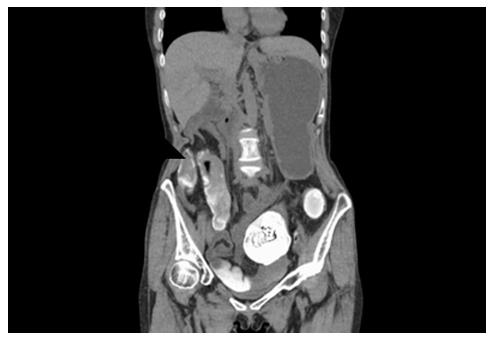
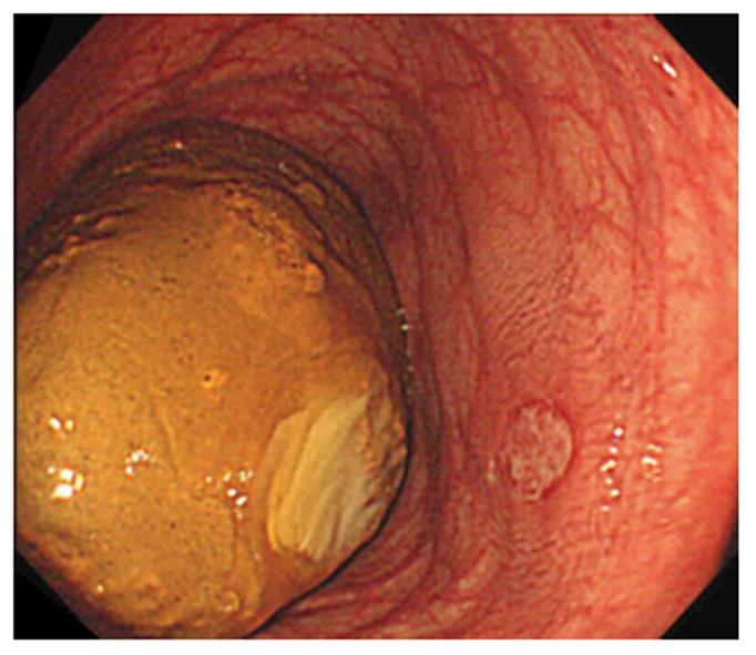
Abdominal computed tomography scans showed a highly absorbing round substance, which was 7 cm in size, with layered calcification in the transverse colon, resulting in a diagnosis of fecaloma (Figure 1). A colonoscopy revealed that a giant brown fecaloma occupied the lumen of the dilated transverse colon. A shallow 3-cm ulcer covered with a white coat was present near the fecaloma. The white coat was adherent to the fecaloma, suggesting it to be the cause of the ulcer (Figure 2).
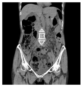
As the fecaloma was huge and actually occupied the colonic lumen, endoscopic extraction was attempted without laxatives because it was thought that the oral administration of laxatives, such as polyethylene glycol, might cause an intestinal obstruction. The surface of the fecaloma was hard, large and slippery, and fragmentation was not possible despite the use of biopsy forceps (Radial Jaw 4 Biopsy Forceps Standard Capacity, Boston Scientific, United States), an injection needle for endoscopic treatment (Impact Flow, Top, Japan), a needle knife (KD-10Q-1-A, Olympus, Japan), and a snare for endoscopic mucosal resection (EMR) (Snare Master, Olympus, Japan). Consequently, the surface of the fecaloma was shaved using jumbo forceps (Radial Jaw 4 Jumbo Cold Polypectomy Forceps, Boston Scientific, Japan) with about ten passes, which scraped the surface and made it possible to advance the forceps into the fecaloma. The same procedure was then repeated several hundred times, aiming for the center of the fecaloma and resulting in gradual fragmentation (Figure 3). A total of 6 h over 2 d were required to break the fecaloma into fragments of a size that could pass through the anus. We used the midazolam as a sedative and the pentazocine as an analgesic. Midazolam was administered intravenously according to the degree of affliction and administered 5 mg per time of the procedure. Pentazocine was administered 15 mg per each procedure. The fecaloma was then eliminated using laxatives (Figure 4).
Fecalomas, or hardened masses of food, discarded gastrointestinal cells, and digestive juice in the gastrointestinal tract, exceed fecal impactions in hardness[1]. Fecalomas commonly develop in the sigmoid colon and the rectum, which have a narrower lumen than that of the right colon[2]. Underlying diseases reported to result in fecalomas include chronic fecal impaction, Hirschsprung's disease, psychiatric disorders, and intestinal tuberculosis[3,4]. Generally, fecalomas cause intestinal obstruction, megacolon, and gastrointestinal perforation, and can even result in deep vein thrombosis and urinary tract compression on rare occassions[5-7]. Fecalomas must be treated to prevent the onset of these complications. Initial treatments include administration of laxatives, enemas, and stool extraction. When patients do not respond to these treatments, surgical extraction becomes necessary[8].
Our patient had MCTD and presented mainly with systemic scleroderma as the underlying disease. The frequency of gastrointestinal lesions is the highest among internal organ lesions associated with scleroderma. With the combined atrophy of smooth muscle in the intrinsic muscle layer, replacement by collagen fibers, and a nervous system disorder, dilatation of the gastrointestinal tract and reduced peristaltic activity occur. The percentage of gastrointestinal tract lesions in patients with scleroderma listed as the cause of death reportedly range from 6% to 12%[9]. In our patient, reduced segmental movement and peristaltic activity of the colon attributable to MCTD led to fecal impaction that progressed to a fecaloma in the transverse colon before reaching the left colon. Her small amount of water intake, likely associated with the SMA-like syndrome due to a loss of peristasis with the scleroderma, may also have contributed to the hardened fecaloma formation.
Our patient also had pulmonary hypertension associated with MCTD and used steroids. Therefore, she was at high risk of respiratory failure associated with surgery under general anesthesia and at an increased risk of anastomotic leakage in the intestinal tract. Endoscopic extraction of the fecaloma is an ideal treatment to avoid surgery for such patients with serious comorbidities. However, there are few reports describing endoscopic extraction of a large fecaloma. In previous reports, endoscopic extraction was successfully performed using conventional forceps and a snare for polypectomy[2,4]. In our patient, however, the fecaloma could not be fragmented with forceps or a snare, presumably due to its hardness and the slipperiness of its surface.
One case of fecaloma that was too slippery and hard to grasp with forceps has been reported. That fecaloma was dissolved by a cola injection and was sufficiently softened to be removed[10]. Cola injection are often reported as the method for the removal of gastric bezoars. The mechanism of this method is considered the contribution of carbon dioxide bubble and the secretolic activity of sodium hydrogen carbonate. Jumbo forceps combined with a cola injection may be more efficient therapy for the removal of fecaloma endoscopically.
The fecaloma in this patient, which could not be fragmented with standard capacity forceps or a snare for EMR, was successfully grasped and fragmented with jumbo forceps. Jumbo forceps, which have larger cups than the standard capacity biopsy forceps, can collect large samples. Therefore, they are used in the resection of the colonic adenomas. When the routine biopsy forceps used at our institution are compared with the jumbo cold polypectomy forceps used in fecaloma fragmentation in the present patient, there are large differences in the dimensions of the outer diameter of the cup (2.2 mm and 2.8 mm, respectively), the maximum opening width (7.1 mm and 8.8 mm), and the quantities which can be removed (5.3 mm3 and 12.4 mm3). The size and shape of the jumbo forceps, for the collection of large quantities of fecaloma contents, was appropriate for achieving fragmentation of a giant fecaloma in our patient.
In summary, Jumbo forceps might be a useful device for fragmenting hardened fecalomas.
A 59-year-old woman with abdominal pain, which had persisted for 3 mo.
Queasiness and peri-umbilical abdominal pain.
Superior mesenteric artery syndrome, ileus, constipation, colonic cancer, volvulus of sigmoid colon.
All labs were within normal limits.
Abdominal computed tomography scans showed a highly absorbing round substance, which was 7 cm in size, with layered calcification in the transverse colon.
Endoscopic jumbo forceps break the fecaloma by repeated biting.
Underlying diseases reported to result in fecalomas include chronic fecal impaction, Hirschsprung’s disease, psychiatric disorders, and intestinal tuberculosis. Most of these cases needed the surgical treatments.
Jumbo forceps, which have larger cups than the standard capacity biopsy forceps, can collect large samples.
Jumbo forceps might be a useful device for fragmenting hardened fecalomas.
This is a case report of a large faecaloma that was successfully fragmented by jumbo forceps.
Manuscript source: Unsolicited manuscript
Specialty type: Gastroenterology and hepatology
Country of origin: Japan
Peer-review report classification
Grade A (Excellent): 0
Grade B (Very good): 0
Grade C (Good): C
Grade D (Fair): D
Grade E (Poor): 0
P- Reviewer: Pinho R, Tham TCK S- Editor: Qi Y L- Editor: A E- Editor: Li D
| 1. | Garisto JD, Campillo L, Edwards E, Harbour M, Ermocilla R. Giant fecaloma in a 12-year-old-boy: a case report. Cases J. 2009;2:127. [PubMed] [DOI] [Cited in This Article: ] |
| 2. | Sakai E, Inokuchi Y, Inamori M, Uchiyama T, Iida H, Takahashi H, Akiyama T, Akimoto K, Sakamoto Y, Fujita K. Rectal fecaloma: successful treatment using endoscopic removal. Digestion. 2007;75:198. [PubMed] [DOI] [Cited in This Article: ] |
| 3. | Campbell JB, Robinson AE. Hirschsprung’s disease presenting as calcified fecaloma. Pediatr Radiol. 1973;1:161-163. [PubMed] [Cited in This Article: ] |
| 4. | Kim SM, Ryu KH, Kim YS, Lee TH, Im EH, Huh KC, Choi YW, Kang YW. Cecal fecaloma due to intestinal tuberculosis: endoscopic treatment. Clin Endosc. 2012;45:174-176. [PubMed] [DOI] [Cited in This Article: ] |
| 5. | Alvarez C, Hernández MA, Quintano A. Clinical challenges and images in GI: Image 2: Deep venous thrombosis due to idiopathic megarectum and giant fecaloma. Gastroenterology. 2006;131:702-703, 983. [PubMed] [Cited in This Article: ] |
| 6. | Chute DJ, Cox J, Archer ME, Bready RJ, Reiber K. Spontaneous rupture of urinary bladder associated with massive fecal impaction (fecaloma). Am J Forensic Med Pathol. 2009;30:280-283. [PubMed] [DOI] [Cited in This Article: ] |
| 7. | Aguilera JF, Lugo RE, Luna AW. [Stercoral perforation of the colon: case report and literature review]. Medwave. 2015;15:e6108. [PubMed] [Cited in This Article: ] |
| 8. | Yucel AF, Akdogan RA, Gucer H. A giant abdominal mass: fecaloma. Clin Gastroenterol Hepatol. 2012;10:e9-e10. [PubMed] [DOI] [Cited in This Article: ] |
| 9. | Schubert GC, Setka CS. [Diagnostic difficulties in large, hardened stercoroma of the colon (fecaloma)]. Arch Geschwulstforsch. 1966;27:307-314. [PubMed] [Cited in This Article: ] |
| 10. | Kang JH, Lim YJ. Can Fecaloma be Dissolved by Cola Injection in a Similar Way to Bezoars? Intest Res. 2014;12:333-334. [PubMed] [DOI] [Cited in This Article: ] |


