Erschienen in: Cancer and Metastasis Reviews 4/2010

Open Access 01.12.2010 | NON-THEMATIC REVIEW

Malignant melanoma as a target malignancy for the study of the anti-metastatic properties of the heparins

verfasst von: Anthony Maraveyas, Miriam J. Johnson, Yu Pei Xiao, Simon Noble

Erschienen in: Cancer and Metastasis Reviews | Ausgabe 4/2010

Abstract

The outlook for metastatic melanoma to the brain is dismal. New therapeutic avenues are therefore needed. The anti-metastatic mechanisms that may underpin the effects of low molecular weight heparins (LMWHs) in in vitro and preclinical melanoma models warrant translating to a clinical setting. This review outlines a rationale that supports our proposal that metastatic melanoma to the brain is a clinical setting in which to study the anti-metastatic potential of LMWHs. Prevention or delay of brain metastases in melanoma is a clinically relevant and measurable target. Studies to explore the effect of anticoagulants on cancer survival are underway in other malignancies such as lung, pancreas, ovary, breast, and stomach cancer. However, no study to our knowledge has a methodology that could produce clinical evidence in support of a mechanism for whatever benefit may be seen. The setting we propose would allow translation of the molecular knowledge of the metastatic pathways mediated by platelets and the selectins—all potential targets of heparin—in a “time to appearance” of brain metastases endpoint. Since brain metastases are so common and they have a singularly adverse impact on survival, the “biological neuroprotection” model we propose in metastatic melanoma could provide the translational evidence to support the benefit of LMWHs in melanoma. More significantly, this would open the door to a wider “anti-metastatic” approach that could have much greater impact in patients with minimal disease being treated in adjuvant settings for the more common malignancies such as breast and colon cancer.

1 Introduction

The benefits from using anticoagulants in the treatment of patients with cancer may arise from two clinically different but biologically intertwined routes: direct effects (treatment or prevention) on thrombotic complications of malignancy and postulated anti-metastatic cancer effects.
From early laboratory and preclinical studies, melanoma may be a malignancy that would benefit from anticoagulant agents. Unlike pancreatic cancer [1], melanoma is not highly thrombogenic, and the clinical benefit suggested is unlikely to be solely due to the prevention of venous thromboembolism [2]. Preclinical experiments in the 1980s of anticoagulants in cancer treatment led to some poorly designed underpowered clinical trials which suggested improved survival in small cell lung cancer (SCLC) and melanoma. These two malignancies have three things in common: a relatively low incidence of thrombosis [2, 3], neuroectodermal origin, and early metastasis, often to the brain. These two malignancies, however, are very different in terms of sensitivity to conventional treatments. The exquisite radiosensitivity of SCLC has led to survival gains through the neuroprotective strategy of prophylactic cranial irradiation [4] whereas melanoma is relatively radioresistant. However, if a method of neuroprotection can be found for melanoma, similar survival benefits to those in SCLC may result.
These almost unique clinical properties of malignant melanoma—a tendency for indiscriminate and multiple metastases and lethal consequences of brain metastases, coupled to the lack of confounding parameters of thrombophilia or a neuroprotective strategy—suggest melanoma as a model malignancy to study the anti-metastatic potential of low molecular weight heparins (LMWHs). This paper outlines the rationale supporting this proposal.

2 Brain metastases in melanoma

The high mortality in melanoma patients relates to the high incidence of brain metastases; the median survival of unselected melanoma patients with brain metastases is only 2–4 months [5]. Melanoma cells appear to have substantial neurotropism; it is the fourth most common primary site to metastasize to the brain [6]. Melanoma accounts for 10% of all brain metastases and has the second highest incident proportion percentage [7]; up to 13% of patients with regional disease (AJCC stage III) [8], 18–46% of stage IV patients [6, 8], with a prevalence of up to 75% at autopsy [811].
It was hoped that two cytotoxic agents that cross the blood–brain barrier (BBB) (temozolomide and/or fotemustine) would improve survival. Unfortunately, neither drug provided any statistical advantage compared with the current gold standard, dacarbazine (DTIC) [12, 13]. This probably reflects the poor overall chemosensitivity of melanoma, although fotemustine [12] did fare somewhat better than dacarbazine in demonstrating a delay in the onset of brain metastases; the median time to brain metastases was 22.7 months (95% confidence interval (CI), 9.62–23.33) in the fotemustine arm versus 7.2 months (95% CI, 6.28–11.70) in the DTIC arm (P = .059). This just failed to translate into an overall survival advantage; 7.3 months (95% CI, 6.01–8.84) in the fotemustine arm versus 5.6 months (95% CI, 5.03–6.54) in the DTIC arm (P = .067 in the intent-to-treat population) which may reflect an underpowered study. Nevertheless, it strongly suggests that a survival benefit could accompany better CNS control of melanoma metastases and that a strategy of “biological interference” with the metastatic process to the brain may result in survival benefit for patients with advanced melanoma.

3 Elements of neuroprotective strategies

The data from the above mentioned phase III trial [12] show that 20% of patients had brain metastases at study entry and 20% developed them over the assessment period of around 6 months. It is unlikely that all the brain metastases that develop post-randomization result from pre-implanted melanoma cells. It is more likely that a proportion of these have resulted from an ongoing process of melanoma cells shed from other metastases continuously arriving in the microcirculation of the brain—slowing down, attaching to endothelium, and finally, successfully implanting.
A neuroprotection model of “biological interference” would assume that BBB invasion by melanoma cells is a stochastic event, i.e., occurs randomly on a background of a predictable continuous process, thus requiring continuous prophylaxis. This model can be broken down into three components:
(1)
Cytoreduction—i.e., reducing the burden of BBB implanted cells.
 
(2)
Inactivation of the “metastatic niche”—i.e., interfering with the mechanisms driving invasion.
 
(3)
Prevent implantation—i.e., interfering with the process of endothelial cellular attachment.
 
This model differs to that of SCLC mostly because the conventional modalities of chemotherapy and radiotherapy achieve all the above stated components through the exquisite sensitivity of SCLC to these treatments. Thus, cytoreduction and inactivation are achieved by the chemotherapy and the radiotherapy while further implantation is reduced by the systemic response to chemotherapy; the efficacy of this strategy is mostly seen in patients who have achieved complete or consolidated partial response with radiotherapy [4].
In melanoma, this is not possible as we have no agents that can deliver this “neuroprotective” component through significant cancer cell kill. However, if a strategy of continual “inactivation” and “interference” with the metastatic process can be developed to complement a cytotoxic induction phase, however modest, one may produce a survival benefit.

4 Coagulation pathways and the metastatic process

Cancer cells are continuously shed into the circulation either from the primary or from established metastases. However, the metastatic process is inefficient and cell survival in the circulation is low due to a combination of shear forces, phagocytosis, and obstruction within capillary beds. Only 1.5% of nonhematogenous cells injected into the bloodstream survive for more than 24 h [14].
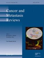
The complex biological pathways of the metastatic process can, simplistically, be categorized as coagulation-dependent and coagulation-independent. These insights have existed for more than 30 years [15]. Although there is some evidence that some of the coagulation factors may also be involved in coagulation-independent promotion of metastasis (such as tissue factor [16]), it appears that the successful negotiation of the intravascular phase of the cancer cell journey is heavily dependent on mechanical and signaling interactions that would promote thrombosis in the physiological state. There is unequivocal evidence of the interactions between disseminating tumor cells and blood cells, mainly platelets [1719]. Initial support for an active role of platelets in metastasis was obtained from a mouse model in which experimental thrombocytopenia led to attenuation of metastasis [19]. The significance of the tumor cell microemboli formation was suggested when the intravenous injection of tumor cells led to a rapid association with platelets, whereas in the absence of this interaction, tumor cells were cleared by natural killer (NK) cells [20]. The intravenous tumor cell injection coincided with a temporal reduction of peripheral platelet counts. Interference with platelet–tumor cell interactions resulted in the attenuation of metastasis, further supporting the protective role of microemboli formation for tumor cell survival [21, 22]. These “transit” metastatic emboli include leucocytes and function as vehicles that shield the cancer cell(s) from shear forces and immunosurveillance (NK cells) [23], maintain viability of the cancer cell(s) through signaling [24, 25], promote retention in capillaries, and provide an in situ readily available repository of signaling molecules that facilitate colonization of distant organs [26, 27] when the microembolic phase is initiated. Propagation–prolongation of this process through maintenance of the micro thrombosis-related microenvironment (activation of cytokines and coagulation factors) promote establishment of the “metastatic niche” (Fig. 1).

4.1 Coagulation factors and melanoma metastases

Coagulation factors, particularly thrombin and tissue factor (TF), play an important role in melanoma metastasis [2831]. Thrombin stimulates platelets, induces tumor angiogenesis, regulates tumor cell adhesion to platelets and endothelial cells, and promotes tumor growth and metastasis [2830]. Thrombin responses in melanoma cell motility and metastasis depend on proteinase-activated receptor-1 (PAR1) [32]; PAR1 is also required in the development of TF-mediated metastasis [33]. The PAR1 expression is significantly higher in metastatic melanoma cells, which however have undetectable levels of endogenous activator protein 2 (AP-2) [30, 32]. The loss of transcription factor AP-2 is a crucial event in the development of malignant melanoma [34] and results in overexpression of PAR1 that regulates the metastatic phenotype of melanoma [35] by induction of cell adhesion molecules, matrix-degrading proteases, stimulating the secretion of angiogenic and invasive factors [30, 3638]. PAR1 and thrombin could be potential targets for antitumor and anti-metastasis therapy for melanoma patients [30, 35, 37]. The data contribute to a mechanistic explanation of the preclinical data that have persistently pointed to potential anti-metastatic properties of anticoagulants [30, 39] via effects on the coagulation cascade.

5 Endothelial accesses and the metastatic process

The arrest of cancer cells in small vessels is an important step in metastasis. Although simple mechanical entrapment has been proposed [40, 41], more complex processes are involved [26, 39, 4244] including the alteration of cell surface glycosylation, which is a common feature of carcinoma progression and metastasis; in particular, high expression of sialylated fucosylated glycans such as sialyl Lewisx/a. A further significant role in these processes is played by integrins [45] and although very recently, anti-metastatic effects of heparins in melanoma have been postulated to be mediated in certain cases though interaction of heparin with integrins [46, 47], the complexity, diversity, and the non-uniform impact of heparins [48] on this pathway put a detailed discussion beyond the scope of this review.

5.1 Selectins and cell “docking”

Selectins are a family of mammalian vascular adhesion molecules: P(latelet)-selectin, E(ndothelial)-selectin, and L(eucocyte)-selectin [49]. Both E- and P-selectins are inducible membrane proteins expressed on activated endothelial cells or platelets following various proinflammatory cytokine stimulations such as TNF and thrombin [50]. Selectins could interact with those sialyl Lewisx/a-containing cell surface glycoconjugates, normally found on mucin-type glycoproteins of leukocytes and endothelium, and mediate tethering, rolling, and adhesion of cells [49, 51, 52]. Thus, “deceleration” to achieve surface purchase is achieved through cellular glycocalyx and selectin-mediated interactions inducing “rolling”, in a manner that is similar to that in the regulated recruitment of leukocytes to tissue sites of damage and inflammation.
P-selectin has been demonstrated to mediate crucial cancer cell interactions with platelets and endothelial cells [39, 49, 51, 53]. The expression of E- and P-selectin on endothelial cells initiates tethering and rolling of the cancer cells and formation of weak adhesions to the endothelial cells within the capillary bed [21]. Rolling is now thought to be a major primary deceleration mechanism used by melanoma cells prior to more definitive integrin-related anchoring to the endothelium [54].

6 Heparins and melanoma metastases

6.1 The in vitro and preclinical evidence

The effect of the heparins on the melanoma metastatic process has been repeatedly demonstrated since in both experimental and spontaneous metastatic models (Table 1). The emergence of the role of selectins regulating this “docking” process of the cancer cell has pinpointed that at least in the experimental tumor cell models the total effect of heparin on the platelet–tumor cloak can be accounted for by its interactions with P-selectin molecules [39, 55]. Moreover, these investigators and others [54, 56] have demonstrated that the interactions of the cancer cell with P- and E-selectins on endothelium are equally important for adhesion and similarly a major therapeutic target of the heparins [55, 57] (Fig. 1).
Table 1
Preclinical and in vitro evidence of molecular targets of heparins in malignant melanoma
Molecular target
Model
Type of Heparin
Mechanisms
Endothelial P-selectin
B16F10 injected mice; murine B16F10, human NW624 and NW1539 cells
UFH
Heparin not only inhibits endothelial P-selectin-mediated melanoma cell rolling on microvasculature, but also attenuates melanoma metastasis formation in vivo [61].
P-selectin
Human A375 cells
Modified heparin
Heparin can block P-selectin-mediated A375 human melanoma cell adhesion [67, 68].
P-selectin
B16 injected mice; murine B16, human NW624 and NW1539 cells
UFH; LMWHs (nadroparin, enoxaparin); synthetic pentasaccharide fondaparinux
UFH and LMWHs inhibit P-selectin mediated rolling of melanoma cells in vitro and in vivo [56].
P-selectin
B16-BL6 injected mice; murine B16-BL6 cell
Heparin derivatives
Modified non-anticoagulant species of heparin specifically inhibit selectin-mediated cell–cell interactions, heparanase enzymatic activity, or both [69].
Selectins
B16F10 injected mice; murine B16F10 cell
Orally absorbable heparin derivative (LHD)
Orally active LHD could have anti-metastatic effect via inhibition of cell–cell interaction between melanoma cells and platelets or HUVECs by interrupting selectin-mediated interactions [54].
Integrin α4β1 (VLA-4)
Murine B16F10, human MV3 cells
UFH; LMWH (tinzaparin), a fraction of tinzaparin, fondaparinux.
Heparin is shown to interfere with the VLA-4/VCAM-1 interaction and inhibit integrin VLA-4-mediated interactions [46, 70].
Integrin αIIbβ3
B16F10 or A375 injected mice; murine B16F10 and Human A375 cells
Modified heparins
Modified heparins can inhibit melanoma tumor cell–platelet interaction mediated by platelet integrin αIIbβ3 [47].
PAR1and CD24
B16 and K1735 injected mice; murine B16 and K1735 cells
LMWH (nadroparin)
LMWH inhibits thrombin-dependent PAR1 activation, thus, further inhibiting the platelet or endothelial cell activation and L- and P- selectin-mediated cell–cell adhesion; cancer cell express CD24 takes part in metastasis as a ligand of P-selectin. LMWH inhibits P-selectin/CD24 interaction prevent binding of platelets to cancer cells [71].
UFH unfractionated sodium heparin, LMWHs low molecular weight heparins

6.2 The proposed mechanism of the heparin–selectin interaction

P-selectin binds a large range of heparin sulfate and heparin fragments [57]. L-selectin may require a more specific sequence for recognition. Although binding to Sialyl-Lewis X motif on mucin-like molecules is thought to be a shared ligand between the selectins, this interaction is weak [42, 58], and there is no clear superior single structural motif. The suggested mechanism is that the recognition involves a “clustered anionic patch”, rather than a linear-defined oligosaccharide sequence. The most convincing high affinity interaction discovered is that of P-selectin glycoprotein ligand-1 (PSGL-1) with P-selectin. This sialomucin polypeptide is expressed on all leucocytes but becomes specialized for selectin recognition on neutrophils and monocytes. The most interesting biophysical aspect of interaction of these molecules is that it involves novel “catch bonds” which help to explain the rollin g behavior shown by leucocytes coming into contact with endothelium [59, 60].
Unrelated glycans (e.g., the heparins) can also act as ligands for L- and P-selectins and the clinically used molecules (highly sulfated) are potent inhibitors, causing complete inhibition of both L- and P-selectins in vitro assay within recommended clinical anticoagulation target levels, with unfractionated heparin being the most potent (Fig. 1). Ludwig et al. [56, 61], employing the murine B16 melanoma lung metastasis model, revealed that heparin treatment inhibited lung colonization (by >80%) if given prior to, but not after intravenous inoculation of melanoma cells. They showed that heparin inhibits melanoma rolling in a P-selectin-dependent fashion. Whereas in P- selectin−/− mice rolling remained unaffected by heparin, in wild-type mice heparin treatment reduced rolling of three different melanoma cell lines (Mel1539, Mel624, and B16F10) in a dose-dependent manner. Together, these data support the assumption that both platelet and endothelial P-selectin are potential targets of heparin action in melanoma metastasis formation. These effects have been reproduced using an orally administered heparin (a chemical conjugate of LMWH and deoxycholic acid) in a murine melanoma lung metastases model [54]. The LMWHs have similar effects but IC50 values of these compounds against selectin–PSGL-1 interactions were closer to the recommended target range for therapeutic anticoagulation. This difference between unfractionated and LMW heparins is likely to be due to the reduced efficacy of smaller-sized fragments interacting with the “clustered anionic patch”. Nevertheless, even these agents, dosed at therapeutic (rather than prophylactic) levels, are highly effective selectin inhibitors [56, 61].

6.3 The clinical evidence

There has been a recent resurgence in the interest of the potential effect of anticoagulants in cancer. Unfortunately, the studies reported to date [62, 63], although hinting at potential survival benefits from the use of LMWH, are difficult to interpret as the populations studied are usually mixed in terms of histology type and stage of cancer. It is difficult to determine whether the effect is through prevention of thrombosis (e.g., in mucin-producing adenocarcinomas) or through potential non-anticoagulant-mediated anti-malignancy effects. Therefore, it is interesting to consider the historical studies which, while smaller and less robust, focused on specific cancers.
At least two studies in SCLC demonstrated a survival benefit for those receiving unfractionated sodium heparin (UFH) or LMWH [3, 64]. The thrombosis incidence in the control groups was negligible but the benefit was significant. This was difficult to explain and although it was attributed to a better response rate at least in one study, we suggest that other mechanisms relating to modification of the natural history of this disease (such as reducing incidence or delaying onset of brain metastases) may have played a role. There are no data from these trials to support or refute this hypothesis.
The data suggesting clinical role and effect of anticoagulants on melanoma have been previously summarized [65, 66]. All the studies reviewed pointed to survival benefit with anticoagulants although warfarin was the only anticoagulant used in these early studies. Mechanisms suggested were those of thrombin inhibition and possibly beneficial effects on TF generation.
We therefore suggest that heparin or LMWH strategies employed for their capacity to interfere with the metastatic process have reasonable data in support and that the model of prevention or delay of melanoma brain metastases could be one that could elicit this benefit in a clinically measurable sense (brain metastases-free interval) that could also result in a patient relevant benefit in terms of survival.

7 Summary

The anti-metastatic mechanisms that may underpin the effects of heparin–LMWH in in vitro and preclinical melanoma models warrant testing in the clinical setting as prevention or delay of brain metastases in melanoma appears to be a clinically relevant and measurable target. Studies to explore the effect of anticoagulants on cancer survival are underway in other malignancies such as lung, pancreas, ovary, breast, and stomach cancer. However, no study to our knowledge has a methodology that could produce clinical evidence in support of a mechanism for whatever benefit may be seen. The “biological neuroprotection” model we propose in metastatic melanoma could provide this evidence, supporting the benefit of LMWH in melanoma, but more significantly, opening the door to a wider “anti-metastatic” approach that could have much greater impact in patients with minimal disease being treated in adjuvant settings for the more common malignancies such as breast and colon cancer.

Acknowledgments

The authors wish to thank Jan Sharp for the technical support with the photographic images.

Open Access

This article is distributed under the terms of the Creative Commons Attribution Noncommercial License which permits any noncommercial use, distribution, and reproduction in any medium, provided the original author(s) and source are credited.
Open AccessThis is an open access article distributed under the terms of the Creative Commons Attribution Noncommercial License (https://​creativecommons.​org/​licenses/​by-nc/​2.​0), which permits any noncommercial use, distribution, and reproduction in any medium, provided the original author(s) and source are credited.

Unsere Produktempfehlungen

e.Med Interdisziplinär

Kombi-Abonnement

Für Ihren Erfolg in Klinik und Praxis - Die beste Hilfe in Ihrem Arbeitsalltag

Mit e.Med Interdisziplinär erhalten Sie Zugang zu allen CME-Fortbildungen und Fachzeitschriften auf SpringerMedizin.de.

e.Med Innere Medizin

Kombi-Abonnement

Mit e.Med Innere Medizin erhalten Sie Zugang zu CME-Fortbildungen des Fachgebietes Innere Medizin, den Premium-Inhalten der internistischen Fachzeitschriften, inklusive einer gedruckten internistischen Zeitschrift Ihrer Wahl.

Literatur
1.
Zurück zum Zitat Sgouros, J., & Maraveyas, A. (2008). Excess premature (3-month) mortality in advanced pancreatic cancer could be related to fatal vascular thromboembolic events. A hypothesis based on a systematic review of phase III chemotherapy studies in advanced pancreatic cancer. Acta Oncológica, 47(3), 337–346.CrossRefPubMed Sgouros, J., & Maraveyas, A. (2008). Excess premature (3-month) mortality in advanced pancreatic cancer could be related to fatal vascular thromboembolic events. A hypothesis based on a systematic review of phase III chemotherapy studies in advanced pancreatic cancer. Acta Oncológica, 47(3), 337–346.CrossRefPubMed
2.
Zurück zum Zitat Chew, H. K., Wun, T., Harvey, D., Zhou, H., & White, R. H. (2006). Incidence of venous thromboembolism and its effect on survival among patients with common cancers. Archives of Internal Medicine, 166(4), 458–464.CrossRefPubMed Chew, H. K., Wun, T., Harvey, D., Zhou, H., & White, R. H. (2006). Incidence of venous thromboembolism and its effect on survival among patients with common cancers. Archives of Internal Medicine, 166(4), 458–464.CrossRefPubMed
3.
Zurück zum Zitat Altinbas, M., Coskun, H. S., Er, O., Ozkan, M., Eser, B., Unal, A., et al. (2004). A randomized clinical trial of combination chemotherapy with and without low-molecular-weight heparin in small cell lung cancer. Journal of Thrombosis and Haemostasis, 2(8), 1266–1271.CrossRefPubMed Altinbas, M., Coskun, H. S., Er, O., Ozkan, M., Eser, B., Unal, A., et al. (2004). A randomized clinical trial of combination chemotherapy with and without low-molecular-weight heparin in small cell lung cancer. Journal of Thrombosis and Haemostasis, 2(8), 1266–1271.CrossRefPubMed
4.
Zurück zum Zitat Slotman, B., Faivre-Finn, C., Kramer, G., Rankin, E., Snee, M., Hatton, M., et al. (2007). Prophylactic cranial irradiation in extensive small-cell lung cancer. The New England Journal of Medicine, 357(7), 664–672.CrossRefPubMed Slotman, B., Faivre-Finn, C., Kramer, G., Rankin, E., Snee, M., Hatton, M., et al. (2007). Prophylactic cranial irradiation in extensive small-cell lung cancer. The New England Journal of Medicine, 357(7), 664–672.CrossRefPubMed
5.
Zurück zum Zitat Douglas, J. G., & Margolin, K. (2002). The treatment of brain metastases from malignant melanoma. Seminars in Oncology, 29(5), 518–524.CrossRefPubMed Douglas, J. G., & Margolin, K. (2002). The treatment of brain metastases from malignant melanoma. Seminars in Oncology, 29(5), 518–524.CrossRefPubMed
6.
Zurück zum Zitat Wen, P. Y., Black, P. M., & Joeffler, S. L. (2001). Treatment of metastatic cancer. In V. T. Devita, S. Hellman, & S. A. Rosenberg (Eds.), Cancer: principles and practice of oncology (6th ed., pp. 2655–2670). Philadelphia: Lippincott, Williams & Wilkins. Wen, P. Y., Black, P. M., & Joeffler, S. L. (2001). Treatment of metastatic cancer. In V. T. Devita, S. Hellman, & S. A. Rosenberg (Eds.), Cancer: principles and practice of oncology (6th ed., pp. 2655–2670). Philadelphia: Lippincott, Williams & Wilkins.
7.
Zurück zum Zitat Barnholtz-Sloan, J. S., Sloan, A. E., Davis, F. G., Vigneau, F. D., Lai, P., & Sawaya, R. E. (2004). Incidence proportions of brain metastases in patients diagnosed (1973–2001) in the Metropolitan Detroit Cancer Surveillance System. Journal of Clinical Oncology, 22(14), 2865–2872.CrossRefPubMed Barnholtz-Sloan, J. S., Sloan, A. E., Davis, F. G., Vigneau, F. D., Lai, P., & Sawaya, R. E. (2004). Incidence proportions of brain metastases in patients diagnosed (1973–2001) in the Metropolitan Detroit Cancer Surveillance System. Journal of Clinical Oncology, 22(14), 2865–2872.CrossRefPubMed
8.
Zurück zum Zitat Amer, M. H., Al-Sarraf, M., Baker, L. H., & Vaitkevicius, V. K. (1978). Malignant melanoma and central nervous system metastases: incidence, diagnosis, treatment and survival. Cancer, 42(2), 660–668.CrossRefPubMed Amer, M. H., Al-Sarraf, M., Baker, L. H., & Vaitkevicius, V. K. (1978). Malignant melanoma and central nervous system metastases: incidence, diagnosis, treatment and survival. Cancer, 42(2), 660–668.CrossRefPubMed
9.
Zurück zum Zitat Budman, D. R., Camacho, E., & Wittes, R. E. (1978). The current causes of death in patients with malignant melanoma. European Journal of Cancer, 14(4), 327–330.PubMed Budman, D. R., Camacho, E., & Wittes, R. E. (1978). The current causes of death in patients with malignant melanoma. European Journal of Cancer, 14(4), 327–330.PubMed
10.
Zurück zum Zitat Bullard, D. E., Cox, E. B., & Seigler, H. F. (1981). Central nervous system metastases in malignant melanoma. Neurosurgery, 8(1), 26–30.CrossRefPubMed Bullard, D. E., Cox, E. B., & Seigler, H. F. (1981). Central nervous system metastases in malignant melanoma. Neurosurgery, 8(1), 26–30.CrossRefPubMed
11.
Zurück zum Zitat Patel, J. K., Didolkar, M. S., Pickren, J. W., & Moore, R. H. (1978). Metastatic pattern of malignant melanoma. A study of 216 autopsy cases. American Journal of Surgery, 135(6), 807–810.CrossRefPubMed Patel, J. K., Didolkar, M. S., Pickren, J. W., & Moore, R. H. (1978). Metastatic pattern of malignant melanoma. A study of 216 autopsy cases. American Journal of Surgery, 135(6), 807–810.CrossRefPubMed
12.
Zurück zum Zitat Avril, M. F., Aamdal, S., Grob, J. J., Hauschild, A., Mohr, P., Bonerandi, J. J., et al. (2004). Fotemustine compared with dacarbazine in patients with disseminated malignant melanoma: a phase III study. Journal of Clinical Oncology, 22(6), 1118–1125.CrossRefPubMed Avril, M. F., Aamdal, S., Grob, J. J., Hauschild, A., Mohr, P., Bonerandi, J. J., et al. (2004). Fotemustine compared with dacarbazine in patients with disseminated malignant melanoma: a phase III study. Journal of Clinical Oncology, 22(6), 1118–1125.CrossRefPubMed
13.
Zurück zum Zitat Middleton, M. R., Grob, J. J., Aaronson, N., Fierlbeck, G., Tilgen, W., Seiter, S., et al. (2000). Randomized phase III study of temozolomide versus dacarbazine in the treatment of patients with advanced metastatic malignant melanoma. Journal of Clinical Oncology, 18(1), 158–166.PubMed Middleton, M. R., Grob, J. J., Aaronson, N., Fierlbeck, G., Tilgen, W., Seiter, S., et al. (2000). Randomized phase III study of temozolomide versus dacarbazine in the treatment of patients with advanced metastatic malignant melanoma. Journal of Clinical Oncology, 18(1), 158–166.PubMed
14.
Zurück zum Zitat Partridge, M. A., & Marcantonio, E. E. (2006). Initiation of attachment and generation of mature focal adhesions by integrin-containing filopodia in cell spreading. Molecular Biology of the Cell, 17(10), 4237–4248.CrossRefPubMed Partridge, M. A., & Marcantonio, E. E. (2006). Initiation of attachment and generation of mature focal adhesions by integrin-containing filopodia in cell spreading. Molecular Biology of the Cell, 17(10), 4237–4248.CrossRefPubMed
15.
Zurück zum Zitat Fidler, I. J. (1970). Metastasis: quantitative analysis of distribution and fate of tumor emboli labeled with 125i-5-iodo-2′- deoxyuridine. Journal of the National Cancer Institute, 45(4), 773–782.PubMed Fidler, I. J. (1970). Metastasis: quantitative analysis of distribution and fate of tumor emboli labeled with 125i-5-iodo-2′- deoxyuridine. Journal of the National Cancer Institute, 45(4), 773–782.PubMed
16.
Zurück zum Zitat Bromberg, M. E., Konigsberg, W. H., Madison, J. F., Pawashe, A., & Garen, A. (1995). Tissue factor promotes melanoma metastasis by a pathway independent of blood coagulation. Proceedings of the National Academy of Sciences of the United States of America, 92(18), 8205–8209.CrossRefPubMed Bromberg, M. E., Konigsberg, W. H., Madison, J. F., Pawashe, A., & Garen, A. (1995). Tissue factor promotes melanoma metastasis by a pathway independent of blood coagulation. Proceedings of the National Academy of Sciences of the United States of America, 92(18), 8205–8209.CrossRefPubMed
17.
Zurück zum Zitat Honn, K. V., Tang, D. G., & Crissman, J. D. (1992). Platelets and cancer metastasis: a causal relationship? Cancer and Metastasis Reviews, 11(3–4), 325–351.CrossRefPubMed Honn, K. V., Tang, D. G., & Crissman, J. D. (1992). Platelets and cancer metastasis: a causal relationship? Cancer and Metastasis Reviews, 11(3–4), 325–351.CrossRefPubMed
18.
Zurück zum Zitat Gasic, G. J. (1984). Role of plasma, platelets, and endothelial cells in tumor metastasis. Cancer and Metastasis Reviews, 3(2), 99–114.CrossRefPubMed Gasic, G. J. (1984). Role of plasma, platelets, and endothelial cells in tumor metastasis. Cancer and Metastasis Reviews, 3(2), 99–114.CrossRefPubMed
19.
Zurück zum Zitat Karpatkin, S., & Pearlstein, E. (1981). Role of platelets in tumor cell metastases. Annals of Internal Medicine, 95(5), 636–641.PubMed Karpatkin, S., & Pearlstein, E. (1981). Role of platelets in tumor cell metastases. Annals of Internal Medicine, 95(5), 636–641.PubMed
20.
Zurück zum Zitat Palumbo, J. S., Talmage, K. E., Massari, J. V., La Jeunesse, C. M., Flick, M. J., Kombrinck, K. W., et al. (2005). Platelets and fibrin(ogen) increase metastatic potential by impeding natural killer cell-mediated elimination of tumor cells. Blood, 105(1), 178–185.CrossRefPubMed Palumbo, J. S., Talmage, K. E., Massari, J. V., La Jeunesse, C. M., Flick, M. J., Kombrinck, K. W., et al. (2005). Platelets and fibrin(ogen) increase metastatic potential by impeding natural killer cell-mediated elimination of tumor cells. Blood, 105(1), 178–185.CrossRefPubMed
21.
Zurück zum Zitat Nash, G. F., Turner, L. F., Scully, M. F., & Kakkar, A. K. (2002). Platelets and cancer. The Lancet Oncology, 3(7), 425–430.CrossRefPubMed Nash, G. F., Turner, L. F., Scully, M. F., & Kakkar, A. K. (2002). Platelets and cancer. The Lancet Oncology, 3(7), 425–430.CrossRefPubMed
22.
Zurück zum Zitat Jurasz, P., Alonso-Escolano, D., & Radomski, M. W. (2004). Platelet–cancer interactions: mechanisms and pharmacology of tumour cell-induced platelet aggregation. British Journal of Pharmacology, 143(7), 819–826.CrossRefPubMed Jurasz, P., Alonso-Escolano, D., & Radomski, M. W. (2004). Platelet–cancer interactions: mechanisms and pharmacology of tumour cell-induced platelet aggregation. British Journal of Pharmacology, 143(7), 819–826.CrossRefPubMed
23.
Zurück zum Zitat Nieswandt, B., Hafner, M., Echtenacher, B., & Mannel, D. N. (1999). Lysis of tumor cells by natural killer cells in mice is impeded by platelets. Cancer Research, 59(6), 1295–1300.PubMed Nieswandt, B., Hafner, M., Echtenacher, B., & Mannel, D. N. (1999). Lysis of tumor cells by natural killer cells in mice is impeded by platelets. Cancer Research, 59(6), 1295–1300.PubMed
24.
Zurück zum Zitat Varon, D., & Brill, A. (2001). Platelets cross-talk with tumor cells. Haemostasis, 31(Suppl 1), 64–66.PubMed Varon, D., & Brill, A. (2001). Platelets cross-talk with tumor cells. Haemostasis, 31(Suppl 1), 64–66.PubMed
25.
Zurück zum Zitat Baj-Krzyworzeka, M., Majka, M., Pratico, D., Ratajczak, J., Vilaire, G., Kijowski, J., et al. (2002). Platelet-derived microparticles stimulate proliferation, survival, adhesion, and chemotaxis of hematopoietic cells. Experimental Hematology, 30(5), 450–459.CrossRefPubMed Baj-Krzyworzeka, M., Majka, M., Pratico, D., Ratajczak, J., Vilaire, G., Kijowski, J., et al. (2002). Platelet-derived microparticles stimulate proliferation, survival, adhesion, and chemotaxis of hematopoietic cells. Experimental Hematology, 30(5), 450–459.CrossRefPubMed
26.
Zurück zum Zitat Varki, N. M., & Varki, A. (2002). Heparin inhibition of selectin-mediated interactions during the hematogenous phase of carcinoma metastasis: rationale for clinical studies in humans. Seminars in Thrombosis and Hemostasis, 28(1), 53–66.CrossRefPubMed Varki, N. M., & Varki, A. (2002). Heparin inhibition of selectin-mediated interactions during the hematogenous phase of carcinoma metastasis: rationale for clinical studies in humans. Seminars in Thrombosis and Hemostasis, 28(1), 53–66.CrossRefPubMed
27.
Zurück zum Zitat Browder, T., Folkman, J., & Pirie-Shepherd, S. (2000). The hemostatic system as a regulator of angiogenesis. The Journal of Biological Chemistry, 275(3), 1521–1524.CrossRefPubMed Browder, T., Folkman, J., & Pirie-Shepherd, S. (2000). The hemostatic system as a regulator of angiogenesis. The Journal of Biological Chemistry, 275(3), 1521–1524.CrossRefPubMed
28.
Zurück zum Zitat Fischer, E. G., Ruf, W., & Mueller, B. M. (1995). Tissue factor-initiated thrombin generation activates the signaling thrombin receptor on malignant melanoma cells. Cancer Research, 55(8), 1629–1632.PubMed Fischer, E. G., Ruf, W., & Mueller, B. M. (1995). Tissue factor-initiated thrombin generation activates the signaling thrombin receptor on malignant melanoma cells. Cancer Research, 55(8), 1629–1632.PubMed
29.
Zurück zum Zitat Melnikova, V. O., Villares, G. J., & Bar-Eli, M. (2008). Emerging roles of PAR-1 and PAFR in melanoma metastasis. Cancer Microenvironment, 1(1), 103–111.CrossRefPubMed Melnikova, V. O., Villares, G. J., & Bar-Eli, M. (2008). Emerging roles of PAR-1 and PAFR in melanoma metastasis. Cancer Microenvironment, 1(1), 103–111.CrossRefPubMed
30.
Zurück zum Zitat Tellez, C., & Bar-Eli, M. (2003). Role and regulation of the thrombin receptor (PAR-1) in human melanoma. Oncogene, 22(20), 3130–3137.CrossRefPubMed Tellez, C., & Bar-Eli, M. (2003). Role and regulation of the thrombin receptor (PAR-1) in human melanoma. Oncogene, 22(20), 3130–3137.CrossRefPubMed
31.
Zurück zum Zitat Mueller, B. M., Reisfeld, R. A., Edgington, T. S., & Ruf, W. (1992). Expression of tissue factor by melanoma cells promotes efficient hematogenous metastasis. Proceedings of the National Academy of Sciences of the United States of America, 89(24), 11832–11836.CrossRefPubMed Mueller, B. M., Reisfeld, R. A., Edgington, T. S., & Ruf, W. (1992). Expression of tissue factor by melanoma cells promotes efficient hematogenous metastasis. Proceedings of the National Academy of Sciences of the United States of America, 89(24), 11832–11836.CrossRefPubMed
32.
Zurück zum Zitat Shi, X., Gangadharan, B., Brass, L. F., Ruf, W., & Mueller, B. M. (2004). Protease-activated receptors (PAR1 and PAR2) contribute to tumor cell motility and metastasis. Molecular Cancer Research, 2(7), 395–402.PubMed Shi, X., Gangadharan, B., Brass, L. F., Ruf, W., & Mueller, B. M. (2004). Protease-activated receptors (PAR1 and PAR2) contribute to tumor cell motility and metastasis. Molecular Cancer Research, 2(7), 395–402.PubMed
33.
Zurück zum Zitat Bromberg, M. E., Bailly, M. A., & Konigsberg, W. H. (2001). Role of protease-activated receptor 1 in tumor metastasis promoted by tissue factor. Thrombosis and Haemostasis, 86(5), 1210–1214.PubMed Bromberg, M. E., Bailly, M. A., & Konigsberg, W. H. (2001). Role of protease-activated receptor 1 in tumor metastasis promoted by tissue factor. Thrombosis and Haemostasis, 86(5), 1210–1214.PubMed
34.
Zurück zum Zitat Bar-Eli, M. (1999). Role of AP-2 in tumor growth and metastasis of human melanoma. Cancer and Metastasis Reviews, 18(3), 377–385.CrossRefPubMed Bar-Eli, M. (1999). Role of AP-2 in tumor growth and metastasis of human melanoma. Cancer and Metastasis Reviews, 18(3), 377–385.CrossRefPubMed
35.
Zurück zum Zitat Silini, A., Ghilardi, C., Ardinghi, C., Bernasconi, S., Oliva, P., Carraro, F., et al. (2010). Protease-activated receptor-1 (PAR-1) promotes the motility of human melanomas and is associated to their metastatic phenotype. Clinical & Experimental Metastasis, 27(1), 43–53.CrossRef Silini, A., Ghilardi, C., Ardinghi, C., Bernasconi, S., Oliva, P., Carraro, F., et al. (2010). Protease-activated receptor-1 (PAR-1) promotes the motility of human melanomas and is associated to their metastatic phenotype. Clinical & Experimental Metastasis, 27(1), 43–53.CrossRef
36.
Zurück zum Zitat Massi, D., Naldini, A., Ardinghi, C., Carraro, F., Franchi, A., Paglierani, M., et al. (2005). Expression of protease-activated receptors 1 and 2 in melanocytic nevi and malignant melanoma. Human Pathology, 36(6), 676–685.CrossRefPubMed Massi, D., Naldini, A., Ardinghi, C., Carraro, F., Franchi, A., Paglierani, M., et al. (2005). Expression of protease-activated receptors 1 and 2 in melanocytic nevi and malignant melanoma. Human Pathology, 36(6), 676–685.CrossRefPubMed
37.
Zurück zum Zitat Villares, G. J., Zigler, M., Wang, H., Melnikova, V. O., Wu, H., Friedman, R., et al. (2008). Targeting melanoma growth and metastasis with systemic delivery of liposome-incorporated protease-activated receptor-1 small interfering RNA. Cancer Research, 68(21), 9078–9086.CrossRefPubMed Villares, G. J., Zigler, M., Wang, H., Melnikova, V. O., Wu, H., Friedman, R., et al. (2008). Targeting melanoma growth and metastasis with systemic delivery of liposome-incorporated protease-activated receptor-1 small interfering RNA. Cancer Research, 68(21), 9078–9086.CrossRefPubMed
38.
Zurück zum Zitat Melnikova, V. O., & Bar-Eli, M. (2009). Inflammation and melanoma metastasis. Pigment Cell & Melanoma Research, 22(3), 257–267.CrossRef Melnikova, V. O., & Bar-Eli, M. (2009). Inflammation and melanoma metastasis. Pigment Cell & Melanoma Research, 22(3), 257–267.CrossRef
39.
Zurück zum Zitat Borsig, L., Wong, R., Feramisco, J., Nadeau, D. R., Varki, N. M., & Varki, A. (2001). Heparin and cancer revisited: mechanistic connections involving platelets, P-selectin, carcinoma mucins, and tumor metastasis. Proceedings of the National Academy of Sciences of the United States of America, 98(6), 3352–3357.CrossRefPubMed Borsig, L., Wong, R., Feramisco, J., Nadeau, D. R., Varki, N. M., & Varki, A. (2001). Heparin and cancer revisited: mechanistic connections involving platelets, P-selectin, carcinoma mucins, and tumor metastasis. Proceedings of the National Academy of Sciences of the United States of America, 98(6), 3352–3357.CrossRefPubMed
40.
Zurück zum Zitat Koop, S., MacDonald, I. C., Luzzi, K., Schmidt, E. E., Morris, V. L., Grattan, M., et al. (1995). Fate of melanoma cells entering the microcirculation: over 80% survive and extravasate. Cancer Research, 55(12), 2520–2523.PubMed Koop, S., MacDonald, I. C., Luzzi, K., Schmidt, E. E., Morris, V. L., Grattan, M., et al. (1995). Fate of melanoma cells entering the microcirculation: over 80% survive and extravasate. Cancer Research, 55(12), 2520–2523.PubMed
41.
Zurück zum Zitat Weiss, L. (1994). Inefficiency of metastasis from colorectal carcinomas. Relationship to local therapy for hepatic metastasis. Cancer Treatment and Research, 69, 1–11.PubMed Weiss, L. (1994). Inefficiency of metastasis from colorectal carcinomas. Relationship to local therapy for hepatic metastasis. Cancer Treatment and Research, 69, 1–11.PubMed
42.
Zurück zum Zitat Stevenson, J. L., Choi, S. H., & Varki, A. (2005). Differential metastasis inhibition by clinically relevant levels of heparins—correlation with selectin inhibition, not antithrombotic activity. Clinical Cancer Research, 11(19 Pt 1), 7003–7011.CrossRefPubMed Stevenson, J. L., Choi, S. H., & Varki, A. (2005). Differential metastasis inhibition by clinically relevant levels of heparins—correlation with selectin inhibition, not antithrombotic activity. Clinical Cancer Research, 11(19 Pt 1), 7003–7011.CrossRefPubMed
43.
Zurück zum Zitat Koenig, A., Norgard-Sumnicht, K., Linhardt, R., & Varki, A. (1998). Differential interactions of heparin and heparan sulfate glycosaminoglycans with the selectins. Implications for the use of unfractionated and low molecular weight heparins as therapeutic agents. Journal of Clinical Investigation, 101(4), 877–889.CrossRefPubMed Koenig, A., Norgard-Sumnicht, K., Linhardt, R., & Varki, A. (1998). Differential interactions of heparin and heparan sulfate glycosaminoglycans with the selectins. Implications for the use of unfractionated and low molecular weight heparins as therapeutic agents. Journal of Clinical Investigation, 101(4), 877–889.CrossRefPubMed
44.
Zurück zum Zitat Stevenson, J. L., Varki, A., & Borsig, L. (2007). Heparin attenuates metastasis mainly due to inhibition of P- and L-selectin, but non-anticoagulant heparins can have additional effects. Thrombosis Research, 120(Suppl 2), S107–S111.CrossRefPubMed Stevenson, J. L., Varki, A., & Borsig, L. (2007). Heparin attenuates metastasis mainly due to inhibition of P- and L-selectin, but non-anticoagulant heparins can have additional effects. Thrombosis Research, 120(Suppl 2), S107–S111.CrossRefPubMed
45.
Zurück zum Zitat Ramsay, A. G., Marshall, J. F., & Hart, I. R. (2007). Integrin trafficking and its role in cancer metastasis. Cancer and Metastasis Reviews, 26(3–4), 567–578.CrossRefPubMed Ramsay, A. G., Marshall, J. F., & Hart, I. R. (2007). Integrin trafficking and its role in cancer metastasis. Cancer and Metastasis Reviews, 26(3–4), 567–578.CrossRefPubMed
46.
Zurück zum Zitat Fritzsche, J., Simonis, D., & Bendas, G. (2008). Melanoma cell adhesion can be blocked by heparin in vitro: suggestion of VLA-4 as a novel target for antimetastatic approaches. Thrombosis and Haemostasis, 100(6), 1166–1175.PubMed Fritzsche, J., Simonis, D., & Bendas, G. (2008). Melanoma cell adhesion can be blocked by heparin in vitro: suggestion of VLA-4 as a novel target for antimetastatic approaches. Thrombosis and Haemostasis, 100(6), 1166–1175.PubMed
47.
Zurück zum Zitat Zhang, C., Liu, Y., Gao, Y., Shen, J., Zheng, S., Wei, M., et al. (2009). Modified heparins inhibit integrin alpha(IIb)beta(3) mediated adhesion of melanoma cells to platelets in vitro and in vivo. International Journal of Cancer, 125(9), 2058–2065.CrossRef Zhang, C., Liu, Y., Gao, Y., Shen, J., Zheng, S., Wei, M., et al. (2009). Modified heparins inhibit integrin alpha(IIb)beta(3) mediated adhesion of melanoma cells to platelets in vitro and in vivo. International Journal of Cancer, 125(9), 2058–2065.CrossRef
48.
Zurück zum Zitat Lydolph, M. C., Morgan-Fisher, M., Hoye, A. M., Couchman, J. R., Wewer, U. M., & Yoneda, A. (2009). Alpha9beta1 integrin in melanoma cells can signal different adhesion states for migration and anchorage. Experimental Cell Research, 315(19), 3312–3324.CrossRefPubMed Lydolph, M. C., Morgan-Fisher, M., Hoye, A. M., Couchman, J. R., Wewer, U. M., & Yoneda, A. (2009). Alpha9beta1 integrin in melanoma cells can signal different adhesion states for migration and anchorage. Experimental Cell Research, 315(19), 3312–3324.CrossRefPubMed
49.
Zurück zum Zitat Witz, I. P. (2008). The selectin–selectin ligand axis in tumor progression. Cancer and Metastasis Reviews, 27(1), 19–30.CrossRefPubMed Witz, I. P. (2008). The selectin–selectin ligand axis in tumor progression. Cancer and Metastasis Reviews, 27(1), 19–30.CrossRefPubMed
50.
Zurück zum Zitat Fu, J., Naren, A. P., Gao, X., Ahmmed, G. U., & Malik, A. B. (2005). Protease-activated receptor-1 activation of endothelial cells induces protein kinase Calpha-dependent phosphorylation of syntaxin 4 and Munc18c: Role in signaling p-selectin expression. The Journal of Biological Chemistry, 280(5), 3178–3184.CrossRefPubMed Fu, J., Naren, A. P., Gao, X., Ahmmed, G. U., & Malik, A. B. (2005). Protease-activated receptor-1 activation of endothelial cells induces protein kinase Calpha-dependent phosphorylation of syntaxin 4 and Munc18c: Role in signaling p-selectin expression. The Journal of Biological Chemistry, 280(5), 3178–3184.CrossRefPubMed
51.
Zurück zum Zitat Kansas, G. S. (1996). Selectins and their ligands: current concepts and controversies. Blood, 88(9), 3259–3287.PubMed Kansas, G. S. (1996). Selectins and their ligands: current concepts and controversies. Blood, 88(9), 3259–3287.PubMed
53.
Zurück zum Zitat Kim, Y. J., Borsig, L., Varki, N. M., & Varki, A. (1998). P-selectin deficiency attenuates tumor growth and metastasis. Proceedings of the National Academy of Sciences of the United States of America, 95(16), 9325–9330.CrossRefPubMed Kim, Y. J., Borsig, L., Varki, N. M., & Varki, A. (1998). P-selectin deficiency attenuates tumor growth and metastasis. Proceedings of the National Academy of Sciences of the United States of America, 95(16), 9325–9330.CrossRefPubMed
54.
Zurück zum Zitat Lee, D. Y., Park, K., Kim, S. K., Park, R. W., Kwon, I. C., Kim, S. Y., et al. (2008). Antimetastatic effect of an orally active heparin derivative on experimentally induced metastasis. Clinical Cancer Research, 14(9), 2841–2849.CrossRefPubMed Lee, D. Y., Park, K., Kim, S. K., Park, R. W., Kwon, I. C., Kim, S. Y., et al. (2008). Antimetastatic effect of an orally active heparin derivative on experimentally induced metastasis. Clinical Cancer Research, 14(9), 2841–2849.CrossRefPubMed
55.
Zurück zum Zitat Ludwig, R. J., Schon, M. P., & Boehncke, W. H. (2007). P-selectin: a common therapeutic target for cardiovascular disorders, inflammation and tumour metastasis. Expert Opinion on Therapeutic Targets, 11(8), 1103–1117.CrossRefPubMed Ludwig, R. J., Schon, M. P., & Boehncke, W. H. (2007). P-selectin: a common therapeutic target for cardiovascular disorders, inflammation and tumour metastasis. Expert Opinion on Therapeutic Targets, 11(8), 1103–1117.CrossRefPubMed
56.
Zurück zum Zitat Ludwig, R. J., Alban, S., Bistrian, R., Boehncke, W. H., Kaufmann, R., Henschler, R., et al. (2006). The ability of different forms of heparins to suppress P-selectin function in vitro correlates to their inhibitory capacity on bloodborne metastasis in vivo. Thrombosis and Haemostasis, 95(3), 535–540.PubMed Ludwig, R. J., Alban, S., Bistrian, R., Boehncke, W. H., Kaufmann, R., Henschler, R., et al. (2006). The ability of different forms of heparins to suppress P-selectin function in vitro correlates to their inhibitory capacity on bloodborne metastasis in vivo. Thrombosis and Haemostasis, 95(3), 535–540.PubMed
57.
Zurück zum Zitat Kim, Y. J., Borsig, L., Han, H. L., Varki, N. M., & Varki, A. (1999). Distinct selectin ligands on colon carcinoma mucins can mediate pathological interactions among platelets, leukocytes, and endothelium. The American Journal of Pathology, 155(2), 461–472.PubMed Kim, Y. J., Borsig, L., Han, H. L., Varki, N. M., & Varki, A. (1999). Distinct selectin ligands on colon carcinoma mucins can mediate pathological interactions among platelets, leukocytes, and endothelium. The American Journal of Pathology, 155(2), 461–472.PubMed
58.
Zurück zum Zitat Fuster, M. M., & Esko, J. D. (2005). The sweet and sour of cancer: glycans as novel therapeutic targets. Nature Reviews. Cancer, 5(7), 526–542.CrossRefPubMed Fuster, M. M., & Esko, J. D. (2005). The sweet and sour of cancer: glycans as novel therapeutic targets. Nature Reviews. Cancer, 5(7), 526–542.CrossRefPubMed
59.
Zurück zum Zitat Marshall, B. T., Long, M., Piper, J. W., Yago, T., McEver, R. P., & Zhu, C. (2003). Direct observation of catch bonds involving cell-adhesion molecules. Nature, 423(6936), 190–193.CrossRefPubMed Marshall, B. T., Long, M., Piper, J. W., Yago, T., McEver, R. P., & Zhu, C. (2003). Direct observation of catch bonds involving cell-adhesion molecules. Nature, 423(6936), 190–193.CrossRefPubMed
60.
Zurück zum Zitat Evans, E., Leung, A., Heinrich, V., & Zhu, C. (2004). Mechanical switching and coupling between two dissociation pathways in a P-selectin adhesion bond. Proceedings of the National Academy of Sciences of the United States of America, 101(31), 11281–11286.CrossRefPubMed Evans, E., Leung, A., Heinrich, V., & Zhu, C. (2004). Mechanical switching and coupling between two dissociation pathways in a P-selectin adhesion bond. Proceedings of the National Academy of Sciences of the United States of America, 101(31), 11281–11286.CrossRefPubMed
61.
Zurück zum Zitat Ludwig, R. J., Boehme, B., Podda, M., Henschler, R., Jager, E., Tandi, C., et al. (2004). Endothelial P-selectin as a target of heparin action in experimental melanoma lung metastasis. Cancer Research, 64(8), 2743–2750.CrossRefPubMed Ludwig, R. J., Boehme, B., Podda, M., Henschler, R., Jager, E., Tandi, C., et al. (2004). Endothelial P-selectin as a target of heparin action in experimental melanoma lung metastasis. Cancer Research, 64(8), 2743–2750.CrossRefPubMed
62.
Zurück zum Zitat Hejna, M., Raderer, M., & Zielinski, C. C. (1999). Inhibition of metastases by anticoagulants. Journal of the National Cancer Institute, 91(1), 22–36.CrossRefPubMed Hejna, M., Raderer, M., & Zielinski, C. C. (1999). Inhibition of metastases by anticoagulants. Journal of the National Cancer Institute, 91(1), 22–36.CrossRefPubMed
63.
Zurück zum Zitat Akl, E. A., van Doormaal, F. F., Barba, M., Kamath, G., Kim, S. Y., Kuipers, S., et al. (2007). Parenteral anticoagulation for prolonging survival in patients with cancer who have no other indication for anticoagulation. Cochrane Database of Systematic Reviews, 3, CD006652. Akl, E. A., van Doormaal, F. F., Barba, M., Kamath, G., Kim, S. Y., Kuipers, S., et al. (2007). Parenteral anticoagulation for prolonging survival in patients with cancer who have no other indication for anticoagulation. Cochrane Database of Systematic Reviews, 3, CD006652.
64.
Zurück zum Zitat Lebeau, B., Chastang, C., Brechot, J. M., Capron, F., Dautzenberg, B., Delaisements, C., et al. (1994). Subcutaneous heparin treatment increases survival in small cell lung cancer. “Petites Cellules” Group. Cancer, 74(1), 38–45.CrossRefPubMed Lebeau, B., Chastang, C., Brechot, J. M., Capron, F., Dautzenberg, B., Delaisements, C., et al. (1994). Subcutaneous heparin treatment increases survival in small cell lung cancer. “Petites Cellules” Group. Cancer, 74(1), 38–45.CrossRefPubMed
65.
Zurück zum Zitat Zacharski, L. R., Henderson, W. G., Rickles, F. R., Forman, W. B., Cornell, C. J., Jr., Forcier, R. J., et al. (1984). Effect of warfarin anticoagulation on survival in carcinoma of the lung, colon, head and neck, and prostate. Final report of VA Cooperative Study #75. Cancer, 53(10), 2046–2052.CrossRefPubMed Zacharski, L. R., Henderson, W. G., Rickles, F. R., Forman, W. B., Cornell, C. J., Jr., Forcier, R. J., et al. (1984). Effect of warfarin anticoagulation on survival in carcinoma of the lung, colon, head and neck, and prostate. Final report of VA Cooperative Study #75. Cancer, 53(10), 2046–2052.CrossRefPubMed
66.
Zurück zum Zitat Ornstein, D. L., & Zacharski, L. R. (2001). Treatment of cancer with anticoagulants: rationale in the treatment of melanoma. International Journal of Hematology, 73(2), 157–161.CrossRefPubMed Ornstein, D. L., & Zacharski, L. R. (2001). Treatment of cancer with anticoagulants: rationale in the treatment of melanoma. International Journal of Hematology, 73(2), 157–161.CrossRefPubMed
67.
Zurück zum Zitat Ma, Y. Q., & Geng, J. G. (2000). Heparan sulfate-like proteoglycans mediate adhesion of human malignant melanoma a375 cells to P-selectin under flow. Journal of Immunology, 165(1), 558–565. Ma, Y. Q., & Geng, J. G. (2000). Heparan sulfate-like proteoglycans mediate adhesion of human malignant melanoma a375 cells to P-selectin under flow. Journal of Immunology, 165(1), 558–565.
68.
Zurück zum Zitat Wei, M., Gao, Y., Tian, M., Li, N., Hao, S., & Zeng, X. (2005). Selectively desulfated heparin inhibits P-selectin-mediated adhesion of human melanoma cells. Cancer Letters, 229(1), 123–126.CrossRefPubMed Wei, M., Gao, Y., Tian, M., Li, N., Hao, S., & Zeng, X. (2005). Selectively desulfated heparin inhibits P-selectin-mediated adhesion of human melanoma cells. Cancer Letters, 229(1), 123–126.CrossRefPubMed
69.
Zurück zum Zitat Hostettler, N., Naggi, A., Torri, G., Ishai-Michaeli, R., Casu, B., Vlodavsky, I., et al. (2007). P-selectin- and heparanase-dependent antimetastatic activity of non-anticoagulant heparins. The FASEB Journal, 21(13), 3562–3572.CrossRefPubMed Hostettler, N., Naggi, A., Torri, G., Ishai-Michaeli, R., Casu, B., Vlodavsky, I., et al. (2007). P-selectin- and heparanase-dependent antimetastatic activity of non-anticoagulant heparins. The FASEB Journal, 21(13), 3562–3572.CrossRefPubMed
70.
Zurück zum Zitat Schlesinger, M., Simonis, D., Schmitz, P., Fritzsche, J., & Bendas, G. (2009). Binding between heparin and the integrin VLA-4. Thrombosis and Haemostasis, 102(5), 816–822.PubMed Schlesinger, M., Simonis, D., Schmitz, P., Fritzsche, J., & Bendas, G. (2009). Binding between heparin and the integrin VLA-4. Thrombosis and Haemostasis, 102(5), 816–822.PubMed
71.
Zurück zum Zitat Niers, T. M., Bruggemann, L. W., Klerk, C. P., Muller, F. J., Buckle, T., Reitsma, P. H., et al. (2009). Differential effects of anticoagulants on tumor development of mouse cancer cell lines b16, k1735 and ct26 in lung. Clinical & Experimental Metastasis, 26(3), 171–178.CrossRef Niers, T. M., Bruggemann, L. W., Klerk, C. P., Muller, F. J., Buckle, T., Reitsma, P. H., et al. (2009). Differential effects of anticoagulants on tumor development of mouse cancer cell lines b16, k1735 and ct26 in lung. Clinical & Experimental Metastasis, 26(3), 171–178.CrossRef
Metadaten
Titel
Malignant melanoma as a target malignancy for the study of the anti-metastatic properties of the heparins
verfasst von
Anthony Maraveyas
Miriam J. Johnson
Yu Pei Xiao
Simon Noble
Publikationsdatum
01.12.2010
Verlag
Springer US
Erschienen in
Cancer and Metastasis Reviews / Ausgabe 4/2010
Print ISSN: 0167-7659
Elektronische ISSN: 1573-7233
DOI
https://doi.org/10.1007/s10555-010-9263-y

Weitere Artikel der Ausgabe 4/2010

Cancer and Metastasis Reviews 4/2010 Zur Ausgabe

Update Onkologie

Bestellen Sie unseren Fach-Newsletter und bleiben Sie gut informiert.