Nodular glomerulosclerosis, when seen on light microscopy, should be followed up with Congo red staining and examination under polarized light to assess for “apple-green birefringence” diagnostic of amyloidosis. Subsequent electron microscopy helps to confirm the diagnosis, and immunohistochemistry for kappa and lambda light chains and serum amyloid A protein differentiates between primary and secondary amyloidosis.
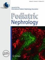
The light micrograph (Fig. 1) shows nodular glomerulosclerosis in 13 of 18 non-obsolescent glomeruli. Nineteen of 37 total glomeruli were obsolescent. The differential diagnosis of nodular glomerulosclerosis includes diabetic nephropathy (that may be associated with cystic fibrosis after years of pancreatic insufficiency), chronic membranoproliferative glomerulonephritis, dysproteinemias (amyloidosis, monoclonal immunoglobulin deposition), organized glomerular deposition diseases (fibrillary glomerulonephritis, immunotactoid glomerulonephritis, fibronectin glomerulopathy, collagen III glomerulopathy), chronic hypoxic or ischemic conditions or idiopathic nodular glomerulosclerosis, often associated with smoking and chronic hypertension [1, 2]. In this patient, the nodular lesions demonstrated apple-green birefringence in sections stained with Congo red and examined with polarized light. The nodules stained positively with monoclonal antibodies directed against AA amyloid protein using histochemical techniques. Immunofluorescence revealed 3+ staining intensity for kappa and lambda light chains and immunoglobulin M. Electron microscopy showed haphazardly arranged fibrils characteristic of amyloid A protein amyloidosis (AA amyloidosis).
Fig. 1
Light photomicrograph of renal biopsy sample stained with hematoxylin and eosin
×
2.
In secondary amyloidosis, evaluation for underlying chronic inflammatory and infectious disorders should be pursued.
AA amyloidosis is rare in children and young adults, and is typically associated with chronic underlying infectious or inflammatory conditions, including juvenile idiopathic arthritis (JIA), inflammatory bowel disease (IBD) and Familial Mediterranean fever (FMF). Our patient did not have clinical symptoms or pathologic findings of JIA or IBD, but biopsies obtained during esophagoduodenoscopy and colonoscopy revealed gastric active Helicobacter pylori infection (which was treated). Genetic testing for familial inflammatory conditions (MEFV and TNFRSF1A genes) were normal. His kidney biopsy included chronic lymphocytic infiltrate, suggesting that chronic pyelonephritis may be the cause of his amyloidosis, although this is rare. The underlying etiology is not identified in approximately 7 % of patients with confirmed renal AA amyloidosis [3].
3.
Treatment options for secondary amyloidosis include treatment of the underlying condition if present, colchicine and eprodisate.
Treatment for AA amyloidosis hinges on quiescence of the underlying inflammatory condition to reduce production of the serum AA protein. The treatment regimen requires antibiotics in chronic infections and immune modulation in inflammatory conditions. Colchicine is used in FMF to decrease amyloid production, and interleukin-1 inhibitors are often used to treat underlying autoinflammatory conditions, when one is identified. Data on an investigational medication, eprodisate, which prevents polymerization of amyloid fibrils and prevents progression of renal AA amyloidosis is promising [4].
Our patient has had persistent nephrotic range proteinuria with fluctuating levels of renal function (serum creatinine 1.3–1.6 mg/dL). He was treated with renin–angiotensin system inhibition, oral antibiotics followed by topical gentamicin to sterilize his urinary tract, and colchicine empirically to reduce serum amyloid A (SAA) production. Enrollment for treatment with eprodisate was offered, but his family declined. His urine total protein to creatinine ratio has been persistently elevated, but serum albumin has normalized at 4.6 mg/dL. His C-reactive protein has normalized, but C3 remains persistently low for unclear reasons.
…
Anzeige
Bitte loggen Sie sich ein, um Zugang zu diesem Inhalt zu erhalten