Erschienen in: Der Pathologe 2/2020

21.02.2020 | Knochentumoren | Schwerpunkt: Tumoren des Knochens und der Gelenke

Riesenzelltumor des Knochens

Morphologie, molekulare Pathogenese und Differenzialdiagnose

verfasst von: Albert Roessner, Maria Smolle, Johannes Haybäck

Erschienen in: Die Pathologie | Ausgabe 2/2020

Einloggen, um Zugang zu erhalten

Zusammenfassung

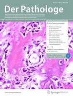
Das histologische Bild des Riesenzelltumors des Knochens ist charakterisiert durch die zahlreichen osteoklastenähnlichen Riesenzellen. Diese sind aber nicht die eigentlichen Tumorzellen, sondern ein reaktives Infiltrat. Die Tumorzellen sind vielmehr mononukleäre mesenchymale Zellen, die sogar eine osteoblastische Liniendifferenzierung erkennen lassen. Die zweite Gruppe unter den mononukleären Zellen sind reaktive CD68-positive Makrophagen. An der Aktivierung der Riesenzellen ist entscheidend das RANK/RANKL-System („receptor activator of nuclear factor-kappa B ligand“) beteiligt, das zu der Tumor-Necrosis-Factor-Cytokine-Familie gehört. Es ist allgemein akzeptiert, dass eine RANKL-Expression der mononukleären Stromazellen für die Entstehung und Differenzierung der osteoklastenähnlichen Riesenzellen verantwortlich ist. Daher ist verständlich, dass der RANKL-Inhibitor Denosumab in den letzten Jahren ein wesentliches Element für die Therapie des Riesenzelltumors dargestellte, da er die Reifung der Osteoklasten und damit die osteolytische Aktivität und die Ausbreitung des Tumors blockiert. Allerdings hat sich gezeigt, dass die nicht ganz nebenwirkungsfreie Therapie mit Denosumab bei Absetzen derselben zu einem ausgeprägten Rezidiv führen kann, weshalb in neuester Zeit von dieser Therapie eher Abstand genommen wird.
Molekulargenetisch sind die Riesenzelltumoren des Knochens durch Punktmutationen im H3F3A-Gen charakterisiert. Der Nachweis dieser Mutation dient zur diagnostischen Abgrenzung von anderen riesenzellhaltigen Knochenläsionen. Das riesenzellhaltige Osteosarkom enthält nur sehr selten H3F3A-Mutationen. Das Chondroblastom ist durch Mutationen im H3F3B-Gen charakterisiert.
Literatur
1.
Zurück zum Zitat Allen CE, Li L, Peters TL et al (2010) Cell-specific gene expression in Langerhans cell histiocytosis lesions reveals a distinct profile compared with epidermal Langerhans cells. J Immunol 284:4557–4567 Allen CE, Li L, Peters TL et al (2010) Cell-specific gene expression in Langerhans cell histiocytosis lesions reveals a distinct profile compared with epidermal Langerhans cells. J Immunol 284:4557–4567
2.
Zurück zum Zitat Akpalo H, Lange C, Zustin J et al (2012) Discovered on gastrointestinal stromal tumour 1 (DOG1): a useful immunohistochemical marker for diagnosing chondroblastoma. Histopathology 60:1099–1106PubMed Akpalo H, Lange C, Zustin J et al (2012) Discovered on gastrointestinal stromal tumour 1 (DOG1): a useful immunohistochemical marker for diagnosing chondroblastoma. Histopathology 60:1099–1106PubMed
3.
Zurück zum Zitat Amary MF, Berisha F, Mozela R et al (2016) The H3F3 K36M mutant antibody is a sensitive and specific marker for the diagnosis of chondroblastoma. Histopathology 69:121–127PubMed Amary MF, Berisha F, Mozela R et al (2016) The H3F3 K36M mutant antibody is a sensitive and specific marker for the diagnosis of chondroblastoma. Histopathology 69:121–127PubMed
4.
Zurück zum Zitat Amary F, Berisha F, Ye H et al (2017) H3F3A (Histone 3.3) G34W immunohistochemistry. Am J Surg Pathol 41:1059–1068PubMedPubMedCentral Amary F, Berisha F, Ye H et al (2017) H3F3A (Histone 3.3) G34W immunohistochemistry. Am J Surg Pathol 41:1059–1068PubMedPubMedCentral
5.
Zurück zum Zitat Annels NE, Da Costa CE, Prins FA et al (2003) Aberrant chemokine receptor expression and chemokine production by Langerhans cells underlies the pathogenesis of Langerhans cell histiocytosis. J Exp Med 197:1385–1390PubMedPubMedCentral Annels NE, Da Costa CE, Prins FA et al (2003) Aberrant chemokine receptor expression and chemokine production by Langerhans cells underlies the pathogenesis of Langerhans cell histiocytosis. J Exp Med 197:1385–1390PubMedPubMedCentral
6.
Zurück zum Zitat Atkins GJ, Haynes DR, Graves SE et al (2010) Expression of osteoclast differentiation signals by stromal elements of giant cell tumors. J Bone Miner Res 15:640–649 Atkins GJ, Haynes DR, Graves SE et al (2010) Expression of osteoclast differentiation signals by stromal elements of giant cell tumors. J Bone Miner Res 15:640–649
7.
Zurück zum Zitat Badalian-Very G, Vergilio J, Degar BA et al (2019) Recurrent BRAF mutations in Langerhans cell histiocytosis. Blood 116:1919–1923 Badalian-Very G, Vergilio J, Degar BA et al (2019) Recurrent BRAF mutations in Langerhans cell histiocytosis. Blood 116:1919–1923
8.
Zurück zum Zitat Balke M, Schremper L, Gebert C et al (2008) Giant cell tumor of bone: treatment and outcome of 214 cases. J Cancer Res Clin Oncol 134:969–978PubMed Balke M, Schremper L, Gebert C et al (2008) Giant cell tumor of bone: treatment and outcome of 214 cases. J Cancer Res Clin Oncol 134:969–978PubMed
9.
Zurück zum Zitat Baruffi MR, Neto JB, Barbieri CH et al (2001) Aneurysmal bone cyst with chromosomal changes involving 7q and 16p. Cancer Genet Cytogenet 129:177–180PubMed Baruffi MR, Neto JB, Barbieri CH et al (2001) Aneurysmal bone cyst with chromosomal changes involving 7q and 16p. Cancer Genet Cytogenet 129:177–180PubMed
10.
Zurück zum Zitat Baumhoer D, Kovac M, Sperveslage J, Ameline B et al (2019) Activating mutations in the MAP-kinase pathway define non-ossifying fibroma of bone. J Pathol 248:116–122PubMed Baumhoer D, Kovac M, Sperveslage J, Ameline B et al (2019) Activating mutations in the MAP-kinase pathway define non-ossifying fibroma of bone. J Pathol 248:116–122PubMed
11.
Zurück zum Zitat Behjati S, Tarpey PS, Presneau N et al (2013) Distinct H3F3A and H3F3B driver mutations define chondroblastoma and giant cell tumor of bone. Nat Genet 45:1479–1482PubMed Behjati S, Tarpey PS, Presneau N et al (2013) Distinct H3F3A and H3F3B driver mutations define chondroblastoma and giant cell tumor of bone. Nat Genet 45:1479–1482PubMed
12.
Zurück zum Zitat Bernstrand C, Sandscedt B, Ahstrom L et al (2005) Long-term follow-up of Langerhans cell histiocytosis: 39 years’ experience at a single centre. Acta Paediatr 94:1073–1084PubMed Bernstrand C, Sandscedt B, Ahstrom L et al (2005) Long-term follow-up of Langerhans cell histiocytosis: 39 years’ experience at a single centre. Acta Paediatr 94:1073–1084PubMed
13.
Zurück zum Zitat Berres M, Phaik K, Lim H et al (2014) BRAF-V600E expression in precursor versus differentiated dendritic cells defines clinically distinct LCH risk group. J Exp Med 211:669–683PubMedPubMedCentral Berres M, Phaik K, Lim H et al (2014) BRAF-V600E expression in precursor versus differentiated dendritic cells defines clinically distinct LCH risk group. J Exp Med 211:669–683PubMedPubMedCentral
14.
Zurück zum Zitat Bertoni F, Bacchini P, Staats EL (2003) Malignancy in giant cell tumor of bone. Cancer 97:2520–2529PubMed Bertoni F, Bacchini P, Staats EL (2003) Malignancy in giant cell tumor of bone. Cancer 97:2520–2529PubMed
15.
Zurück zum Zitat Biesecker JL, Marcove RC, Huvos AG et al (1970) Aneurysmal bone cyst. A clinicopathologic study of 66 cases. Cancer 26:615–625PubMed Biesecker JL, Marcove RC, Huvos AG et al (1970) Aneurysmal bone cyst. A clinicopathologic study of 66 cases. Cancer 26:615–625PubMed
16.
Zurück zum Zitat Bowers LM, Cohen DM, Bhattacharyya I et al (2013) The non-ossifying fibroma: a case report and review of the literature. Head Neck Pathol 7:203–210PubMed Bowers LM, Cohen DM, Bhattacharyya I et al (2013) The non-ossifying fibroma: a case report and review of the literature. Head Neck Pathol 7:203–210PubMed
17.
Zurück zum Zitat Boyce BF, Xing L (2008) Functions of RANKL/RANK/OPG in bone modeling and remodeling. Arch Biochem Biophys 473:139–146PubMedPubMedCentral Boyce BF, Xing L (2008) Functions of RANKL/RANK/OPG in bone modeling and remodeling. Arch Biochem Biophys 473:139–146PubMedPubMedCentral
18.
Zurück zum Zitat Chen W, DiFrancesco LM (2017) Chondroblastoma an update. Arch Pathol Lab Med 141:867–871PubMed Chen W, DiFrancesco LM (2017) Chondroblastoma an update. Arch Pathol Lab Med 141:867–871PubMed
19.
Zurück zum Zitat Collin M, Bigley V, McClain KL et al (2015) Cells of origin o f Langerhans cell histiocytosis. Hematol Oncol Clin North Am 29:825–838PubMedPubMedCentral Collin M, Bigley V, McClain KL et al (2015) Cells of origin o f Langerhans cell histiocytosis. Hematol Oncol Clin North Am 29:825–838PubMedPubMedCentral
20.
Zurück zum Zitat de Groot AF, Appelman-Dijkstra NM, van der Burg SH et al (2018) The anti-tumor effect of RANKL inhibition in malignant solid tumors—a systematic review. Cancer Treat Rev 62:18–28PubMed de Groot AF, Appelman-Dijkstra NM, van der Burg SH et al (2018) The anti-tumor effect of RANKL inhibition in malignant solid tumors—a systematic review. Cancer Treat Rev 62:18–28PubMed
21.
Zurück zum Zitat Dahlin DC, Ivins JC (1972) Benign chondroblastoma. A study of 125 cases. Cancer 30:401–413PubMed Dahlin DC, Ivins JC (1972) Benign chondroblastoma. A study of 125 cases. Cancer 30:401–413PubMed
22.
Zurück zum Zitat de Silva MV, Reid R (2003) Chondroblastoma: varied histologic appearance, potential diagnostic pitfalls, and clinicopathologic features associated with local recurrence. Ann Diagn Pathol 7:2052013 de Silva MV, Reid R (2003) Chondroblastoma: varied histologic appearance, potential diagnostic pitfalls, and clinicopathologic features associated with local recurrence. Ann Diagn Pathol 7:2052013
23.
Zurück zum Zitat Fadare O (2002) Benign metastasizing giant cell tumor. Arch Pathol Lab Med 126:1133–1134PubMed Fadare O (2002) Benign metastasizing giant cell tumor. Arch Pathol Lab Med 126:1133–1134PubMed
24.
Zurück zum Zitat Girolami I, Mancini I, Simoni A et al (2016) Denosumab treated giant cell tumour of bone: a morphological, immunohistochemical and molecular analysis of a series. J Clin Pathol 69:240–247PubMed Girolami I, Mancini I, Simoni A et al (2016) Denosumab treated giant cell tumour of bone: a morphological, immunohistochemical and molecular analysis of a series. J Clin Pathol 69:240–247PubMed
25.
Zurück zum Zitat Golding SR, Roelke MS, Petrison KK et al (1987) Human giant cell tumor of bone: identification and characterisation of cell types. J Clin Invest 79:483–491 Golding SR, Roelke MS, Petrison KK et al (1987) Human giant cell tumor of bone: identification and characterisation of cell types. J Clin Invest 79:483–491
26.
Zurück zum Zitat Gomes CC, Gayden T, Bajic A et al (2018) TRPV4 and KRAS and FGFR1 gain-of-function mutations drive giant cell lesions of the jaw. Nat Commun 9:4572PubMedPubMedCentral Gomes CC, Gayden T, Bajic A et al (2018) TRPV4 and KRAS and FGFR1 gain-of-function mutations drive giant cell lesions of the jaw. Nat Commun 9:4572PubMedPubMedCentral
27.
Zurück zum Zitat Gomes CC, Gomez R (2019) MAPK pathway-activating mutations drive giant cell lesions of the jaws and non-ossifying fibromas of bone. J Pathol 248:123–124PubMed Gomes CC, Gomez R (2019) MAPK pathway-activating mutations drive giant cell lesions of the jaws and non-ossifying fibromas of bone. J Pathol 248:123–124PubMed
28.
Zurück zum Zitat Hakozaki M, Tajino T, Yamada H (2014) Radiological and pathological characteristics of giant cell tumor of bone treated with denosumab. Diagn Pathol 9:2–7 Hakozaki M, Tajino T, Yamada H (2014) Radiological and pathological characteristics of giant cell tumor of bone treated with denosumab. Diagn Pathol 9:2–7
29.
Zurück zum Zitat Haroche J, Cohen-Aubart F, Emile JF et al (2013) Dramatic efficacy of vemurafenib in both multisystemic and refractory Erdheim-Chester disease and Langerhans cell histiocytosis harboring the BRAF V600E mutation. Blood 121:1495–1501PubMed Haroche J, Cohen-Aubart F, Emile JF et al (2013) Dramatic efficacy of vemurafenib in both multisystemic and refractory Erdheim-Chester disease and Langerhans cell histiocytosis harboring the BRAF V600E mutation. Blood 121:1495–1501PubMed
30.
Zurück zum Zitat Héritier S, Emile J, Barkaoui M et al (2016) BRAF mutation correlates with high-risk Langerhans cell histiocytosis and increased resistance to first-line therapy. J Clin Oncol 34:3023–3030PubMedPubMedCentral Héritier S, Emile J, Barkaoui M et al (2016) BRAF mutation correlates with high-risk Langerhans cell histiocytosis and increased resistance to first-line therapy. J Clin Oncol 34:3023–3030PubMedPubMedCentral
31.
Zurück zum Zitat Héritier S, Hélias-Rodzewicz Z, Chakraborty R et al (2017) New somatic BRAF splicing mutation in Langerhans cell histiocytosis. Mol Cancer 16:115PubMedPubMedCentral Héritier S, Hélias-Rodzewicz Z, Chakraborty R et al (2017) New somatic BRAF splicing mutation in Langerhans cell histiocytosis. Mol Cancer 16:115PubMedPubMedCentral
33.
Zurück zum Zitat James IE, Dodds RA, Olivera DL et al (1996) Human osteoclastoma-derived stromal cells: correlation of the ability to form mineralized nodules in vitro with formation of bone in vivo. J Bone Miner Res 11:1453–1460PubMed James IE, Dodds RA, Olivera DL et al (1996) Human osteoclastoma-derived stromal cells: correlation of the ability to form mineralized nodules in vitro with formation of bone in vivo. J Bone Miner Res 11:1453–1460PubMed
34.
Zurück zum Zitat Kapoor SK, Jain V, Agrawal M et al (2007) Primary malignant giant cell tumor of bone: a series of three rare cases. J Surg Orthop Adv 16:89–92PubMed Kapoor SK, Jain V, Agrawal M et al (2007) Primary malignant giant cell tumor of bone: a series of three rare cases. J Surg Orthop Adv 16:89–92PubMed
35.
Zurück zum Zitat Kato I, Furuya M, Matsuo K et al (2018) Giant cell tumours of bone treated with denosumab: histological, immunohistochemical and H3F3A mutation analyses. Histopathology 72:914–922PubMed Kato I, Furuya M, Matsuo K et al (2018) Giant cell tumours of bone treated with denosumab: histological, immunohistochemical and H3F3A mutation analyses. Histopathology 72:914–922PubMed
36.
Zurück zum Zitat Kauzman A, Li SQ, Bradley G et al (2003) Cyclin alterations in giant cell tumor of bone. Mod Pathol 16:210–218PubMed Kauzman A, Li SQ, Bradley G et al (2003) Cyclin alterations in giant cell tumor of bone. Mod Pathol 16:210–218PubMed
37.
Zurück zum Zitat Kilpatrick SE, Wenger DE, Gilchrist GS et al (1995) Langerhans’ cell histiocytosis (histiocytosis X) of bone—a clinicopathologic analysis of 263 pediatric and adult cases. Cancer 76:2471–2484PubMed Kilpatrick SE, Wenger DE, Gilchrist GS et al (1995) Langerhans’ cell histiocytosis (histiocytosis X) of bone—a clinicopathologic analysis of 263 pediatric and adult cases. Cancer 76:2471–2484PubMed
38.
Zurück zum Zitat Kobayashi M (2018) Langerhans cell histiocytosis in adults : advances in pathophysiology and treatment. Cancer Sci 109:3707–3713PubMedPubMedCentral Kobayashi M (2018) Langerhans cell histiocytosis in adults : advances in pathophysiology and treatment. Cancer Sci 109:3707–3713PubMedPubMedCentral
39.
Zurück zum Zitat Koelsche C, Schrimpf D, Tharun L et al (2017) Histone 3.3 hotspot mutations in conventional osteosarcomas: a comprehensive clinical and molecular characterization of six H3F3A mutated cases. Clin Sarcoma Res 7:9PubMedPubMedCentral Koelsche C, Schrimpf D, Tharun L et al (2017) Histone 3.3 hotspot mutations in conventional osteosarcomas: a comprehensive clinical and molecular characterization of six H3F3A mutated cases. Clin Sarcoma Res 7:9PubMedPubMedCentral
40.
Zurück zum Zitat Konishi E, Nakashima Y, Iwasa Y et al (2010) Immunohistochemical analysis for Sox9 reveals the cartilaginous character of chondroblastoma and chondromyxoid fibroma of bone. Hum Pathol 41:208–213PubMed Konishi E, Nakashima Y, Iwasa Y et al (2010) Immunohistochemical analysis for Sox9 reveals the cartilaginous character of chondroblastoma and chondromyxoid fibroma of bone. Hum Pathol 41:208–213PubMed
41.
Zurück zum Zitat Kumar R, Angelini S, Snellman E et al (2004) BRAF mutations are common somatic events in melanocytic nevi. J Invest Dermatol 122:342–348PubMed Kumar R, Angelini S, Snellman E et al (2004) BRAF mutations are common somatic events in melanocytic nevi. J Invest Dermatol 122:342–348PubMed
42.
Zurück zum Zitat Li HR, Tai CF, Huang HY et al (2018) USP6 gene rearrangement differentiates primary paranasal sinus solid aneurysmal bone cyst from other giant cell–rich lesions: report of a rare case. Hum Pathol 76:117–121PubMed Li HR, Tai CF, Huang HY et al (2018) USP6 gene rearrangement differentiates primary paranasal sinus solid aneurysmal bone cyst from other giant cell–rich lesions: report of a rare case. Hum Pathol 76:117–121PubMed
43.
Zurück zum Zitat Lowe BR, Maxham LA, Hamey JJ et al (2019) Histone H3 mutations: an updated view of their role in chromatin deregulation and cancer. Cancers 11:1–24 Lowe BR, Maxham LA, Hamey JJ et al (2019) Histone H3 mutations: an updated view of their role in chromatin deregulation and cancer. Cancers 11:1–24
44.
Zurück zum Zitat Lu C, Ramirez D, Hwang S et al (2019) Histone H3K36M mutation and trimethylation patterns in chondroblastoma. Histopathology 74:291–299PubMed Lu C, Ramirez D, Hwang S et al (2019) Histone H3K36M mutation and trimethylation patterns in chondroblastoma. Histopathology 74:291–299PubMed
45.
Zurück zum Zitat Matsuzaki K, Udagawa N, Takahashi N et al (1998) Osteoclast differentiation factor (ODF) induces osteoclast-like cell formation in human peripheral blood mononuclear cell cultures. Biochem Biophys Res Commun 246:199–204PubMed Matsuzaki K, Udagawa N, Takahashi N et al (1998) Osteoclast differentiation factor (ODF) induces osteoclast-like cell formation in human peripheral blood mononuclear cell cultures. Biochem Biophys Res Commun 246:199–204PubMed
46.
Zurück zum Zitat Monda L, Wick M (1985) S‑100 protein immunostaining in the differential diagnosis of chondroblastoma. Hum Pathol 16:287–293PubMed Monda L, Wick M (1985) S‑100 protein immunostaining in the differential diagnosis of chondroblastoma. Hum Pathol 16:287–293PubMed
47.
Zurück zum Zitat Morgan T, Atkins GJ, Trivett MK et al (2005) Molecular profiling of giant cell tumor of bone and the osteoclastic localization of ligand for receptor activator of nuclear factor κB. Am J Pathol 167:117–128PubMedPubMedCentral Morgan T, Atkins GJ, Trivett MK et al (2005) Molecular profiling of giant cell tumor of bone and the osteoclastic localization of ligand for receptor activator of nuclear factor κB. Am J Pathol 167:117–128PubMedPubMedCentral
48.
49.
Zurück zum Zitat Muheremu A, Huang Z, Niu X (2015) Treatment for giant cell tumor of the spine metastasizing to the lung: a report of two cases and a literature review. Oncol Lett 9:1321–1326PubMed Muheremu A, Huang Z, Niu X (2015) Treatment for giant cell tumor of the spine metastasizing to the lung: a report of two cases and a literature review. Oncol Lett 9:1321–1326PubMed
50.
Zurück zum Zitat Noh BJ, Park YK (2018) Giant cell tumor of bone: updated molecular pathogenesis and tumor biology. Hum Pathol 81:1–8PubMed Noh BJ, Park YK (2018) Giant cell tumor of bone: updated molecular pathogenesis and tumor biology. Hum Pathol 81:1–8PubMed
51.
Zurück zum Zitat Nohr E, Lee LH, Cates JM et al (2017) Diagnostic value of histone 3 mutations in osteoclast-rich bone tumors. Hum Pathol 68:119–127PubMed Nohr E, Lee LH, Cates JM et al (2017) Diagnostic value of histone 3 mutations in osteoclast-rich bone tumors. Hum Pathol 68:119–127PubMed
52.
Zurück zum Zitat Oliveira AM, Perez-Atayde AR, Inwards CY et al (2004a) USP6 and CDH11 oncogenes identify the neoplastic cell in primary aneurysmal bone cysts and are absent in so-called secondary aneurysmal bone cysts. Am J Pathol 165:1773–1780PubMedPubMedCentral Oliveira AM, Perez-Atayde AR, Inwards CY et al (2004a) USP6 and CDH11 oncogenes identify the neoplastic cell in primary aneurysmal bone cysts and are absent in so-called secondary aneurysmal bone cysts. Am J Pathol 165:1773–1780PubMedPubMedCentral
53.
Zurück zum Zitat Oliveira AM, Hsi B, Weremowicz S et al (2004b) USP6 (Tre2) fusion oncogenes in aneurysmal bone cyst. Cancer Res 64:1920–1923PubMed Oliveira AM, Hsi B, Weremowicz S et al (2004b) USP6 (Tre2) fusion oncogenes in aneurysmal bone cyst. Cancer Res 64:1920–1923PubMed
54.
Zurück zum Zitat Oliveira AM, Perez-Atayde AR, Cin PD et al (2005) Aneurysmal bone cyst variant translocations upregulate USP6 transcription by promoter swapping with the ZNF9, COL1A1, TRAP150, and OMD genes. Oncogene 24:3419–3342PubMed Oliveira AM, Perez-Atayde AR, Cin PD et al (2005) Aneurysmal bone cyst variant translocations upregulate USP6 transcription by promoter swapping with the ZNF9, COL1A1, TRAP150, and OMD genes. Oncogene 24:3419–3342PubMed
55.
Zurück zum Zitat Oliveira AM, Chou MM (2014) USP6-induced neoplasms: the biologic spectrum of aneurysmal bone cyst and nodular fasciitis. Hum Pathol 45:1–11PubMed Oliveira AM, Chou MM (2014) USP6-induced neoplasms: the biologic spectrum of aneurysmal bone cyst and nodular fasciitis. Hum Pathol 45:1–11PubMed
56.
Zurück zum Zitat Oliveira AM, Chou MM (2012) The TRE17/USP6 oncogene: a riddle wrapped in a mystery inside an enigma. Front Biosci 4:321–334 Oliveira AM, Chou MM (2012) The TRE17/USP6 oncogene: a riddle wrapped in a mystery inside an enigma. Front Biosci 4:321–334
57.
Zurück zum Zitat Palmerini E, Picci P, Reichardt P et al (2019) Malignancy in giant cell tumor of bone: a review of the literature. Technol Cancer Res Treat 18:1–9 Palmerini E, Picci P, Reichardt P et al (2019) Malignancy in giant cell tumor of bone: a review of the literature. Technol Cancer Res Treat 18:1–9
58.
Zurück zum Zitat Panoutsakopoulos G, Pandis N, Kyriazoglou I et al (1999) Recurrent t(16;17)(q22;p13) in aneurysmal bone cysts. Genes Chromosomes Cancer 126:265–276 Panoutsakopoulos G, Pandis N, Kyriazoglou I et al (1999) Recurrent t(16;17)(q22;p13) in aneurysmal bone cysts. Genes Chromosomes Cancer 126:265–276
59.
Zurück zum Zitat Presneau N, Baumhoer D, Behjati S et al (2015) Diagnostic value of H3F3A mutations in giant cell tumour of bone compared to osteoclast-rich mimics. J Pathol Clin Res 1:113–123PubMedPubMedCentral Presneau N, Baumhoer D, Behjati S et al (2015) Diagnostic value of H3F3A mutations in giant cell tumour of bone compared to osteoclast-rich mimics. J Pathol Clin Res 1:113–123PubMedPubMedCentral
60.
Zurück zum Zitat Raskin KA, Schwab JH, Mankin HJ et al (2013) Giant cell tumor of bone. J Am Acad Orthop Surg 21:118–126PubMed Raskin KA, Schwab JH, Mankin HJ et al (2013) Giant cell tumor of bone. J Am Acad Orthop Surg 21:118–126PubMed
61.
Zurück zum Zitat Rehkämper J, Steinestel K, Jeiler B et al (2018) Diagnostic tools in the differential diagnosis of giant cell-rich lesions of bone at biopsy. Oncotarget 9:30106–30114PubMedPubMedCentral Rehkämper J, Steinestel K, Jeiler B et al (2018) Diagnostic tools in the differential diagnosis of giant cell-rich lesions of bone at biopsy. Oncotarget 9:30106–30114PubMedPubMedCentral
62.
Zurück zum Zitat Righi A, Mancini I, Gambarotti M et al (2017) Histone 3.3 mutations in giant cell tumor and giant cell–rich sarcomas of bone. Hum Pathol 68:128–135PubMed Righi A, Mancini I, Gambarotti M et al (2017) Histone 3.3 mutations in giant cell tumor and giant cell–rich sarcomas of bone. Hum Pathol 68:128–135PubMed
63.
Zurück zum Zitat Ritschl P, Karnel F, Hajek P (1988) Fibrous metaphyseal defects—determination of their origin and natural history using a radiomorphological study. Skeletal Radiol 17:8–15PubMed Ritschl P, Karnel F, Hajek P (1988) Fibrous metaphyseal defects—determination of their origin and natural history using a radiomorphological study. Skeletal Radiol 17:8–15PubMed
64.
Zurück zum Zitat Rock MG, Sim FH, Unni KK et al (1986) Secondary malignant giant-cell tumor of bone. Clinicopathological assessment of nineteen patients. J Bone Joint Surg Am 68:1073–1079PubMed Rock MG, Sim FH, Unni KK et al (1986) Secondary malignant giant-cell tumor of bone. Clinicopathological assessment of nineteen patients. J Bone Joint Surg Am 68:1073–1079PubMed
65.
Zurück zum Zitat Roessner A, von Bassewitz DB, Schlake W et al (1984) Biologic characterization of human bone tumors. III. Giant cell tumor of bone. A combined electron microscopical, histochemical, and autoradiographical study. Pathol Res Pract 178:431–440PubMed Roessner A, von Bassewitz DB, Schlake W et al (1984) Biologic characterization of human bone tumors. III. Giant cell tumor of bone. A combined electron microscopical, histochemical, and autoradiographical study. Pathol Res Pract 178:431–440PubMed
66.
Zurück zum Zitat Sangle NA, Layfield LJ (2012) Telangiectatic osteosarcoma. Arch Pathol Lab Med 136:572–576PubMed Sangle NA, Layfield LJ (2012) Telangiectatic osteosarcoma. Arch Pathol Lab Med 136:572–576PubMed
67.
Zurück zum Zitat Santini D, Perrone G, Roato I et al (2011) Expression pattern of receptor activator of NFκB (RANK) in a series of primary solid tumors and related bone metastases. J Cell Physiol 226:780–784PubMed Santini D, Perrone G, Roato I et al (2011) Expression pattern of receptor activator of NFκB (RANK) in a series of primary solid tumors and related bone metastases. J Cell Physiol 226:780–784PubMed
68.
Zurück zum Zitat Sasaki H, Nagano S, Shimada H et al (2017) Diagnosing and discriminating between primary and secondary aneurysmal bone cysts. Oncol Lett 13:2290–2296PubMedPubMedCentral Sasaki H, Nagano S, Shimada H et al (2017) Diagnosing and discriminating between primary and secondary aneurysmal bone cysts. Oncol Lett 13:2290–2296PubMedPubMedCentral
69.
Zurück zum Zitat Scheil-Bertram S, Hartwig E, Bruderlein S et al (2004) Metachronous and multiple aneurysmal bone cysts: a rare variant of primary aneurysmal bone cysts. Virchows Arch 444:293–299PubMed Scheil-Bertram S, Hartwig E, Bruderlein S et al (2004) Metachronous and multiple aneurysmal bone cysts: a rare variant of primary aneurysmal bone cysts. Virchows Arch 444:293–299PubMed
70.
Zurück zum Zitat Szuhai K, Cleton-Jansen AM, Hoendoorn PCW et al (2012) Molecular pathology and its diagnostic use in bone tuors. Cancer Genet 205:193–204PubMed Szuhai K, Cleton-Jansen AM, Hoendoorn PCW et al (2012) Molecular pathology and its diagnostic use in bone tuors. Cancer Genet 205:193–204PubMed
71.
Zurück zum Zitat Winnepenninckx V, Debiec-Rychter M, Jorissen M et al (2001) Aneurysmal bone cyst of the nose with 17p13 involvement. Virchows Arch 439:636–639PubMed Winnepenninckx V, Debiec-Rychter M, Jorissen M et al (2001) Aneurysmal bone cyst of the nose with 17p13 involvement. Virchows Arch 439:636–639PubMed
72.
Zurück zum Zitat van der Heijden L, Dijkstra PDS, van de Sande MAJ et al (2014) The clinical approach toward giant cell tumor of bone. Oncologist 19:550–561PubMedPubMedCentral van der Heijden L, Dijkstra PDS, van de Sande MAJ et al (2014) The clinical approach toward giant cell tumor of bone. Oncologist 19:550–561PubMedPubMedCentral
73.
Zurück zum Zitat van der Heijden L, Dijkstra PDS, Blay JY et al (2017) Giant cell tumour of bone in the denosumab era. Eur J Cancer 77:75–83PubMed van der Heijden L, Dijkstra PDS, Blay JY et al (2017) Giant cell tumour of bone in the denosumab era. Eur J Cancer 77:75–83PubMed
74.
Zurück zum Zitat Varadi Z, Kertész G, Csóka M (2017) Effective BRAF inhibitor vemurafenib therapy in a 2-year-old patient with sequentially diagnosed Langerhans cell histiocytosis and Erdheim—Chester disease. Onco Targets Ther 10:521–526PubMedPubMedCentral Varadi Z, Kertész G, Csóka M (2017) Effective BRAF inhibitor vemurafenib therapy in a 2-year-old patient with sequentially diagnosed Langerhans cell histiocytosis and Erdheim—Chester disease. Onco Targets Ther 10:521–526PubMedPubMedCentral
75.
Zurück zum Zitat Wada T, Nakashima T, Hiroshi N et al (2006) RANKL-RANK signaling in osteoclastogenesis and bone disease. Trends Mol Med 12:17–25PubMed Wada T, Nakashima T, Hiroshi N et al (2006) RANKL-RANK signaling in osteoclastogenesis and bone disease. Trends Mol Med 12:17–25PubMed
76.
Zurück zum Zitat Wick MR, McDermott MB, Swanson PE (2014) Proliferative, reparative, and reactive benign bone lesions that may be confused diagnostically with true osseous neoplasms. Semin Diagn Pathol 31:66–88PubMed Wick MR, McDermott MB, Swanson PE (2014) Proliferative, reparative, and reactive benign bone lesions that may be confused diagnostically with true osseous neoplasms. Semin Diagn Pathol 31:66–88PubMed
77.
Zurück zum Zitat Wülling M, Delling G, Kaiser E (2003) The origin of the neoplastic stromal cell in giant cell tumor of bone. Hum Pathol 34:983–993PubMed Wülling M, Delling G, Kaiser E (2003) The origin of the neoplastic stromal cell in giant cell tumor of bone. Hum Pathol 34:983–993PubMed
78.
Zurück zum Zitat Yamamoto H, Iwasaki T, Yamada Y (2018) Diagnostic utility of histone H3.3 G34W, G34R, and G34V mutant-specific antibodies for giant cell tumors of bone. Hum Pathol 73:41–50PubMed Yamamoto H, Iwasaki T, Yamada Y (2018) Diagnostic utility of histone H3.3 G34W, G34R, and G34V mutant-specific antibodies for giant cell tumors of bone. Hum Pathol 73:41–50PubMed
79.
Zurück zum Zitat Yayan Y (2019) Increased risk of lung metastases in patients with giant cell bone tumors: a systematic review. Adv Exp Med Biol 1176:1–17PubMed Yayan Y (2019) Increased risk of lung metastases in patients with giant cell bone tumors: a systematic review. Adv Exp Med Biol 1176:1–17PubMed
Metadaten
Titel
Riesenzelltumor des Knochens
Morphologie, molekulare Pathogenese und Differenzialdiagnose
verfasst von
Albert Roessner
Maria Smolle
Johannes Haybäck
Publikationsdatum
21.02.2020
Verlag
Springer Medizin
Erschienen in
Die Pathologie / Ausgabe 2/2020
Print ISSN: 2731-7188
Elektronische ISSN: 2731-7196
DOI
https://doi.org/10.1007/s00292-020-00760-5

Weitere Artikel der Ausgabe 2/2020

Der Pathologe 2/2020 Zur Ausgabe

Mitteilungen der Österreichischen Gesellschaft für Pathologie

Mitteilungen der Österreichischen Gesellschaft für Pathologie

CME Zertifizierte Fortbildung

Benigne Lebertumoren

Schwerpunkt: Tumoren des Knochens und der Gelenke

Chordome: Molekulargenetische Grundlagen für Diagnostik und Therapie

Schwerpunkt: Tumoren des Knochens und der Gelenke

Die Rolle von Interleukin-11 beim Osteosarkom

Schwerpunkt: Tumoren des Knochens und der Gelenke

Knorpeltumoren: Morphologie, Genetik und Basisaspekte der Targettherapie

Leitlinien kompakt für die Innere Medizin

Mit medbee Pocketcards sicher entscheiden.

Seit 2022 gehört die medbee GmbH zum Springer Medizin Verlag

Update Innere Medizin

Bestellen Sie unseren Fach-Newsletter und bleiben Sie gut informiert.