

Abstract
Recent developments in radiation therapy aimed at more precise dose delivery along with higher dose gradients (dose painting) and more efficient dose delivery with higher dose rates e.g. flattening filter free (FFF) irradiation.
Magnetic-resonance-imaging based polymer gel dosimetry offers 3D information for precise dose delivery techniques. Many of the proposed polymer gels have been reported to exhibit a dose response, measured as relaxation rate ΔR2(D), which is dose rate dependent. A lack of or a reduced dose-rate sensitivity is very important for dosimetric accuracy, especially with regard to the increasing clinical use of FFF irradiation protocols with LINACs at high dose rates. Some commonly used polymer gels are based on Methacrylic-Acid-Gel-Initiated-by-Copper (MAGIC). Here, we report on the dose sensitivity (ΔR2/ΔD) of MAGIC-type gels with different oxygen scavenger concentration for their specific dependence on the applied dose rate in order to improve the dosimetric performance, especially for high dose rates. A preclinical x-ray machine ('Yxlon', E = 200 kV) was used for irradiation to cover a range of dose rates from low  min = 0.6 Gy min−1 to high
  max = 18 Gy min−1. The dose response was evaluated using R2-imaging of the gel on a human high-field (7T) MR-scanner. The results indicate that all of the investigated dose rates had an impact on the dose response in polymer gel dosimeters, being strongest in the high dose region and less effective for low dose levels. The absolute dose rate dependence
  of the dose response in MAGIC-type gel is significantly reduced using higher concentrations of oxygen scavenger at the expense of reduced dose sensitivity. For quantitative dose evaluations the relative dose rate dependence of a polymer gel, normalized to its sensitivity is important. Based on this normalized sensitivity the dose rate sensitivity was reduced distinctly using an increased oxygen scavenger concentration with reference to standard MAGIC-type gel formulation at high dose rate levels. The proposed gel composition with high oxygen scavenger concentration exhibits a larger linear active dose response and might be used especially in FFF-radiation applications and preclinical dosimetry at high dose rates. We propose in general to use high dose rates for calibration and evaluation as the change in relative dose sensitivity is reduced at higher dose rates in all of the investigated gel types.
Export citation and abstract BibTeX RIS
Abbreviations
| Caa | Ascorbic acid concentration |
| D | Dose |
 | Dose rate |
| FFF | Flattening filter free |
| IMRT | Intensity modulated radiation therapy |
| LINAC | Linear accelerator |
| MA | Methacrylic acid |
| MAGIC | Methacrylic acid gel initiated by copper |
| MAGAT | Methacrylic acid gel and THPC |
| MRI | Magnetic resonance imaging |
| MRPD | Magnetic resonance imaging based polymer gel dosimetry |
| SSD | Source to surface distance |
| THPC | Tetrakis-hydroxy methyl phosphonium chloride |
| ROI | Region of interest |
| R2 | Transverse relaxation rate (R2 = 1/T2) |
| α | Dose sensitivity = ΔR2/ΔD |
1. Introduction
Today's advanced radiotherapy technology enables the delivery of very high dose levels in complex beam shapes with very steep dose gradients (Schreiner 2009, Wong et al 2012, Keshtkar et al 2014). The underlying technology advancements have prompted the development of pre-clinical precision irradiators, which can mimic clinically advanced radiotherapy treatments in an experimental setting, including image guidance (Verhaegen et al 2011, Lindsay et al 2014, Tillner et al 2016). Both clinical and pre-clinical precision radiotherapy technology and techniques challenge dosimetry with respect to small fields and high dose rates.
Conventional dosimeters such as ionization chambers, thermal luminescence devices, diodes, and film dosimeters are unable to measure small dose distributions in three dimensions (3D) within a single experiment. Hence, polymer gel dosimeters became an interesting alternative for measuring the 3D-dose distribution not only at voxel size of 1 × 1 × 1 mm3 but also at sub-mm spatial resolution (Berg et al 2001, 2004, Bayreder et al 2008, Pappas et al 2008, Babic et al 2009). Moreover, gel dosimeters also have the advantage of tissue equivalence and energy independent properties (Maryanski et al 1994, De Deene et al 1998, De Deene 2004, Pappas et al 2008, Baldock et al 2010).
Different gel dosimeters of various compositions and ingredients have been studied (Baldock et al 1998, Fong et al 2001, Senden et al 2006, Baldock 2009). The polymerization due to irradiation is very sensitive to oxygen exposure in the gel (De Deene et al 2001, 2002, 2006, Fong et al 2001, Bayreder et al 2006, Senden et al 2006). The idea of adding oxygen scavenging ingredients to the polymer gel led to the development of a new type of polymer gel: Methacrylic Acid and ascorbic acid in Gelatin Initiated by Copper (MAGIC) in 2001 (Fong et al 2001). Oxygen scavenging is therefore considered to be one of the most important developments in polymer gel dosimetry due to the possibility to have the gels manufactured in a minimally-equipped laboratory at atmospheric conditions. Various studies are available on different compositions and formulations of normoxic polymer gel dosimeters (De Deene et al 2006, Senden et al 2006, Luci et al 2007, Baldock 2009). The Magnetic resonance measurement parameter R2 = 1/T2 is sensitive to the polymerization level dependent on the initial radical production by ionizing radiation and allows for 3D-dose imaging after performing a calibration procedure. (Maryanski et al 1994, Courbon et al 2006). In many different gel compositions, the calibration curve exhibits a rather linear dose response relationship at least in the low dose range (Maryanski et al 1993, Baldock 2009, Crescenti et al 2009, Sedaghat et al 2009). Methacrylic acid-based polymer gels have been proposed for their high sensitivity and less toxicity, but it has been reported that there is a strong dose-rate dependence of the dose response (Bayreder et al 2006, De Deene et al 2006, Karlsson et al 2007).
Polymer gels, as other detectors, need to show stability against dose rate (Baldock et al 2010). The clinical use of gel dosimeters depends on their dose-rate independence and sensitivity (De Deene et al 1998, De Deene 2004). A reduction by 34% in dose response was observed for gels with tetrakis hydroxymethyl phosphonium chloride (THPC) (nMAG), if the dose rate was changed from 0.2 Gy min−1 to 4 Gy min−1 (De Deene et al 2006). Such a dose-rate effect might strongly distort the dose distributions in radiation fields. Corresponding errors might be observed in absolute dose quantification, if data is used from low dose rate calibration irradiation and applied to high dose rate regions in dosimeters. In this context, we would like to point to the fact that the dose rate in a collimation based irradiation set-up, e.g. a Gamma Knife® system or Brachytherapy, the local dose rate might vary strongly, being proportional to the lateral dose decay within one irradiation procedure.
Some studies reported on polymer gel types with low-dose-rate effect, e.g. for acrylamide/Bis-acrylamide based polymer gels (Novotny et al 2001, De Deene et al 2006, Jirasek et al 2015). However, the existing literature on the sensitivity of MAGIC-type gel does not offer quantitative proposals for improvements of the dose rate dependence (Fong et al 2001, Bayreder et al 2006, De Deene et al 2006, Luci et al 2007, Zhu et al 2010, Wong et al 2012, Masoumi et al 2016). MAGIC-type polymer gels based on methacrylic acid feature higher sensitivity in comparison to PAG type gels and reduced safety hazards of compounds.
In this study, we present data on the impact of different concentrations of oxygen scavengers on the dose rate dependence of the dose response. The dose response was determined over a large dose range (D = 0 Gy to D = 55 Gy) using parameter selective MR-imaging based on the relaxation rate R2 = 1/T2. The dose sensitivity, defined as slope ΔR2/ΔD within the range of linear dose response, was evaluated. In order to verify our results, we investigated two different manufacturing batches, the second one investigating high dose rates from 8 Gy min−1 to 18 Gy min−1 in more detail. The results can be used to improve quantitative dosimetric results in polymer gel dosimetry based on MAGIC-type polymer gels and also offer recommendations for polymer gels with similar dose rate dependence.
2. Materials and methods
2.1. Manufacturing of polymer gels
Polymer gel dosimeters were manufactured using gelatine, water, methacrylic acid and ascorbic acid together with copper-sulfate as an oxygen scavenger in a chemical laboratory with standard equipment under normal atmospheric conditions (Fong et al 2001). The different gel compositions are summarized in table 1. Three types of MAGIC gel (further referred to as MA1, MA2, and MA3) with different concentrations of oxygen scavenger were manufactured. Gelatine was slowly added to distilled water at 50 °C under continuous magnetic stirring. After approximately 45 min the gelatine had been completely dissolved, leaving a clear solution. After that, methacrylic acid and finally, ascorbic acid and copper sulfate were added to the gel. The solution was mixed at high blending speed. The milky foam mixture was then put into a water bath and kept at 54 °C until a homogeneous clear solution was obtained. The liquid polymer gel was poured into BAREX™ flacon type containers of approximately 28 ml volume. The outer diameter was about 26 mm (thickness of the wall container: ≅1 mm). We did not observe any indication of oxygen penetration for this container material. One of the key features of BAREX™ material is low oxygen permeability (Ibbott et al 2002). The containers were completely filled and a layer of oxygen-dense Saran™ film was additionally used to prevent oxygen penetration through the opening at the top. All the containers were left at room temperature in a dark area to cool down very slowly with the opening positioned upwards to minimize possible air bubbles in the vicinity of the target area for dose delivery. To analyze reproducibility, a second batch (batch #2) of MA1 and MA3 was prepared which featured identical composition as in batch #1 (table 1).
Table 1. Gel composition(1120 g). The relative weight of the chemicals is indicated (w/w) unless otherwise mentioned.
| Ingredients | MA1 | MA2 | MA3 |
|---|---|---|---|
| Distilled water | 81% | 81% | 81% |
| Gelatin | 10% | 10% | 10% |
| Methacrylic acid | 9% | 9% | 9% |
| Ascorbic acid | 2 mmol kg−1 | 6 mmol kg−1 | 18 mmol kg−1 |
| Copper(II) sulfate pentahydrate | 80 µmol kg−1 | 80 µmol kg−1 | 80 µmol kg−1 |
2.2. Irradiation
An x-ray machine for preclinical research (YXLON International GmbH, Hamburg, Germany) with x-ray tube Y.TU/320-D03 was used for irradiation (Eph = 200 kV) filtered with 3 mm Be and 3 mm Al to cover a large dose range and different dose rates (Kuess et al 2014).
The irradiation was performed in two different batches in three subsections (figure 1): the first irradiation sequence was used for information on the range of the linear dose response and calibration (see section 2.2.1) for a standard dose rate  = 5 Gy min−1 in clinical routine. The second irradiation set-up was aiming on the quantitative dependence of the dose response on dose rate and in addition, as a second parameter, the influence of the oxygen scavenger concentration (figure 1, section 2.2.2). The third irradiation-section represented mainly a reproducibility check of results for a separate manufacturing batch using the same manufacturing ingredients and procedure but increasing the maximum applied dose rate from 12.87 to 18 Gy min−1.
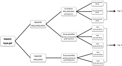
Figure 1. Scheme of irradiation procedure for calibration and evaluation of the dose-rate effect. A second batch is prepared for checking reproducibility and investigating more accurately the high dose rate range extending the dose rate to 18 Gy min−1.
Download figure:
Standard image High-resolution imageGel calibration and dose-rate dependence experiments, were performed with a beam perpendicular to the surface of the gel container. The applied dose levels and dose rates were verified using a calibrated ionization chamber (type 31013, PTW-Freiburg, Freiburg, Germany) with a sensitive volume of 0.3 cm3. The ionization chamber was placed in the container, which was filled with water, at the same reference position (1 cm depth from the bottom of the container) as used for the polymer gel evaluation. The gel containers were brought to the irradiation room approximately 1 h before irradiation in order to approach thermal equilibration.
The flacon-type gel containers were positioned with the bottom side directed towards the irradiation source in a home-made perspex device with holes for fixing the polymer gel containers in place (figure 2). After irradiation, all the gel containers were kept at room temperature for at least 36 h until the gels were scanned on the MR-tomograph, in order to ensure that an apparently stable state of polymerization has been achieved.
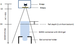
Figure 2. Sketch of gel irradiation set-up (non-scaled) indicating the position of the gel container with reference to the radiation source without collimation. The source to surface distance (SSD) was adjusted here to a distance of 34 cm for the calibration set-up. For investigations on the dose rate effect the SSD and tube current was adjusted differently (SSD = 18, 22 and 36 cm).
Download figure:
Standard image High-resolution image2.2.1. Calibration of gels
A set of three different gel types (MA1, MA2 and MA3) from batch #1 was irradiated 1 d after manufacturing with various dose levels (0, 3, 6, 12, 24 and 40 Gy) (18 gels). A dose rate of 5 Gy min−1 as typically used in LINACs was used, to obtain the dose response as calibration curve for each type of gel (figures 1 and 3). One gel container per dose level from each type was positioned at a fixed SSD of 34 cm (distance from the x-ray focal spot) within an area size of 10 × 10 cm2 (figure 2).
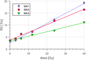
Figure 3. (Batch #1) Dose response for the reference polymer gels for calibration ( stand = 5 Gy min−1) with different concentrations of oxygen scavenger. MA1 featured minimum (caa = 2 mmol kg−1) and MA3 (caa = 18 mmol kg−1) maximum concentration of ascorbic acid. Error bars were obtained as the standard deviation of R2 in ROI. They are usually smaller than the symbol for measurement data.
Download figure:
Standard image High-resolution image2.2.2. Dose-rate effect
Another set of polymer gels with three different oxygen scavenger concentrations from batch #1 were exposed to six different dose rates ( ) ranging from
  min = 0.6 Gy min−1 to
  max = 12.87 Gy min−1. The results were taken into account for slightly modifying the protocol for batch #2 for a more detailed investigation in the high dose rate range. The high dose rate range was investigated more densely and the maximum dose rate was extended (
 min = 0.6 Gy min−1,
  max = 18 Gy min−1; figures 1 and 4). The dose rates were adjusted by varying (a) the current of the x-ray tube using an external control unit and (b) the SSD. We investigated three different dose levels: low, medium and high, with a maximum dose of 40, 45 and 55 Gy, respectively assuming the response of the gels remains linear (figure 4). The gels were positioned in small sub-groups in the centre below the 200 kV x-ray source within a maximum area of 10 × 10 cm2. The position for reference dosimetry has been set 1 cm distant from the bottom of the gel container. The homogeneity in the area of 10 × 10 cm2 was checked for spatial dose rate variations with an ionization chamber in water and the deviations were found to be less than 4%.
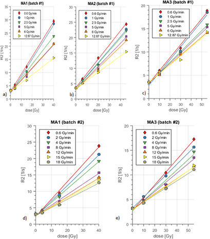
Figure 4. (a) Batch #1, dose response R2 to different dose rates ranging up to  max = 12.87 Gy min−1 for MA1 type gel (lowest concentration of oxygen scavenger). Error bars, usually smaller than the symbol for measurement data, were obtained as standard deviation of R2 in the selected ROI. Results of linear regression analysis are plotted. Note the strong reduction in absolute R2 with dose rate for MA1. (b) Batch #1, MA2 dose response for different dose rates. (c) Batch #1, dose response for MA3 with the highest concentration of ascorbic acid. (d) Batch #2: MA1 dose response for different dose rates ranging up to
  max = 18 Gy min−1. (e) Batch #2: dose response for MA3. The variation in slope with dose rate is the lowest for MA3. Note the difference in maximum dose levels for the different polymer gels. These were used due to the changing in sensitivity and linear range with oxygen scavenger concentration.
Download figure:
Standard image High-resolution image2.3. MRI measurements
The gel containers were stored in the MR scanner room for at least 2 h before measurements to reduce temperature variation with impact on T2. They were scanned on a 7T human MR scanner (Siemens, Magnetom) using a head-coil about 36 h after irradiation. The reference (non-irradiated) polymer gels (D = 0 Gy) were treated in the same way as the irradiated ones (from manufacturing—including storage in the radiation room—till measurement). Also parameter selective T2 imaging was performed at the same time. A multiple spin echo sequence with 15 echoes with echo time spacing ΔTE = 10 ms (10–150 ms), allowed for differently T2-weighted imaging (repetition time TR = 10 s, field of view = 100 mm, matrix size = 128 × 128, slice thickness = 5 mm, number of slices = 5). For quantitative T2-evaluation, a mono-exponential fitting tool was used, which was developed for ImageJ (National Institute of Health, USA) by Karl Schmidt (Harvard, USA). Before processing the first echo was removed.
3. Results
3.1. Polymer gels calibration at  = 5 Gy min−1

The results of the non-irradiated control gel served as a baseline readout. The irradiation of gel containing low oxygen scavenger concentrations (MA1) yielded an R2 value of 19.2/s in the high dose region at 40 Gy, reflecting high sensitivity (figure 3). Increasing the scavenger concentrations resulted in lowered MRI relaxation rates at 40 Gy (R2MA2 = 16.4/s and R2MA3 = 11.1/s) (figure 3).
3.2. Impact of oxygen scavenger concentration on dose-rate effect of different polymer gels
For batch #1 the dose rates varied from 0.6 to 12.87 Gy min−1. The results of batch #1 on dose response are shown in figures 4(a)–(c). Calculated dose sensitivities (evaluated as slope α = ΔR2/ΔD) obtained for the different dose rates are summarized in table 2. Also, absolute values and sensitivities αn, normalized to the sensitivity of the lowest dose rate of the specific polymer gel, are indicated including corresponding errors, evaluated from the linear fitting routine.
Table 2. (a) (top) (Batch #1): absolute and normalized sensitivities, calculated as slope α = ΔR2/ΔD for different polymer gel types and varying dose rates. Errors are calculated from fitting routine. The normalization was performed referring to the dose sensitivity of the lowest dose rate (0.6 Gy min−1) for each type of polymer gel. (b) (bottom) (Batch #2): absolute and normalized sensitivities. Note the small relative change of 6% in sensitivity for MA3 in the high dose rate region between  = 12 Gy min−1 and
  = 18 Gy min−1 for batch #2.
| Dose rate (Gy min−1) | Sensitivities α | ||||||||
|---|---|---|---|---|---|---|---|---|---|
| MA1 | MA2 | MA3 | |||||||
| Absolute (Gy−1 s−1) | Normalized | Absolute (Gy−1 s−1) | Normalized | Absolute (Gy−1 s−1) | Normalized | ||||
| (a) Batch#1 | 0.6 | 0.660 ± 0.02 | 1 ± 0.03 | 0.462 ± 0.01 | 1 ± 0.022 | 0.277 ± 0.01 | 1 ± 0.036 | ||
| 1 | 0.648 ± 0.04 | 0.982 ± 0.06 | 0.423 ± 0.02 | 0.916 ± 0.043 | 0.275 ± 0.02 | 0.993 ± 0.072 | |||
| 2.5 | 0.528 ± 0.02 | 0.80 ± 0.03 | 0.414 ± 0.01 | 0.896 ± 0.022 | 0.226 ± 0.01 | 0.816 ± 0.036 | |||
| 5 | 0.451 ± 0.03 | 0.683 ± 0.045 | 0.379 ± 0.02 | 0.82 ± 0.043 | 0.216 ± 0.01 | 0.78 ± 0.036 | |||
| 6 | 0.448 ± 0.02 | 0.679 ± 0.03 | 0.351 ± 0.02 | 0.76 ± 0.043 | 0.196 ± 0.01 | 0.708 ± 0.036 | |||
| 12.87 | 0.313 ± 0.01 | 0.474 ± 0.02 | 0.262 ± 0.004 | 0.567 ± 0.009 | 0.189 ± 0.02 | 0.682 ± 0.072 | |||
| (b) Batch#2 | 0.6 | 0.53 ± 0.03 | 1 ± 0.057 | 0.26 ± 0.01 | 1 ± 0.038 | ||||
| 2 | 0.47 ± 0.02 | 0.887 ± 0.038 | 0.22 ± 0.003 | 0.846 ± 0.012 | |||||
| 4 | 0.41 ± 0.02 | 0.774 ± 0.038 | 0.21 ± 0.02 | 0.808 ± 0.077 | |||||
| 8 | 0.32 ± 0.02 | 0.604 ± 0.038 | 0.19 ± 0.01 | 0.731 ± 0.038 | |||||
| 12 | 0.29 ± 0.02 | 0.547 ± 0.038 | 0.16 ± 0.005 | 0.615 ± 0.019 | |||||
| 15 | 0.27 ± 0.02 | 0.509 ± 0.038 | 0.16 ± 0.004 | 0.615 ± 0.015 | |||||
| 18 | 0.25 ± 0.02 | 0.472 ± 0.038 | 0.15 ± 0.01 | 0.577 ± 0.038 | |||||
Considering the differences in dose sensitivity in the high dose rate region for MA3 compared to MA1, the experimental set-up for batch #2 was slightly changed: dose rates varying from 0.6 to 18 Gy min−1 with more data points in the high dose rate region and increased maximum dose rate were investigated (figures 5(a) and (b)). Calculated dose sensitivities including normalized data and errors for batch #2 are listed in table 2.
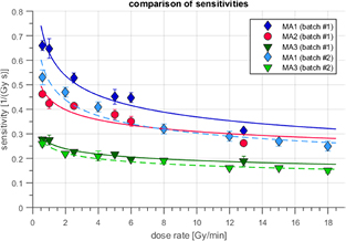
Figure 5. Absolute sensitivities versus dose rate for the different gel types with increasing oxygen scavenger concentration from MA1 to MA3. Continuous lines indicate the fitting curve (2-parameter power law) for batch #1. Dashed lines indicate fitting data for batch #2. Note the higher absolute sensitivity for the standard type of MAGIC gel (MA1) and reduced variation of sensitivity with different dose rates for MA3, especially in the region between 6 to 12.87 Gy min−1 and 6 to 18 Gy min−1 for batch #1 and batch #2 respectively.
Download figure:
Standard image High-resolution image3.2.1. Standard (low-level) oxygen scavenger concentration gel (MA1)
The dose response curves of different gel compositions (MA1-3) of batch #1 and batch #2 with respect to different dose rates are shown in figures 4(a)–(e). MA1 featured the lowest concentration of ascorbic acid (2 mmol kg−1), being close to the standard recipe of the MAGIC gel as proposed by Fong et al (2001). For MA1 (batch #1) at the highest dose level ( = 40 Gy), the dose response (R2max = (29.4/s) for the lowest dose rate (
 min = 0.6 Gy min−1) dropped significantly by about 47% (R2min = 15.6/s) with increasing dose rate to
  max = 12.87 Gy min−1 (figure 4(a)). The reduction in dose response (R2) at medium and low dose regions amounted to about 41% and 23% respectively. The reduction in dose response R2 for MA1 of batch #2 ranged in similar values as batch #1 but appeared to be even slightly stronger for the overall dose rate range. The slight differences in absolute values of R2 were likely due to the higher maximum dose rate (
 max = 18 Gy min−1) and the different manufacturing batch and MR-scanning conditions. The relaxation rate reduced with changing dose rate from the lowest to the highest from R2max = 24/s down to R2 = 12.6/s by about 47% (figures 4(a) and (d)).
For a quantitative description of the impact of the dose rate on the dose response, the sensitivity, defined here as the change of slope α = ΔR2/ΔD, was calculated. The dependence of the dose sensitivity on dose rate is shown in figure 5. The dose sensitivity was obtained by linear regression analysis of the data of figure 4, assuming a linear dose response across the whole investigated dose range. Systematic deviations from the linear fitting curve and statistical errors in data were estimated from the regression analysis and evaluated as an expected error in slope (err_α). They are indicated in figure 5 and table 2 (batch #1 and #2). With changing dose rate from the lowest to the highest ( max = 18 Gy min−1 for batch #2), the sensitivity dropped by about 53% (figure 5).
3.2.2. Impact of medium-level oxygen scavenger concentration gel (MA2)
When the scavenger concentration was increased to 6 mmol kg−1 (MA2), the R2 values were reduced for all dose rates (figure 4(b)). R2 values dropped from about R2 = 24/s to R2 = 15/s while changing the dose rate from 0.6 Gy min−1 to 12.87 Gy min−1 at the high dose level (45 Gy). The corresponding relative dose sensitivity (slope) was reduced by about 43%. Sensitivity was continuously decreasing from the lowest to the highest dose rates (figure 5). Increasing the scavenger concentrations from 2 mmol kg−1 (MA1) to 6 mmol kg−1 (MA2) resulted in a lowered percentage drop in R2 for all dose levels.
3.2.3. Impact of highest level oxygen scavenger concentration gel (MA3)
The relaxation rate R2 of batch #1 reduced by about 24% when changing dose rates from 0.6 Gy min−1 to 12.87 Gy min−1 at the high dose level (55 Gy) (figure 4(c)). The change in absolute values of sensitivity with dose rate (ΔR2/ΔD) is shown in figure 5.
Also MA3 featured a reduction in sensitivity from the lowest dose rate (0.6 Gy min−1) to the highest dose rate (12.87 Gy min−1). However, the corresponding change with dose rate (≅32% (figure 5)) appeared to be clearly less than that for the standard recipe (MA1: ≅53%).
The absolute change in dose response for MA1 from minimum dose rate (0.6 Gy min−1) to maximum dose rate of 12.87 Gy min−1 (batch #1) amounted to ΔαMA1 = 0.347 Gy−1 s−1 and that for MA3 to ΔαMA3 = 0.088 Gy−1 s−1. The average error on sensitivity was err_α = 0.023 Gy−1 s−1 for MA1 and err_α = 0.013 Gy−1 s−1 for MA3. The difference in absolute change of sensitivities between the lowest and the highest dose rate Δα = 0.259 Gy−1 s−1 for MA1 and MA3 was therefore about a factor 10 larger than the sensitivity errors for MA1 and MA3. The change in absolute values of sensitivity with dose rate for MA3 was therefore reduced significantly in comparison to MA1 (figure 5).
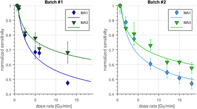
However, this reduction in dose rate dependence is related to a reduction in absolute sensitivity for MA3 and MA2. In order to assess the impact of dose rate in relative polymer gel dosimetry we therefore normalized the dose rate dependent sensitivities to the sensitivity of the specific polymer gel type at the lowest dose rate (0.6 Gy min−1). This normalized sensitivity change with dose rate is plotted in figure 6 for the different polymer gels comparing the results of different batches. Fitting curves based on a three-parameter power law are indicated. Batch #1 and batch #2 show only minor differences below or close to measurement errors of sensitivity as evaluated from one batch (figure 6).

Figure 6. Normalized sensitivities versus dose rate for batch #1 (continuous line) and batch #2 (dashed line). Note the small difference with regard to error bars between the fitted data of the results of the two different batches in context of reproducibility. The dose rate varies for batch #1 from 0.6 Gy min−1 to 12.87 Gy min−1 and for batch #2 from 0.6 Gy min−1 to 18 Gy min−1.
Download figure:
Standard image High-resolution imageA direct comparison of the differences within the same batch in dose rate dependence between MA1 and MA3 is possible using figure 7: the plot of the relative dose response (and thus normalized to the dose sensitivity of each specific polymer gel) with dose rate is indicating a reduction in dose rate change for MA3 in comparison to MA1 for the investigated dose rate range from 0.6 Gy min−1 to 18 Gy min−1. There was a good match between batch #1 and batch #2.
Figure 7. Change of normalized sensitivities with dose rate. A three-parameter power law without initial assumptions on fit parameters was applied. Note the reduced variation of normalized sensitivities with dose rate for MA3, especially in the region between about 5 Gy min−1 to 12.87 Gy min−1 for batch #1 and up to 18 Gy min−1 for batch #2.
Download figure:
Standard image High-resolution image
At low dose rates the differences in sensitivities ranged within errors of normalized dose sensitivities. At about  = 8 Gy min−1 and above the deviation between the normalized sensitivity and dose rate graphs for MA1 and MA3 exceeded the averaged errors. The errors for the specific gel type were estimated by calculating the mean errors for the normalized sensitivities for all of the different dose rates. Thus MA3 featured significant advantages over MA1 also with regard to the normalized sensitivity (p = 0.004 in t-test on MA1 versus MA3 values on dose sensitivity with changing dose rate
  ⩾ 8 Gy min−1, batch #2).
The dose rate dependence of dose sensitivity was substantially reduced to 6% in the dose rate region of 12 Gy min−1 ⩽  ⩽ 18 Gy min−1 for MA3. Thus, MA3 featured also the lowest change in sensitivity with dose rate in the high dose rate region (table 2).
4. Discussion
The impact of the dose rate on the dose response of MAGIC-type polymer gels represents an important restriction for the accuracy of 3D-dosimetric applications.
A dependence of the dose response on the dose rate (dose-rate effect) has been reported mainly for methacrylic acid-based polymer gels with THPC (MAGAT) (De Deene 2004, Bayreder et al 2006, De Deene et al 2006, Karlsson et al 2007). MAGAT-type polymer gels exhibited a stronger variation in the dose response with dose rate in comparison to polyacrylamide-type polymer gels (PAG). The dose sensitivity was reduced by up to 34% when using a dose rate of  max = 4 Gy min−1 instead of
  min = 0.25 Gy min−1 (De Deene 2004).
The influence of oxygen scavengers on the sensitivity of MAG/MAGIC/acrylamide-type gels has been investigated previously (De Deene et al 2002, Jirasek et al 2009).
In this study, we investigated MAGIC-type polymer gels based on methacrylic acid, because of the higher sensitivity in comparison to PAG type gels and reduced safety hazards of compounds. In MAGIC-gels methacrylic acid as monomer features less hazard for genetic defects and cancer and also contains a non-toxic, oxygen scavenger: ascorbic acid (Senden et al 2006). There have been reports, that the dose-rate dependence might be reduced in polymer gels using ascorbic acid as an oxygen scavenger (Bayreder et al 2006). Here we investigated the impact of ascorbic acid concentration on the dose sensitivity and its influence on the dose-rate effect. Moreover, with expansion to previous investigations (De Deene et al 2006) on dose sensitivity in standard type polymer gels, the dose rate range was extended to include the very high dose rates ( ⩾ 8 Gy min−1) as available with modern flattening filter free (FFF) applications (Georg et al 2011, Prendergast 2013) and also progressively available in preclinical animal irradiations. In brachytherapy, dose rates below 1 Gy min−1 are often applied (Farajollahi et al 1999, Pantelis et al 2005, Baldock et al 2010, Zehtabian et al 2012). We therefore varied the dose rates in a wide range from
  = 0.6 up to
  = 18 Gy min−1, comparing polymer gel sensitivity for the medium and high dose rate range to the sensitivity for the lowest dose rate
  = 0.6 Gy min−1.
Low energy photons (E = 200 kV) were used because of accurately adjustable high dose rates at high availability of the animal treatment system. It is reported that no differences to within experimental accuracy have been detected for the dose response between low energy photons (e.g. 300 kV) in comparison to 60Co, 6 and 8 MV beam energies (Farajollahi et al 1999). A hardening effect on the dose rate dependent measurement is not to be expected as all measurements were performed in the same depth and consequently in the same energy spectrum for different dose rates can be assumed.
In general, for all of the investigated MAGIC-type polymer gels we observed an overall reduction in sensitivity when increasing the dose rate from lowest (0.6 Gy min−1) to highest values (18 Gy min−1). This dose-rate effect has been reported in a similar but different extent by De Deene et al (2006) for MAGAT-type gels using THPC as an oxygen scavenger. However, we also observed that the oxygen scavenger concentration exhibited a strong impact on the dose rate dependence measured by the absolute sensitivity change over the whole dose rate interval. We have shown that the change in absolute values in sensitivity with dose rate was significantly less for higher oxygen scavenger concentrations (table 2). However, this reduced absolute dose rate dependence was connected to a reduced dose sensitivity of the polymer gels with higher ascorbic acid concentration and thus the quantitative dose evaluation benefits might be compensated. Therefore, we normalized the dose sensitivities for all of the dose rates to the slope of the lowest dose rate, thus compensating for dose sensitivity reductions (table 2). Our first batch indicated a reduction in the dose-rate effect even on the basis of these normalized dose sensitivities with MA3 in the high dose rate range  > 8 Gy min−1. This was confirmed in batch #2 where the high dose rate region was investigated in more detail. In addition, the reduction in dose rate dependence of the sensitivity for MA3 was even more prominent at the newly applied topmost dose rates above 12.5 Gy min−1 (table 2(b) bottom, figure 5).
The regression plot of the relative change of dose response (figure 7) with dose rate indicates a reduction in the dose-rate effect for MA3 in comparison to MA1 for all of the investigated dose rates from 0.6 Gy min−1 to 18 Gy min−1. However, at low dose rates (<8 Gy min−1) this change ranges within errors of dose sensitivity. At about  = 8 Gy min−1 the reduced dose-rate effect of MA3 (the deviation between the two dose rate graphs of MA1 and MA3) exceeds the averaged errors for MA3 on normalized sensitivities. We therefore conclude, that the dose-rate dependence for MA3 was distinctly lower than for the standard type recipe MA1 and reduced to about 6% (table 2) compared to MA1 (13%) in the high dose rate region (12 Gy min−1 ⩽
  < 18 Gy min−1) even after sensitivity normalization (table 2(a)). The changes (6%) were comparable to the measurement errors in sensitivities (figure 5).
As a drawback, the sensitivity was reduced from MA1 to MA3. However, this reduced sensitivity might be compensated in relative dosimetry by increasing the general dose level in more complex irradiation schemes by a scaling factor.
It has been reported previously, that the dose rate has a larger effect on the absolute R2 response at higher dose regions and less effect at low-dose ranges (Bayreder et al 2006). By increasing the dose rate, we also observed a higher absolute difference in absolute ΔR2 for the high dose region in comparison to low-dose ranges. The absolute ΔR2 difference was strongly reduced for higher oxygen scavenger concentrations (MA1: ΔR2/R2 = 47% at 40 Gy and MA3: ΔR2/R2 = 24% at D = 55 Gy) (figures 4(a) and (c)).
The impact of dose rate on a modified MAGIC-type gel adding formalin (MAGIC-f) has been reported by (Pavoni and Baffa 2012). They reported a reduction in R2 by about 4.7% during increase of the dose rate from 1 Gy min−1 to 6 Gy min−1 at a very low dose of  = 2 Gy. However, they also reported a significant increase (about 23%) of the integrated dose response (slope) on fractionation scheme, if the dose is delivered with 0.25 Gy/fraction instead of 2 Gy/fraction up to 10 Gy, indicating dose rate impact at higher dose levels. We interpret these results as dose-rate effect being low with regard to absolute R2 dose response at low dose (
 = 2 Gy) due to limited recombination of radical carrying molecules, but prominent at higher dose levels and larger fractionation intervals (e.g. ΔD = 2 Gy with high dose rate instead of ΔD = 0.25 Gy/fraction and 70 s pause in between the fractions).
It has been reported previously, that the dose range of quasi-linear dose response of gels with higher sensitivity was less than that of low sensitive gels (Massillon 2010). We also confirmed these results for the higher oxygen scavenger concentrations in gel (MA3), in which we observed a highly linear dose range up to 55 Gy.
The reasons for the reduced dose-rate dependence at higher oxygen scavenger concentrations can be described by the subsequent qualitative physicochemical model: the irradiation of polymer gels is connected to radiolysis of water and subsequent production of different types of free radicals (Baldock et al 2010). The reduction in sensitivity with dose rate is determined by the recombination of radicals, especially present at high dose levels (Bayreder et al 2006, Karlsson et al 2007, Baldock et al 2010). When two radical carrying molecules combine, the reaction is terminated. The rate of this termination is proportional to the square of the radical concentration (Karlsson et al 2007). As a result, the termination rate increases by a factor of four when radical concentration and dose is increased by a factor of 2. At high dose rates, radical generation and subsequently their concentration are high and the recombination rate increases non-linearly with dose. Ascorbic acid plays two important roles in polymer gels. At first, it scavenges the oxygen from the solution to enhance the polymerization process. Secondly, it is one of the strongest reductants and radical scavengers. Therefore, the concentration level affects the sensitivity and dose response via radical scavenging. Beyond a certain level, the dose sensitivity becomes weaker (Jirasek et al 2009) and subsequently the dose-rate effect is reduced. The reduction in dose rate sensitivity at high oxygen scavenger concentration represents a consequence of the non-linear dependence of radical recombination and polymerization termination on radical concentration. At high concentrations, oxygen scavengers reduce oxygen-related and monomer radicals. For a given dose distribution to be dosimetrically measured, a compromise between the sensitivity level and the dose-rate effect has to be found by the decision on the type of the gel.
In addition we would like to point to the relevance of the presented data on dosimetry with MAGIC type and presumably also other types of dose rate sensitive polymer gels in general: The shown reduced dose rate dependence in the high dose rate range, especially prominent for preparations with high oxygen scavenger concentrations, is advantageous for high accuracy in 3D-dosimetry. The presented data on MAGIC type gels lead to a recommendation to calibrate and irradiate polymer gels in the high dose rate range  > 8 Gy min−1 (due to low dose rate dependence there) rather than in the low dose rate range (
 < 2 Gy min−1) for optimized results in relative dosimetry. Intuitively an investigation at low dose rates may be assumed falsely as being advantageous by the applicant of gel dosimeters, known for being sensitive to dose rates.
5. Summary and conclusion
The influence of the dose rate on the dose response for MAGIC-type polymer gels has been investigated ranging from low dose rates  min = 0.6 Gy min−1 as often applied in Brachytherapy up to
  max = 18 Gy min−1, as increasingly used in FFF clinical applications. We assumed an impact of increasing ascorbic acid concentration on the radical-mediated recombination of multimers in the polymer gel. Therefore, three types of polymer gels with increasing oxygen scavenger concentration—up to nine-fold level (MA3) have been investigated for their dose-rate effect—in comparison with a standard recipe. The results revealed that for all of the polymer gel recipes a significant reduction with dose rate in dose response and sensitivity was present. However, the change in absolute values of sensitivity with dose rate was distinctly reduced for higher ascorbic acid concentration, especially, for MA3. This improvement was obtained on the cost of the reduction in absolute values of sensitivity. For quantitative dose resolution the relative change in sensitivity is relevant. Therefore, we also presented sensitivity-normalized quantitative data on the dose-rate effect for different oxygen scavenger concentrations. This sensitivity normalized dose response also indicated a reduction in the dose-rate dependence for MA3 in comparison to MA1 in the high dose rate range from about 8 Gy min−1 up to 18 Gy min−1.
The relative change in sensitivity reported here amounted to only 6.5%, which is comparable to the measurement uncertainties. As a drawback, the dose sensitivity defined as the slope of the corresponding polymer gel (MA3) was reduced by about 50%. In MA3 the R2 to dose response demonstrated an almost linear relationship over a range between 0–55 Gy. In general we propose to operate the calibration and evaluation irradiation in the high dose rate range (due to low dose rate dependence there) rather than in the low dose rate range (<2 Gy min−1) for optimized results in relative dosimetry.
The proposed modifications might be especially useful in dosimetric imaging of FFF LINAC irradiations, and in the application of 3D gel dosimetry in the preclinical treatment of animals, where often also high dose rates are applied for time reasons.
Acknowledgments
M Khan is funded by the higher education commission (HEC) of Pakistan. We also acknowledge the support of the Austrian agency for international mobility and cooperation in education, science, and research (OEAD). Materials were funded by 'Hochschuljubiläumsstiftung' (HSJ) of the City of Vienna (H-1208/2003) and intramural support by the Medical University of Vienna. The financial support by the Austrian Ministry for Digital, Business and Enterprise and the National Foundation for Research, Technology and Development is gratefully acknowledged.