Abstracts
Dermoscopy has being used over the past twenty years as a noninvasive aid in the diagnosis of innumerable skin conditions, including infectious diseases and infestations (Entodermoscopy).Tinea nigra is a superficial phaeohyfomycosis that affects mainly the glabrous skin of palms and soles. We describe a 14 year-old girl with a three-month history of an enlarging brown patch of her hand diagnosed as Tinea Nigra following clinical and dermoscopy examination.These images emphasize the importance of dermoscopy as a diagnostic tool in the daily routine of dermatologists.
Dermatomycoses; Dermoscopy; Hand dermatoses; Skin diseases, infectious; Tinea
Nos últimos anos, a dermatoscopia tem sido utilizada como importante ferramenta auxiliar no diagnóstico de inúmeras dermatoses, incluindo infecções e infestações (Entodermatoscopia). A Tinea nigra é uma feoifomicose rara, que afeta principalmente a pele glabra das palmas e plantas. Descrevemos o caso de doente de 14 anos, com mácula pigmentada de crescimento progressivo na mão esquerda, diagnosticada como Tinea nigra após o exame clínico e dermatoscópico. Estas imagens enfatizam a importância da dermatoscopia na prática dermatológica diária.
Dermatomicoses; Dermatopatias infecciosas; Dermatoses da mão; Dermoscopia; Tinha
The term Entodermoscopy was coined by Zalaudek et al. for the use of dermoscopy as an aid in the in vivo diagnosis of skin infections and infestations.11. Zalaudek I, Giacomel J, Cabo H, Di Stefani A, Ferrara G, Hofmann-Wellenhof, R et al. Entodermoscopy: a new tool for diagnosing skin infections and infestations. Dermatology. 2008;216:14-23. Specific dermoscopic patterns have been recently described for several of these conditions with a view to facilitating their diagnosis.
One of these conditions is Tinea nigra. First identified in 1891 by Alexandre Cerqueira, in Bahia, Brazil, and described in 1916 by his son as Keratomycosis nigricans Palmaris; this superficial phaeohyphomycosis is caused by the mould Hortae werneckii that occurs mainly in tropical or subtropical areas.11. Zalaudek I, Giacomel J, Cabo H, Di Stefani A, Ferrara G, Hofmann-Wellenhof, R et al. Entodermoscopy: a new tool for diagnosing skin infections and infestations. Dermatology. 2008;216:14-23.
2. Cerqueira AG. Keratomycosis nigricans palmaris [tese]. Salvador (BA): Faculdade de Medicina da Bahia; 1916.
3. Negroni R. Historical aspects of dermatomycoses. Clin Dermatol. 2010;28:125-32.
4. Rossetto AL, Cruz RC. Tinea nigra nas formas geográficas em "coração" e "bico do papagaio". An Bras Dermatol. 2011;86:389-90.-55. Piliouras P, Allison S, Rosendahl C, Buettner PG, Weedon D. Dermoscopy improves diagnosis of Tinea nigra: a study of 50 cases. Australas J Dermatol. 2011;52:191-4. The condition is characterized clinically by a gradually enlarging, irregularly pigmented macula on the palms and soles, which can be confused with melanocytic lesions.11. Zalaudek I, Giacomel J, Cabo H, Di Stefani A, Ferrara G, Hofmann-Wellenhof, R et al. Entodermoscopy: a new tool for diagnosing skin infections and infestations. Dermatology. 2008;216:14-23.,66. Rezusta A, Gilaberte Y, Betran A, Gene J, Querol I, Arias M, et al. Tinea nigra: a rare imported infection. J Eur Acad Dermatol Venereol. 2010;24:89-91., 77. Xavier MH, Ribeiro LH, Duarte H, Saraça G, Souza AC. Dermatoscopy in the diagnosis of Tinea nigra. Dermatol Online J. 2008;14:15.
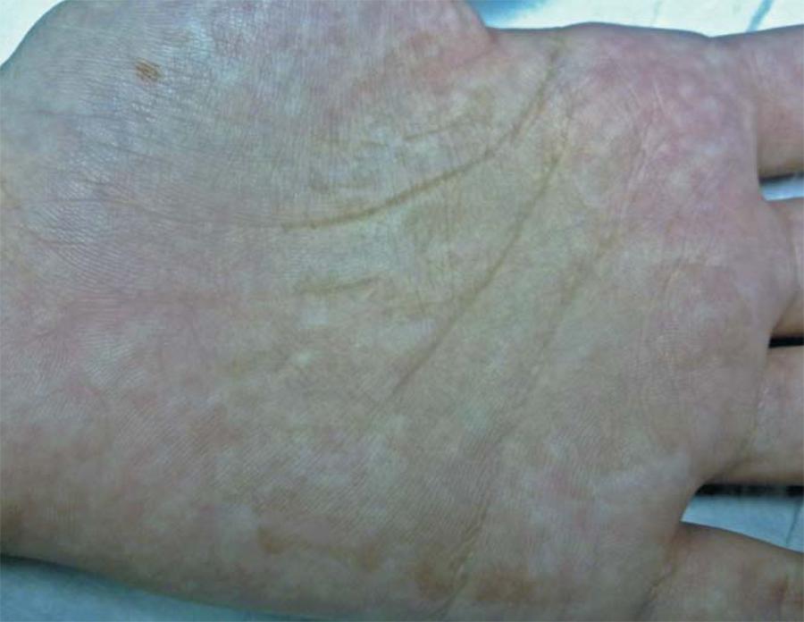
Our report concerns a 14 year-old girl who presented with a 3-month history of a slow-growing asymptomatic pigmentation on her palm. Clinical examination revealed a well-demarcated, nondesquamative, irregularly pigmented macula on her left palm (Figure 1). Dermoscopy of the lesion showed superficial fine, wispy pigmented spicules. These spicules did not respect the dermatoglyphic lines, thus confirming the Tinea nigra diagnosis (Figure 2). The patient was treated with ciclopirox olamine 1% cream, twice daily, with complete clinical resolution after three weeks of treatment.
Tinea nigra: A well-demarcated, nondesquamative, irregularly pigmented macula on the left hand
The characteristic Tinea nigra dermoscopic pattern was first described by Gupta et al. in 1997 as 'pigmented spicules', which form an almost reticulated patch, as in our case described above.88. Gupta G, Burden AD, Shankland GS, Fallowfield ME, Richardson MD. Tinea nigra secondary to Exophiala werneckii responding to Itraconazole. Br J Dermatol. 1997;137:483-4.
Our report emphasizes the importance of dermoscopy in the diagnosis of Tinea nigra.
REFERENCES
-
1Zalaudek I, Giacomel J, Cabo H, Di Stefani A, Ferrara G, Hofmann-Wellenhof, R et al. Entodermoscopy: a new tool for diagnosing skin infections and infestations. Dermatology. 2008;216:14-23.
-
2Cerqueira AG. Keratomycosis nigricans palmaris [tese]. Salvador (BA): Faculdade de Medicina da Bahia; 1916.
-
3Negroni R. Historical aspects of dermatomycoses. Clin Dermatol. 2010;28:125-32.
-
4Rossetto AL, Cruz RC. Tinea nigra nas formas geográficas em "coração" e "bico do papagaio". An Bras Dermatol. 2011;86:389-90.
-
5Piliouras P, Allison S, Rosendahl C, Buettner PG, Weedon D. Dermoscopy improves diagnosis of Tinea nigra: a study of 50 cases. Australas J Dermatol. 2011;52:191-4.
-
6Rezusta A, Gilaberte Y, Betran A, Gene J, Querol I, Arias M, et al. Tinea nigra: a rare imported infection. J Eur Acad Dermatol Venereol. 2010;24:89-91.
-
7Xavier MH, Ribeiro LH, Duarte H, Saraça G, Souza AC. Dermatoscopy in the diagnosis of Tinea nigra. Dermatol Online J. 2008;14:15.
-
8Gupta G, Burden AD, Shankland GS, Fallowfield ME, Richardson MD. Tinea nigra secondary to Exophiala werneckii responding to Itraconazole. Br J Dermatol. 1997;137:483-4.
-
* Study undertaken at the Hospital das Clínicas, Faculty of Medicine, University of São Paulo (HC-FMUSP) - São Paulo (SP), Brazil.
Publication Dates
-
Publication in this collection
Feb 2013
History
-
Received
08 Apr 2012 -
Accepted
21 June 2012