Article

Prevalence of Cancer Predisposition Germline Variants in Male Breast Cancer Patients: Results of the German Consortium for Hereditary Breast and Ovarian Cancer

by
Muriel Rolfes
1,
Julika Borde
1,
Kathrin Möllenhoff
2,
Mohamad Kayali
1,
Corinna Ernst
1,
Andrea Gehrig
3,
Christian Sutter
4,
Juliane Ramser
5,
Dieter Niederacher
6,
Judit Horváth
7,
Norbert Arnold
8,
Alfons Meindl
9,
Bernd Auber
10,
Andreas Rump
11,
Shan Wang-Gohrke
12,
Julia Ritter
13,
Julia Hentschel
14,
Holger Thiele
15,
Janine Altmüller
15,16,17,
Peter Nürnberg
15,
Kerstin Rhiem
1,
Christoph Engel
18,
Barbara Wappenschmidt
1,
Rita K. Schmutzler
1,
Eric Hahnen
1,* and
Jan Hauke
1
add Show full author list remove Hide full author list
1
Center for Familial Breast and Ovarian Cancer, Center for Integrated Oncology (CIO), University of Cologne, Faculty of Medicine and University Hospital Cologne, 50937 Cologne, Germany
2
Mathematisches Institut, Heinrich-Heine-Universität Duesseldorf, 40225 Duesseldorf, Germany
3
Institute of Human Genetics, University Wuerzburg, 97074 Wuerzburg, Germany
4
Institute of Human Genetics, University Hospital Heidelberg, 69120 Heidelberg, Germany
5
Department of Gynecology and Obstetrics, Technical University Munich, 80333 Munich, Germany
6
Department of Gynecology and Obstetrics, University Hospital Duesseldorf, Heinrich-Heine University Duesseldorf, 40225 Duesseldorf, Germany
7
Institute for Human Genetics, University Hospital Muenster, 48149 Muenster, Germany
8
Institute of Clinical Molecular Biology, Department of Gynecology and Obstetrics, University Hospital of Schleswig-Holstein, Campus Kiel, Christian-Albrechts University Kiel, 24105 Kiel, Germany
9
Department of Gynecology and Obstetrics, LMU Munich, University Hospital Munich, 80337 Munich, Germany
10
Department of Human Genetics, Hannover Medical School, 30645 Hannover, Germany
11
Institute for Clinical Genetics, Faculty of Medicine Carl Gustav Carus, TU Dresden, 01062 Dresden, Germany
12
Department of Gynecology and Obstetrics, University of Ulm, 89075 Ulm, Germany
13
Institute of Medical and Human Genetics, Charité-Universitätsmedizin Berlin, 13353 Berlin, Germany
14
Institute of Human Genetics, University of Leipzig Hospitals and Clinics, 04103 Leipzig, Germany
15
Cologne Center for Genomics (CCG) and Center for Molecular Medicine Cologne (CMMC), Faculty of Medicine and University Hospital Cologne, University of Cologne, 50931 Cologne, Germany
16
Core Facility Genomics, Berlin Institute of Health, Charité-Universitätsmedizin Berlin, 10117 Berlin, Germany
17
Max Delbrück Center for Molecular Medicine in the Helmholtz Association (MDC), 13125 Berlin, Germany
18
Institute for Medical Informatics, Statistics and Epidemiology, University of Leipzig, 04107 Leipzig, Germany
*
Author to whom correspondence should be addressed.
Cancers 2022, 14(13), 3292; https://doi.org/10.3390/cancers14133292
Submission received: 9 May 2022 / Revised: 22 June 2022 / Accepted: 30 June 2022 / Published: 5 July 2022
(This article belongs to the Special Issue Risk Assessment for Breast Cancer)

Abstract

:

Simple Summary

Male breast cancer (mBC) is a rare disease associated with a high prevalence of pathogenic germline variants (PVs) in the BRCA2 gene. However, data regarding other breast cancer (BC) predisposition genes are limited or conflicting. We investigated the prevalence of PVs in BRCA1/2 and 23 other cancer predisposition genes using an overall study sample of 614 patients with mBC. A high proportion of patients with mBC carried pathogenic germline variants in BRCA2 (23.0%, 142/614) and BRCA1 (4.6%, 28/614). A BRCA1/2 PV prevalence of 11.0% was identified in patients with mBC without a family history of breast and/or ovarian cancer. Patients with BRCA1/2 PVs did not show an earlier disease onset than those without. Case-control analyses revealed significant associations of protein-truncating variants in BRCA1, BRCA2, CHEK2, PALB2, and ATM with mBC. Our findings support the benefit of multi-gene panel testing in patients with mBC.

Abstract

Male breast cancer (mBC) is associated with a high prevalence of pathogenic variants (PVs) in the BRCA2 gene; however, data regarding other BC predisposition genes are limited. In this retrospective multicenter study, we investigated the prevalence of PVs in BRCA1/2 and 23 non-BRCA1/2 genes using a sample of 614 patients with mBC, recruited through the centers of the German Consortium for Hereditary Breast and Ovarian Cancer. A high proportion of patients with mBC carried PVs in BRCA2 (23.0%, 142/614) and BRCA1 (4.6%, 28/614). The prevalence of BRCA1/2 PVs was 11.0% in patients with mBC without a family history of breast and/or ovarian cancer. Patients with BRCA1/2 PVs did not show an earlier disease onset than those without. The predominant clinical presentation of tumor phenotypes was estrogen receptor (ER)-positive, progesterone receptor (PR)-positive, and HER2-negative (77.7%); further, 10.2% of the tumors were triple-positive, and 1.2% were triple-negative. No association was found between ER/PR/HER2 status and BRCA1/2 PV occurrence. Comparing the prevalence of protein-truncating variants (PTVs) between patients with mBC and control data (ExAC, n = 27,173) revealed significant associations of PTVs in both BRCA1 and BRCA2 with mBC (BRCA1: OR = 17.04, 95% CI = 10.54–26.82, p < 10−5; BRCA2: OR = 77.71, 95% CI = 58.71–102.33, p < 10−5). A case-control investigation of 23 non-BRCA1/2 genes in 340 BRCA1/2-negative patients and ExAC controls revealed significant associations of PTVs in CHEK2, PALB2, and ATM with mBC (CHEK2: OR = 3.78, 95% CI = 1.59–7.71, p = 0.002; PALB2: OR = 14.77, 95% CI = 5.02–36.02, p < 10−5; ATM: OR = 3.36, 95% CI = 0.89–8.96, p = 0.04). Overall, our findings support the benefit of multi-gene panel testing in patients with mBC irrespective of their family history, age at disease onset, and tumor phenotype.

1. Introduction

Male breast cancer (mBC) is a rare disease; less than 1% of all patients with breast cancer (BC) are men [1]. Worldwide, the incidence of mBC is less than 1 per 100,000 man-years [2]. In Germany, approximately 750 men were diagnosed with BC in 2020. In contrast, BC among women is by far the most common tumor entity, with approximately 69.000 newly diagnosed patients annually [3]. Due to its comparative rarity, mBC is routinely excluded from clinical trials on BC. Thus, diagnostic recommendations for mBC have been based on clinical research results primarily focusing on women over the decades. In addition to obvious similarities in the disease course, sex-specific differences reveal, in particular, the need for more specific and separate consideration of mBC [4,5,6]. Examining prospective data has revealed clinically relevant differences in carcinogenesis of mBC, especially the divergent prevalence of germline pathogenic variants (PVs) in the major BC susceptibility genes, BRCA1/2. PVs in BRCA2 represent the most frequent causative gene alteration and have been reported in about 10–16% of patients with mBC [4,7,8]. These are associated with an estimated lifetime risk for mBC of 4–12%, compared with 0.1% in the general male population [9,10]. Barnes et al. demonstrated an average lifetime risk for mBC of 12% for BRCA2 carriers in their polygenic risk score (PRS)-based risk analyses [11]. In contrast to BRCA2, PVs in BRCA1 are underrepresented in male patients compared with those in female patients with BC [12,13]. After initially disproving the involvement of BRCA1 in mBC carcinogenesis [14], several studies subsequently suggested an association between mBC and PVs in BRCA1, although this association is substantially weaker than that with BRCA2 [15]. Li et al. recently confirmed the association of BRCA1 PVs and mBC (risk ratio (RR) = 4.30; 95% confidence interval (CI) = 1.09–16.96) [10].
Recently published guidelines [16,17] concerning managing patients with mBC include, in addition to general therapeutic options, the support of genetic counseling and germline genetic testing for cancer predisposition genes regardless of their family cancer history. Nevertheless, there remains a lack of evidence-based, precise breakdown of possible PVs beyond those in BRCA2. As widely confirmed in female patient cohorts [18], several predisposition genes involved in DNA repair pathways such as ATM, CHEK2, and PALB2 have also been described in mBC cohorts [19,20,21,22,23]. Overall, data regarding these and other suspected mBC predisposition genes remain limited and have revealed controversial results. Pritzlaff et al. [19] investigated the association of 16 BC risk genes in 708 patients with mBC and found that besides BRCA2, PALB2 and CHEK2 were associated with mBC risk. No significant association with increased mBC risk was found for BRCA1 and ATM. A study by Rizzolo et al. [20] investigating 503 BRCA1/2-negative patients with mBC from Italy confirmed the association of PVs in PALB2 and found no significant association of PVs in CHEK2 with increased mBC risk. A study including 102 patients with mBC by Fostira et al. [21] confirmed an association with BRCA2 and identified ATM as the second most frequently mutated risk gene.
As the respective contribution to mBC risk remains controversial, further investigations are needed to evaluate the utility and potential incorporation of multi-gene panel testing in the clinical management of patients with mBC. We performed a multicenter study including 614 patients with mBC recruited for genetic testing through the centers of the German Consortium for Hereditary Breast and Ovarian Cancer (GC-HBOC) to determine PV prevalence in known and suspected BC predisposition genes and to more comprehensively define the genetic predisposition to mBC. We pursued a two-stage approach: first, all patients were screened for PVs in BRCA1/2; second, BRCA1/2-negative patients were further analyzed for PVs in non-BRCA1/2 cancer predisposition genes.

2. Material and Methods

2.1. Study Sample

The overall study sample comprised 614 patients with mBC, diagnosed with unilateral or bilateral BC between 1965 and 2018. The average age at first diagnosis (AAD) was 60 years (range 22–91 years). All patients were recruited from the participating centers of GC-HBOC. For 66.1% of the patients with mBC in the overall study sample (406/614), a positive family history (FH) for BC and/or ovarian cancer (OC) was reported. Positive FH was defined as at least one known relative with BC or OC, irrespective of the AAD of the relative(s) with BC or OC. Among the patients with mBC, 32.6% reported no FH of BC/OC (200/614). Data regarding BC/OC FH were missing for 8/614 of the patients with mBC (1.3%). BRCA1/2 germline analysis was performed in a routine diagnostic setting between 1995 and 2019. All patients were tested for germline mutations after mBC was diagnosed. A high proportion (27.7%) of patients with mBC (170/614) carried PVs in BRCA1/2. Of the 614 patients, 586 were considered index patients, with no prior testing of another family member. The remaining 28 mBC patients, all with a positive FH, were analyzed for a known pathogenic BRCA1/2 family mutation only. Of those, 22 were tested positive. Of the 444 patients with mBC without PVs in BRCA1/2, 104 were excluded from further analyses because of missing DNA samples or lack of patient consent. The remaining 340 patients with mBC who had previously tested negative for PVs in BRCA1/2 were screened for PVs in 23 established or suspected non-BRCA1/2 BC predisposition genes. Genotype and phenotype data were retrieved from the centralized GC-HBOC patient database (BRCA2006/HerediCaRe, accessed on 20 May 2019).

2.2. Gene Selection and Next-Generation-Sequencing (NGS) Analysis

Genomic DNA was isolated from venous blood. Regarding non-BRCA1/2 predisposition gene analyses, approximately one-third (102/340) of the patient genetic data originated from a comprehensive analysis of gene panel testing previously performed at the GC-HBOC centers in a routine diagnostic setting using the customized hybridization capture-based TruRisk® gene panel of the GC-HBOC for target enrichment. Another customized multi-gene panel was used for the remaining 238 patients (Agilent SureSelectXT, Santa Clara, CA, USA). Both multi-gene panels covered the entire coding regions of 23 established or suspected non-BRCA1/2 BC predisposition genes (ATM, NM_000051.3; BARD1, NM_000465.3; BRIP1, NM_032043.2; CDH1, NM_004360.4; CHEK2, NM_007194.3; FAM175A, NM_139076.2; FANCM, NM_020937.3; MLH1, NM_000249.3; MRE11A, NM_005591.3; MSH2, NM_000251.2; MSH6, NM_000179.2; MUTYH, NM_0011428425.1; NBN, NM_002485.4; PALB2, NM_024675.3; PMS2, NM_000535.5; PTEN, NM_000314.4; RAD50, NM_005732.3; RAD51C, NM_058216.2; RAD51D, NM_002878.3; RINT1, NM_021930.4; STK11, NM_000455.4; TP53, NM_000546.5; XRCC2, NM_005431.1). Quantified libraries were sequenced on Illumina NGS devices (HiSeq 4000 or NovaSeq 6000, San Diego, CA, USA) at the Cologne Center for Genomics (CCG). Bioinformatic analyses were performed using the Varbank v.2.26 (Cologne Center for Genomics, Cologne, Germany) pipeline of the CCG.

2.3. Variant Annotation and Classification

The sequencing reads were mapped against human reference genome GRCh38. Mean coverage of at least 100× was chosen as the sequencing quality filter threshold. The Alamut Visual version 2.13 analysis software tool (Interactive Biosoftware, Rouen, France) was used for variant annotation and integration of current ClinVar classifications. Variant classification was performed using the GC-HBOC criteria for the classification of germline sequence variants in risk genes for hereditary BC and OC [24], based on the interpretive guidelines published by the Evidence-Based Network for the Interpretation of Germline Mutant Alleles (ENIGMA) and the American College of Medical Genetics and Genomics (ACMG) [25]. As proposed by the International Agency for Research on Cancer (IARC) [26], a five-tier classification system was applied. This grading system defines variants as pathogenic (class 5), likely pathogenic (class 4), variant of uncertain significance (VUS, class 3), likely benign (class 2), or benign (class 1). Protein-truncating germline variants (PTVs) were defined as nonsense, frameshift, or essential splice site variants affecting the invariant splice sites or the last nucleotide of an exon. PVs (pathogenic variants) included PTVs, (likely) pathogenic missense variants, and (likely) pathogenic copy number variations (CNVs). ExomeDepth v1.1.15 was used for CNV prediction [27] and predicted CNVs were verified by multiplex ligation-dependent probe amplification (MLPA) using specific P041 (ATM), P042 (ATM), and P190 (CHEK2) SALSA® MLPA® kits (MRC Holland, Amsterdam, The Netherlands). All remaining PVs were confirmed by Sanger sequencing.

2.4. Control Sample and Statistical Analysis

To investigate the association of mBC with PTVs in suspected cancer predisposition genes, a case-control analysis was conducted using univariate logistic regression analysis to estimate the odds ratios (ORs) and corresponding 95% confidence intervals (CIs). Publicly available genomic variant data from 27,173 individuals of non-Finnish European ancestry from the Exome Aggregation Consortium (ExAC) [28], excluding samples from The Cancer Genome Atlas (TCGA), were included as control data. Finnish individuals were excluded due to a high prevalence of founder mutations in the Finnish population [29], which may cause a bias in the case-control analysis. Statistical analyses were performed using R v3.6 (R Core Team (2021)). R: A language and environment for statistical computing. R Foundation for Statistical Computing, Vienna, Austria. URL: https://www.R-project.org/ (accessed on 23 February 2022). All statistical tests were two-sided, with p-values < 0.05 considered statistically significant.

3. Results

3.1. Prevalence of BRCA1/2 Pathogenic Variants and Cancer Characteristics in the Overall Study Sample

From the overall study sample of 614 patients with mBC, 170 (27.7%) patients carried a PV in BRCA1 or BRCA2, with a notably higher proportion of BRCA2 PV carriers (142/614, 23.1%) than BRCA1 PV carriers (28/614, 4.6%). In BRCA2, the c.1813dup PV was most frequent (12/142). In BRCA1, the c.5266dup variant accounted for one-quarter of all BRCA1 PVs (7/28). The mean AAD of BRCA1 PV carriers, as well as that of BRCA2 PV carriers, was 62 years (BRCA1: range 33–82 years; BRCA2: range 37–83 years), with a statistically significant difference observed compared with BRCA1/2-negative patients (mean 59 years, range 22–91 years, Welch’s t-test p = 0.005). Among 606 patients with mBC whose BC/OC FH status was known, the prevalence of BRCA1/2 PVs was significantly higher in patients with BC/OC FH than in those without (154/460 (33.5%) vs. 16/146 (11.0%), Fisher’s exact test p < 10−7). A statistically significant association between BC/OC FH and the occurrence of PVs was found the BRCA2 gene (128/460 (27.8%) vs. 14/146 (9.6%), OR = 3.63, 95% CI = 1.99–7.08, Fisher’s exact test p < 10−5) and for the BRCA1 gene (26/460 (5.6%) vs. 2/146 (1.4%), p = 0.04). A high proportion of PVs in the BRCA2 gene was observed in bilaterally affected patients with mBC, though with no statistical significance compared with the prevalence in unilaterally affected patients with mBC (p = 0.112); of the 28 bilaterally affected patients with mBC (Table 1), 10 patients (35.7%) carried PVs in BRCA2 compared with 132/586 (22.5%) in unilaterally affected patients.
Information on tumor estrogen receptor (ER) and progesterone receptor (PR) status was available for 407 patients with mBC (66%) among the overall study sample of 614 patients with mBC. The predominant clinical presentation of tumor phenotypes was ER-positive and PR-positive (359/407, 88.2%); further, 35/407 (8.6%) were ER-positive and PR-negative. Only 13/407 (3.2%) tumors were ER-negative, of which seven were PR-positive and six were PR-negative. Among the 407 patients with mBC with known ER and PR status, human epidermal growth factor receptor 2 (HER2) status was documented in 323 patients. Most patients were ER-positive, PR-positive, and HER2-negative (251/323, 77.7%); further, 33/323 (10.2%) were triple-positive, and 4/323 (1.2%) were triple-negative (Figure S1). Neither AAD nor BRCA1/2 PV carrier status was associated with ER, PR, or HER2 status (Figure S2, and Table S1).

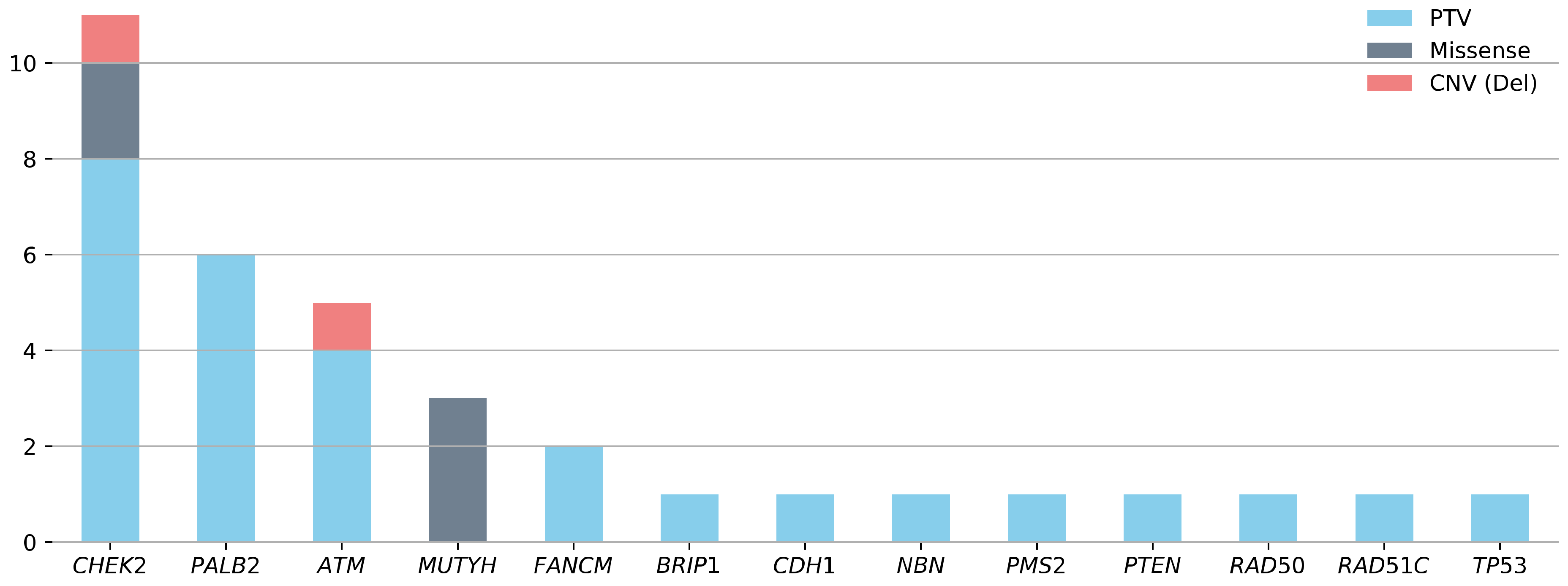
3.2. Prevalence of Pathogenic Variants in BRCA1/2-Negative Patients with mBC

In the overall study sample, 72.3% (444/614) of patients tested negative for PVs in BRCA1/2. Of these patients, 340 were screened for PVs among 23 additional non-BRCA1/2 cancer predisposition genes. This subgroup, with a mean AAD of 60 years (range 27–91), comprised 328 unilaterally and 12 bilaterally affected patients with mBC (Table 1). A BC/OC FH was reported in 69.7% (235/337) of these patients (Table 1, no BC/OC FH data were available for three patients). In the BRCA1/2-negative study sample, 9.4% (32/340) of the patients with mBC, all unilaterally affected, carried at least one PV in the 23 (suspected) non-BRCA1/2 cancer predisposition genes (Figure 1).
Overall, 35 PVs were identified in the 13 genes (Figure 1). Three patients with mBC were double PV carriers (CHEK2/ATM, NBN/RAD50, PALB2/TP53). PVs in CHEK2 were the most frequent and were identified in 3.2% (11/340) of the BRCA1/2-negative patients with mBC. The most prevalent PV in the CHEK2 gene was the c.1100delC variant observed in six of 11 patients. The second highest PV prevalence was in PALB2 (1.8%, 6/340), followed by ATM (1.5%, 5/340). The prevalence of PVs in other investigated genes was less than 1% each (MUTYH (3/340), FANCM (2/340), BRIP1 (1/340), CDH1 (1/340), NBN (1/340), PMS2 (1/340), PTEN (1/340), RAD50 (1/340), RAD51C (1/340), TP53 (1/340)). No PVs were identified in BARD1, FAM175A, MLH1, MRE11A, MSH2, MSH6, RAD51D, RINT1, STK11, or XRCC2 (Table S1).

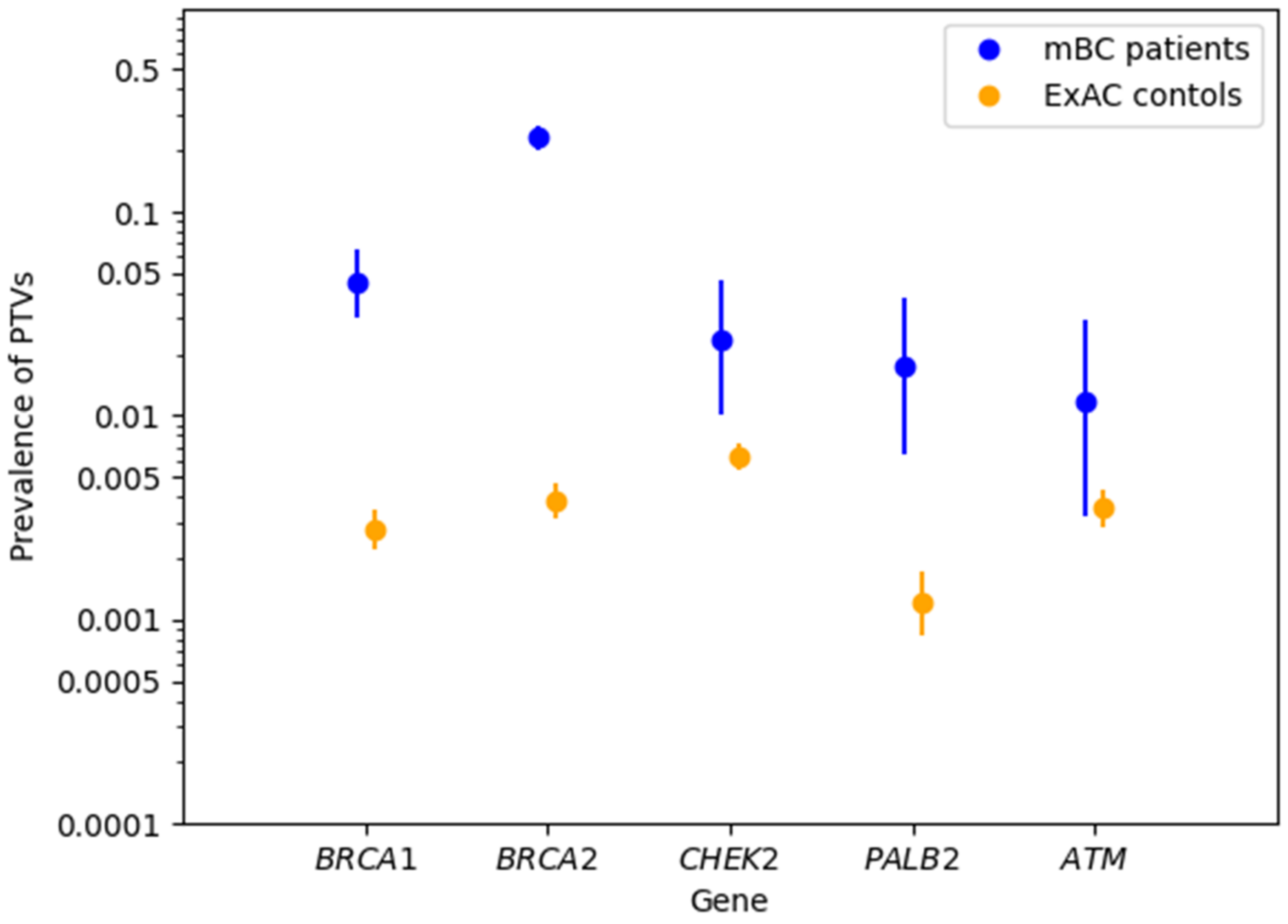
3.3. Associations between mBC and Protein-Truncating Variants in BRCA1/2 and Non-BRCA1/2 Cancer Predisposition Genes

Based on the sequencing results from the publicly available ExAC control dataset [28], case-control analyses were performed to assess associations between mBC and PTVs in BRCA1/2 and selected non-BRCA1/2 genes. PTVs in both BRCA2 and BRCA1 were significantly associated with mBC (BRCA2: OR = 77.41, 95% CI = 58.71–102.33, Fisher’s exact test p < 0.0001; BRCA1: OR = 17.04, 95% CI = 10.54–26.82, p < 0.0001) (Table 2). Among the 340 patients with BRCA1/2-negative mBC, 25 (7.35%) carried PTVs in 10 non-BRCA1/2 genes (Table S1). At a gene-specific level, the prevalence of PTVs in PALB2 was significantly higher in patients with mBC than in the ExAC controls (6/340 (1.76%) vs. 33/27,173 (0.12%); OR = 14.77; 95% CI = 5.02–36.02; p < 0.0001, Table 2). Statistically significant associations were also observed between mBC and PTVs in CHEK2 (OR = 3.78; 95% CI = 1.59–7.71; p = 0.002) and ATM (OR = 3.36; 95% CI = 0.89–8.96; p = 0.04) (Table 2). Notably, the number of identified PTVs was low (≤8) for each of the non-BRCA1/2 genes, and the 95% CIs of the observed PTV prevalence did not reach a distinction for the ATM gene, which only affected four patients with mBC (Figure 2). Beyond PALB2, CHEK2, and ATM, the prevalence of PTVs in the other examined genes was too low for meaningful statistical analysis.

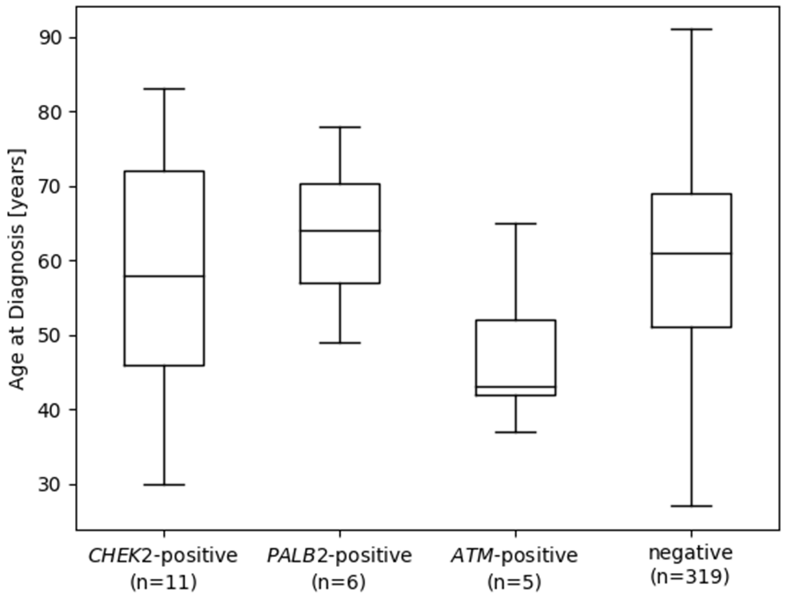
3.4. Pathogenic Variants in Cancer Predisposition Genes according to Cancer Family History, Age at Diagnosis, and Tumor Characteristics

In the subset of 340 BRCA1/2-negative patients with mBC, the overall prevalence of PVs in patients with a BC/OC FH was 9.8% (23/235), and that in patients without a BC/OC FH was 8.8% (9/102) (23/235 vs. 9/102; Fisher’s exact test p = 0.84). When focusing on ATM, CHEK2, and PALB2, the prevalence of PVs in patients with BC/OC FH was 7.7% (18/235), whereas that in patients without BC/OC FH was 2.9% (3/102) (Fisher’s exact test, p = 0.14). The mean AAD of patients with mBC was considerably lower in carriers of a PV in ATM (Figure 3) compared with that in patients without (mean AAD ATM-positive 47.8 years (n = 5) vs. ATM-negative 59.9 years (n = 335), Welch’s t-test p = 0.07). Linear regression analysis with the AAD of mBC in years as the outcome revealed a statistically significant association of PVs in the ATM gene with younger AAD (Table 3) under adjustment for the presence of BC/OC FH, whereas PVs in CHEK2 and PALB2 showed no statistically significant association with AAD.

4. Discussion

Comprehensive germline genetic testing is a prerequisite in targeted risk-adjusted surveillance programs for early cancer detection and targeted therapies in the context of precision oncology. Therefore, germline genetic testing represents a globally established standard; however, it primarily remains limited to women with BC. Research on PARP inhibitors beyond BRCA1/2-mutated carcinomas, for example, is pioneering the field of personalized medicine [30]. Thus, sex-specific consideration of distinct tumor entities in conjunction with their respective predominant germline defects is particularly important. In our multicenter study, we investigated genetic susceptibility to mBC by evaluating the prevalence of PVs in BRCA1/2 using a study sample of 614 patients with mBC and screening their blood-derived DNA samples with multi-gene panel analyses for potentially relevant PVs in 23 non-BRCA1/2 genes. As the largest nationwide sample, with 340 of 614 patients with mBC undergoing comprehensive genetic screening, this study contributes to decoding the genetic predisposition to mBC and reassessing the previously obtained contradictory study results. As expected, the role of BRCA2 as a key risk gene for mBC was highlighted. [12] In the overall study sample, 23.1% (142/614) of patients with mBC carried a PV in BRCA2. Further, as PVs in BRCA1 accounted for 4.6% of the cases, BRCA1 was the second most frequently altered BC predisposition gene. While BRCA2 represents an established BC risk gene for both female and male BC, literature regarding the association between BRCA1 and mBC is limited. The results of our study are in line with those of Li et al. [10], who were among the first to demonstrate a significant association between mBC and PVs in BRCA1 (OR = 17.04, 95% CI 10.54–26.82, p < 10−5).
Consistent with the comprehensive population-specific analyses conducted by Rebbeck et al., we demonstrated that the PVs, BRCA2 c.1813dup and BRCA1 c.5266dup, are the most common variants in the mutational spectrum of mBC. [31] The investigation of available information on ER, PR, and HER2 status revealed predominantly ER-positive, PR-positive, and HER2-negative tumor presentation in the studied sample, consistent with previous findings characterizing the general cancer type in virile breast carcinoma. [32] In this context, Silvestri et al. previously showed that BRCA1/2-positive mBCs were more likely to be ER-positive, PR-positive, and non-triple negative, compared with BC in female BRCA1/2 carriers, suggesting that susceptibility to hereditary BC may be influenced by hormonal background differences between male and female BRCA1/2 mutation carriers [12]. In our study focusing on mBC only, no association of the ER/PR/HER2 status with BRCA1/2 PV occurrence was observed.
Among the non-BRCA1/2 genes, our study confirmed a significantly increased risk association between mBC and PTVs in the BC predisposition genes PALB2, CHEK2, and ATM. This finding may be considered in mBC risk prediction models such as BOADICEA (Breast and Ovarian Analysis of Disease Incidence and Carrier Estimation Algorithm) [33]. We demonstrated the strongest association of mBC in carriers of PVs in PALB2 with an OR of 14.77 (p < 0.0001) compared with that in ExAC controls. As a functional partner and localizer of BRCA2, the tumor suppressor gene PALB2 is critically involved in the homologous recombination (HR) repair mechanism of double-strand breaks [34]. Consistent with previously reported associations between PVs in PALB2 and mBC (with corresponding ORs ranging from 9.63–17.30) [20], our findings contribute to the accumulating evidence for the relevance of PALB2 in genetic testing for hereditary mBC [35,36]. The role of CHEK2 in mBC has been discussed contradictorily in recent studies. A study by Pritzlaff et al. defined CHEK2 as a moderate-risk gene [19], whereas other analyses could not confirm an association of CHEK2 with genetic mBC predisposition [20,21]. In the present investigation, CHEK2 represented the gene most frequently affected by PVs apart from BRCA1/2, with a PV prevalence of 3.2%, and a statistically significant association between PTVs in CHEK2 and mBC risk was confirmed (OR = 3.78, 95% CI = 1.59–7.71, p = 0.002). Furthermore, a statistically significant association was found between PTVs in ATM and mBC (mBC vs. ExAC controls OR = 3.36, 95% CI: 0.89–8.96, p = 0.04), and the data indicate a younger AAD in ATM PV carriers. Future studies with larger sample sizes are required to conclusively assess the role of ATM in mBC development, as our study cohort included only 5 ATM PV carriers. In contrast to the strikingly divergent frequencies of PTVs in BRCA1/2 in male and female patients with BC, the prevalence of PTVs in other predisposition genes, including PALB2, CHEK2, and ATM, were comparable in both genders [18,37]. Patients with BRCA1/2 PVs did not show an earlier disease onset than those without. Except for patients with mBC carrying pathogenic ATM variants, who showed a younger AAD of BC in our study sample, AAD does not seem to be a useful indicator of genetic mBC predisposition. Further, we could not support the results of Pritzlaff et al., who suggested that CHEK2 c.1100delC carriers had a significantly younger AAD compared with that men in carrying PVs in other risk genes [19]. Our results support the NCCN guidelines for male invasive BC version 5.2020, which recommend genetic testing for all men with BC, regardless of their BC/OC FH. Based on this recommendation, we propose to expand the inclusion criteria for testing for genetic to all patients with mBC in Germany, regardless of their AAD, FH, or tumor phenotype. In conclusion, our study results continue to pursue the overall objective of better understanding genetic predisposition to mBC for developing appropriate clinical approaches for sex-specific risk prediction, which could lead to more targeted screening and treatment programs for male PV carriers. It remains to be determined whether the germline mutation status in BRCA1/2, ATM, CHEK2 and PALB2 predicts favourable or unfavourable targeted therapy response in mBC patients, e.g., regarding PARP or CDK4/6 inhibitors [38].
This study has limitations. Our study sample largely comprised patients with mBC who met the GC-HBOC inclusion criteria for germline testing. Therefore, these study results should be validated in a larger study sample of unselected patients with mBC. Further, the prevalence of PTVs in unaffected individuals was retrieved from ExAC non-Finnish Europeans (NFE) under the exclusion of TCGA instead of matched controls, which may have caused bias. The mBC patients who were initially tested for BRCA1/2 mutations only and tested positive were not analyzed further using a more comprehensive gene panel.

5. Conclusions

In addition to PVs in the established mBC risk gene BRCA2, PVs in BRCA1 are particularly associated with an increased risk of mBC. Our results further suggest the role of PALB2, CHEK2, and ATM in mBC predisposition, and support the benefit of multi-gene panel testing in patients with mBC. Due to the high prevalence of BRCA1/2 PVs even in the absence of a BC/OC FH, our data provide a rationale to offer genetic counseling and multi-gene panel testing to all patients with mBC.

Supplementary Materials

The following supporting information can be downloaded at: https://www.mdpi.com/article/10.3390/cancers14133292/s1, Figure S1: Venn diagram of hormone receptor status of 323 indivduals with male breast cancer. ER+/−: Estrogen receptor positive/negative. PR+/−: Progesterone receptor positive/negative. HER2 +/−: human epidermal growth factor receptor 2 positive/negative; Figure S2: Proportion of hormone receptor-positve tumors per age at first diagnosis (AAD) in 323 mBC patients. ER+: Estrogen receptor positive. PR+: Progesterone receptor positive. HER2 +: human epidermal growth factor receptor 2 positive; Table S1: Pathogenic variants (PVs) in non-BRCA1/2 genes detected in 32/340 mBC patients. AAD = age at first diagnosis, CNV = copy number variation, PTV = protein truncating variant, mBC = male breast cancer; Table S2: Binary logistic regression analyses with tumor receptor status as the outcome (1 = positive; 0 = negative) for 323 individuals with male breast cancer. Gene-wise covariates refer to pathogenic variant carrier status (1 = carrier; 0 = non-carrier). CI = confidence interval. SE = standard error.

Author Contributions

Conceptualization: J.H. (Jan Hauke), M.R., J.B., R.K.S., E.H.; Investigation: M.R., M.K., P.N., J.A., H.T.; Methodology: J.H. (Jan Hauke), E.H., M.R., K.M., C.E. (Corinna Ernst), J.B., R.K.S.; Software: C.E. (Corinna Ernst), M.R.; Formal Analysis: M.R., C.E. (Corinna Ernst), J.H. (Jan Hauke); Resources: B.W., J.H. (Jan Hauke), A.G., C.S., J.R. (Juliane Ramser), D.N., J.H. (Judit Horváth), N.A., A.M., B.A., A.R., S.W.-G., J.R. (Julia Ritter), K.R., J.H. (Julia Hentschel), C.E. (Christoph Engel), E.H., R.K.S.; Data curation: M.R., J.B., C.E. (Corinna Ernst), J.H. (Jan Hauke), E.H.; Writing—original draft: M.R., J.B., C.E. (Corinna Ernst), K.M., J.H. (Jan Hauke), E.H.; Writing—review & editing: all authors; Visualization: M.R., J.B., C.E. (Corinna Ernst); Validation: M.R., M.K.; Supervision: E.H., R.K.S., J.H. (Jan Hauke); Project administration: R.K.S., E.H., J.H. (Jan Hauke); Funding acquisition: R.K.S. All authors have read and agreed to the published version of the manuscript.

Funding

The GC-HBOC is supported by the German Cancer Aid (grant no 110837 and grant no 70114178, coordinator: Rita K. Schmutzler, Cologne) and the Federal Ministry of Education and Research (BMBF), Germany (grant no 01GY1901). Genetic analyses were supported by the Köln Fortune Program, Faculty of Medicine, University of Cologne, Germany. The funding sources had no role in the design and conduct of the study; collection, management, analysis, and interpretation of the data; preparation, review, or approval of the manuscript; nor in the decision to submit the manuscript for publication.

Institutional Review Board Statement

Ethical approval was granted by the ethics committee of the University of Cologne (07-048, 22 March 2007).

Informed Consent Statement

Written informed consent was obtained from all patients.

Data Availability Statement

The data presented in this study are available on request from the corresponding author. The data are not publicly available due to privacy.

Acknowledgments

We are thankful to all patients who participated in this study.

Conflicts of Interest

The authors declare no conflict of interest.

Abbreviations

AADage at first diagnosis
ACMGAmerican College of Medical Genetics
BCbreast cancer
CCGCologne Center for Genomics
CIconfidence interval
CNVcopy number variation
ERestrogen receptor
ExACExome Aggregation Consortium
FHfamily history
GC-HBOCGerman Consortium for Hereditary Breast and Ovarian Cancer
HER2human epidermal growth factor receptor 2
IARCInternational Agency for Research on Cancer
mBCmale breast cancer
MLPAmultiplex ligation-dependent probe amplification
NCCNNational Comprehensive Cancer Network
NFEnon-Finnish European
NGSnext-generation sequencing
PRprogesterone receptor
PTVprotein-truncating variant
PVpathogenic variant
OCovarian cancer
ORodds ratio
TCGAThe Cancer Genome Atlas
VUSvariant of uncertain significance

References

  1. Konduri, S.; Singh, M.; Bobustuc, G.; Rovin, R.; Kassam, A. Epidemiology of Male Breast Cancer. Breast 2020, 54, 8–14. [Google Scholar] [CrossRef] [PubMed]
  2. Ly, D.; Forman, D.; Ferlay, J.; Brinton, L.A.; Cook, M.B. An International Comparison of Male and Female Breast Cancer Incidence Rates. Int. J. Cancer 2013, 132, 1918–1926. [Google Scholar] [CrossRef] [PubMed] [Green Version]
  3. Robert Koch-Institut. Krebs in Deutschland 2015/2016; Robert Koch-Institut, und Gesellschaft der Epidemiologischen Krebsregister in Deutschland, E.V.: Berlin, Germany, 2019. [Google Scholar] [CrossRef]
  4. Giordano, S.H. Breast Cancer in Men. N. Engl. J. Med. 2018, 378, 2311–2320. [Google Scholar] [CrossRef] [PubMed]
  5. Leon-Ferre, R.A.; Giridhar, K.V.; Hieken, T.J.; Mutter, R.W.; Couch, F.J.; Jimenez, R.E.; Hawse, J.R.; Boughey, J.C.; Ruddy, K.J. A Contemporary Review of Male Breast Cancer: Current Evidence and Unanswered Questions. Cancer Metastasis Rev. 2018, 37, 599–614. [Google Scholar] [CrossRef]
  6. Campos, F.A.B.; Rouleau, E.; Torrezan, G.T.; Carraro, D.M.; Casali da Rocha, J.C.; Mantovani, H.K.; da Silva, L.R.; Osório, C.A.B.d.T.; Moraes Sanches, S.; Caputo, S.M.; et al. Genetic Landscape of Male Breast Cancer. Cancers 2021, 13, 3535. [Google Scholar] [CrossRef]
  7. Fentiman, I.S. Male Breast Cancer Is Not Congruent with the Female Disease. Crit. Rev. Oncol./Hematol. 2016, 101, 119–124. [Google Scholar] [CrossRef]
  8. De Juan, I.; Palanca, S.; Domenech, A.; Feliubadaló, L.; Segura, Á.; Osorio, A.; Chirivella, I.; de la Hoya, M.; Sánchez, A.B.; Infante, M.; et al. BRCA1 and BRCA2 Mutations in Males with Familial Breast and Ovarian Cancer Syndrome. Results of a Spanish Multicenter Study. Fam. Cancer 2015, 14, 505–513. [Google Scholar] [CrossRef]
  9. Ottini, L. Male Breast Cancer: A Rare Disease That Might Uncover Underlying Pathways of Breast Cancer. Nat. Rev. Cancer 2014, 14, 643–644. [Google Scholar] [CrossRef]
  10. Li, S.; Silvestri, V.; Leslie, G.; Rebbeck, T.R.; Neuhausen, S.L.; Hopper, J.L.; Nielsen, H.R.; Lee, A.; Yang, X.; McGuffog, L.; et al. Cancer Risks Associated With BRCA1 and BRCA2 Pathogenic Variants. J. Clin. Oncol. 2022, 40, JCO2102112. [Google Scholar] [CrossRef]
  11. Barnes, D.R.; Silvestri, V.; Leslie, G.; McGuffog, L.; Dennis, J.; Yang, X.; Adlard, J.; Agnarsson, B.A.; Ahmed, M.; Aittomäki, K.; et al. Breast and Prostate Cancer Risks for Male BRCA1 and BRCA2 Pathogenic Variant Carriers Using Polygenic Risk Scores. JNCI J. Natl. Cancer Inst. 2021, 114, djab147. [Google Scholar] [CrossRef]
  12. kConFab Investigators; Hereditary Breast and Ovarian Cancer Research Group Netherlands (HEBON); EMBRACE; Silvestri, V.; Barrowdale, D.; Mulligan, A.M.; Neuhausen, S.L.; Fox, S.; Karlan, B.Y.; Mitchell, G.; et al. Male Breast Cancer in BRCA1 and BRCA2 Mutation Carriers: Pathology Data from the Consortium of Investigators of Modifiers of BRCA1/2. Breast Cancer Res. 2016, 18, 15. [Google Scholar] [CrossRef] [PubMed] [Green Version]
  13. Ibrahim, M.; Yadav, S.; Ogunleye, F.; Zakalik, D. Male BRCA Mutation Carriers: Clinical Characteristics and Cancer Spectrum. BMC Cancer 2018, 18, 179. [Google Scholar] [CrossRef] [PubMed] [Green Version]
  14. Stratton, M.R.; Ford, D.; Neuhasen, S.; Seal, S.; Wooster, R.; Friedman, L.S.; King, M.-C.; Egilsson, V.; Devilee, P.; McManus, R.; et al. Familial Male Breast Cancer Is Not Linked to the BRCA1 Locus on Chromosome 17q. Nat. Genet. 1994, 7, 103–107. [Google Scholar] [CrossRef] [PubMed]
  15. Tai, Y.C.; Domchek, S.; Parmigiani, G.; Chen, S. Breast Cancer Risk Among Male BRCA1 and BRCA2 Mutation Carriers. JNCI J. Natl. Cancer Inst. 2007, 99, 1811–1814. [Google Scholar] [CrossRef] [PubMed]
  16. Hassett, M.J.; Somerfield, M.R.; Baker, E.R.; Cardoso, F.; Kansal, K.J.; Kwait, D.C.; Plichta, J.K.; Ricker, C.; Roshal, A.; Ruddy, K.J.; et al. Management of Male Breast Cancer: ASCO Guideline. J. Clin. Oncol. 2020, 38, JCO.19.03120. [Google Scholar] [CrossRef]
  17. Arbeitsgemeinschaft der Wissenschaftlichen Medizinischen Fachgesellschaften (AWMF). Interdisziplinäre S3-Leitlinie Für Die Früherkennung, Diagnostik, Therapie Und Nachsorge Des Mammakarzinoms Version 4.3 AWMF-Registernummer: 032-045OL; Version 4.3; Deutsche Krebsgesellschaft: Berlin, Germany, 2020. [Google Scholar]
  18. Hauke, J.; Horvath, J.; Groß, E.; Gehrig, A.; Honisch, E.; Hackmann, K.; Schmidt, G.; Arnold, N.; Faust, U.; Sutter, C.; et al. Gene Panel Testing of 5589 BRCA1/2 -Negative Index Patients with Breast Cancer in a Routine Diagnostic Setting: Results of the German Consortium for Hereditary Breast and Ovarian Cancer. Cancer Med. 2018, 7, 1349–1358. [Google Scholar] [CrossRef]
  19. Pritzlaff, M.; Summerour, P.; McFarland, R.; Li, S.; Reineke, P.; Dolinsky, J.S.; Goldgar, D.E.; Shimelis, H.; Couch, F.J.; Chao, E.C.; et al. Male Breast Cancer in a Multi-Gene Panel Testing Cohort: Insights and Unexpected Results. Breast Cancer Res. Treat. 2017, 161, 575–586. [Google Scholar] [CrossRef] [Green Version]
  20. Rizzolo, P.; Zelli, V.; Silvestri, V.; Valentini, V.; Zanna, I.; Bianchi, S.; Masala, G.; Spinelli, A.M.; Tibiletti, M.G.; Russo, A.; et al. Insight into Genetic Susceptibility to Male Breast Cancer by Multigene Panel Testing: Results from a Multicenter Study in Italy: Multigene Panel Testing in Male Breast Cancer. Int. J. Cancer 2019, 145, 390–400. [Google Scholar] [CrossRef]
  21. Fostira, F.; Saloustros, E.; Apostolou, P.; Vagena, A.; Kalfakakou, D.; Mauri, D.; Tryfonopoulos, D.; Georgoulias, V.; Yannoukakos, D.; Fountzilas, G.; et al. Germline Deleterious Mutations in Genes Other than BRCA2 Are Infrequent in Male Breast Cancer. Breast Cancer Res. Treat. 2018, 169, 105–113. [Google Scholar] [CrossRef]
  22. Szwiec, M.; Tomiczek-Szwiec, J.; Kluźniak, W.; Wokołorczyk, D.; Osowiecka, K.; Sibilski, R.; Wachowiak, M.; Gronwald, J.; Gronwald, H.; Lubiński, J.; et al. Genetic Predisposition to Male Breast Cancer in Poland. BMC Cancer 2021, 21, 975. [Google Scholar] [CrossRef]
  23. Tedaldi, G.; Tebaldi, M.; Zampiga, V.; Cangini, I.; Pirini, F.; Ferracci, E.; Danesi, R.; Arcangeli, V.; Ravegnani, M.; Martinelli, G.; et al. Male Breast Cancer: Results of the Application of Multigene Panel Testing to an Italian Cohort of Patients. Diagnostics 2020, 10, 269. [Google Scholar] [CrossRef] [PubMed]
  24. Wappenschmidt, B.; Hauke, J.; Faust, U.; Niederacher, D.; Wiesmüller, L.; Schmidt, G.; Groß, E.; Gehrig, A.; Sutter, C.; Ramser, J.; et al. Criteria of the German Consortium for Hereditary Breast and Ovarian Cancer for the Classification of Germline Sequence Variants in Risk Genes for Hereditary Breast and Ovarian Cancer. Geburtshilfe Frauenheilkd. 2020, 80, 410–429. [Google Scholar] [CrossRef] [PubMed] [Green Version]
  25. ACMG Laboratory Quality Assurance Committee; Richards, S.; Aziz, N.; Bale, S.; Bick, D.; Das, S.; Gastier-Foster, J.; Grody, W.W.; Hegde, M.; Lyon, E.; et al. Standards and Guidelines for the Interpretation of Sequence Variants: A Joint Consensus Recommendation of the American College of Medical Genetics and Genomics and the Association for Molecular Pathology. Genet. Med. 2015, 17, 405–423. [Google Scholar] [CrossRef] [Green Version]
  26. Plon, S.E.; Eccles, D.M.; Easton, D.; Foulkes, W.D.; Genuardi, M.; Greenblatt, M.S.; Hogervorst, F.B.L.; Hoogerbrugge, N.; Spurdle, A.B.; Tavtigian, S.V.; et al. Sequence Variant Classification and Reporting: Recommendations for Improving the Interpretation of Cancer Susceptibility Genetic Test Results. Hum. Mutat. 2008, 29, 1282–1291. [Google Scholar] [CrossRef] [PubMed] [Green Version]
  27. Plagnol, V.; Curtis, J.; Epstein, M.; Mok, K.Y.; Stebbings, E.; Grigoriadou, S.; Wood, N.W.; Hambleton, S.; Burns, S.O.; Thrasher, A.J.; et al. A Robust Model for Read Count Data in Exome Sequencing Experiments and Implications for Copy Number Variant Calling. Bioinformatics 2012, 28, 2747–2754. [Google Scholar] [CrossRef] [PubMed] [Green Version]
  28. Exome Aggregation Consortium; Lek, M.; Karczewski, K.J.; Minikel, E.V.; Samocha, K.E.; Banks, E.; Fennell, T.; O’Donnell-Luria, A.H.; Ware, J.S.; Hill, A.J.; et al. Analysis of Protein-Coding Genetic Variation in 60,706 Humans. Nature 2016, 536, 285–291. [Google Scholar] [CrossRef] [Green Version]
  29. Kääriäinen, H.; Muilu, J.; Perola, M.; Kristiansson, K. Genetics in an Isolated Population like Finland: A Different Basis for Genomic Medicine? J. Community Genet. 2017, 8, 319–326. [Google Scholar] [CrossRef] [Green Version]
  30. Tomasik, B.; Bieńkowski, M.; Jassem, J. PARP Inhibitors beyond BRCA-Mutated Cancers: Precision Medicine at the Crossroads. Precis. Cancer Med. 2021, 4, 19. [Google Scholar] [CrossRef]
  31. Rebbeck, T.R.; Friebel, T.M.; Friedman, E.; Hamann, U.; Huo, D.; Kwong, A.; Olah, E.; Olopade, O.I.; Solano, A.R.; Teo, S.-H.; et al. Mutational Spectrum in a Worldwide Study of 29,700 Families with BRCA1 or BRCA2 Mutations. Hum. Mutat. 2018, 39, 593–620. [Google Scholar] [CrossRef] [Green Version]
  32. Cardoso, F.; Bartlett, J.M.S.; Slaets, L.; van Deurzen, C.H.M.; van Leeuwen-Stok, E.; Porter, P.; Linderholm, B.; Hedenfalk, I.; Schröder, C.; Martens, J.; et al. Characterization of Male Breast Cancer: Results of the EORTC 10085/TBCRC/BIG/NABCG International Male Breast Cancer Program. Ann. Oncol. 2018, 29, 405–417. [Google Scholar] [CrossRef]
  33. Lee, A.; Yang, X.; Tyrer, J.; Gentry-Maharaj, A.; Ryan, A.; Mavaddat, N.; Cunningham, A.P.; Carver, T.; Archer, S.; Leslie, G.; et al. Comprehensive epithelial tubo-ovarian cancer risk prediction model incorporating genetic and epidemiological risk factors. J. Med. Genet. 2022, 59, 632–643. [Google Scholar] [CrossRef] [PubMed]
  34. Wu, S.; Zhou, J.; Zhang, K.; Chen, H.; Luo, M.; Lu, Y.; Sun, Y.; Chen, Y. Molecular Mechanisms of PALB2 Function and Its Role in Breast Cancer Management. Front. Oncol. 2020, 10, 301. [Google Scholar] [CrossRef] [PubMed] [Green Version]
  35. Silvestri, V.; Zelli, V.; Valentini, V.; Rizzolo, P.; Navazio, A.S.; Coppa, A.; Agata, S.; Oliani, C.; Barana, D.; Castrignanò, T.; et al. Whole-Exome Sequencing and Targeted Gene Sequencing Provide Insights into the Role of PALB2 as a Male Breast Cancer Susceptibility Gene: PALB2 Mutations in Male Breast Cancer. Cancer 2017, 123, 210–218. [Google Scholar] [CrossRef] [PubMed] [Green Version]
  36. Ding, Y.C.; Steele, L.; Kuan, C.-J.; Greilac, S.; Neuhausen, S.L. Mutations in BRCA2 and PALB2 in Male Breast Cancer Cases from the United States. Breast Cancer Res. Treat. 2011, 126, 771–778. [Google Scholar] [CrossRef] [PubMed] [Green Version]
  37. Aloraifi, F.; McCartan, D.; McDevitt, T.; Green, A.J.; Bracken, A.; Geraghty, J. Protein-Truncating Variants in Moderate-Risk Breast Cancer Susceptibility Genes: A Meta-Analysis of High-Risk Case-Control Screening Studies. Cancer Genet. 2015, 208, 455–463. [Google Scholar] [CrossRef] [PubMed]
  38. Zheng, G.; Leone, J.P. Male Breast Cancer: An Updated Review of Epidemiology, Clinicopathology, and Treatment. J. Oncol. 2022, 2022, 1734049. [Google Scholar] [CrossRef] [PubMed]
Figure 1. Gene-specific prevalence of heterozygous pathogenic germline variants (PVs) in 340 patients with BRCA1/2-negative male breast cancer.
Cancers 14 03292 g001
Figure 2. Prevalence of protein-truncating variants (PTVs) with binomial 95% confidence intervals (CIs) per gene in patients with male breast cancer and in ExAC controls.
Cancers 14 03292 g002
Figure 3. Age at diagnosis (AAD) of male breast cancer in BRCA1/2-negative individuals based on germline pathogenic variant (PV) status in the CHEK2, PALB2, and ATM genes. One individual (AAD = 37 years) carried PVs in both CHEK2 and ATM. The term “negative” indicates individuals without PVs in BRCA1/2, CHEK2, PALB2, and ATM.
Cancers 14 03292 g003
Table 1. Patient and cancer characteristics in the overall study sample (n = 614) stratified by pathogenic variant (PV) status, family history (FH), age at first diagnosis (AAD). BC = breast cancer, ER = estrogen receptor, mBC = male breast cancer, PR = progesterone receptor.
SubgroupOverall Study Sample (%)BRCA1/2-
Positive
BRCA1-
Positive
BRCA2-
Positive
BRCA1/2 Negative Patients Further InvestigatedCarriers of Pathogenic Variants in 23 Non-BRCA1/2-Genes (%)
patients with mBC614 (100)1702814234032 (9.4)
unilateral BC586 (95.4)1602813232832 (9.8)
bilateral BC28 (4.6)10010120 (0)
BC/OC FH *460 (75.9)1542612823523 (9.8)
no BC/OC FH *146 (24.1)162141029 (8.8)
mean AAD (range) *60 (22–91)62 (33–83)62 (33–82)62 (37–83)60 (27–91)58 (30–83)
AAD < 40 years30 (4.9)4 (2.4)13182 (11.2)
AAD 40–49 years90 (14.8)18 (10.6)414558 (14.5)
AAD 50–59 years163 (26.8)42 (24.7)537897 (7.9)
AAD 60–69 years183 (30.0)61 (35.9)952977 (7.2)
AAD 70–79 years125 (20.5)40 (23.5)832697 (10.1)
AAD > 80 years18 (3.0)5 [2.9]14121 (8.3)
ER/PR-status available (%)407 (100)107 (100)17 (100)90 (100)243 (100)24 (100)
HER2-status available (%)323 (100)90 (100)15 (100)75 (100)200 (100)21 (100)
ER-positive (%)394 (96.8)105 (98.1)17 (100)88 (97.8)234 (96.3)24 (100)
ER-negative (%)13 (3.2)2 (1.9)0 (0)2 (2.2)9 (3.7)0 (0)
PR-positive (%)366 (89.9)94 (87.9)14 (82.4)80 (88.9)219 (90.1)23 (96.0)
PR-negative (%)41 (10.1)13 (12.1)3 (17.6)10 (11.1)24 (9.9)1 (4.2)
HER2-positive (%)38 (11.7)13 (14.4)2 (13.3)11 (14.7)20 (10.0)4 (19.0)
HER2-negative (%)285 (88.2)77 (85.6)13 (86.7)64 (85.3)180 (90.0)17 (81.0)
* total missing information regarding AAD (n = 5) and missing information regarding FH (n = 8).
Table 2. Prevalence of protein-truncating variants (PTVs) in (suspected) cancer predisposition genes in patients with mBC compared with the control dataset (ExAC). Mutation carrier frequencies are shown in parentheses. Univariate logistic regression analysis was performed to estimate the odds ratios (OR) and corresponding 95% confidence intervals (CI).
GenemBC PTVs (%)Patients with mBCExAC Controls
n = 27,173 (%)
mBC vs. ExAC
OR (95% CI)p *
BRCA2142 (23.13)614105 (0.39)77.41 (58.71–102.33)<10−5
BRCA128 (4.56)61476 (0.28)17.04 (10.54–26.82)<10−5
CHEK28 (2.35)340172 (0.63)3.78 (1.59–7.71)0.002
PALB26 (1.76)34033 (0.12)14.77 (5.02–36.02)<10−5
ATM4 (1.18)34096 (0.35)3.36 (0.89–8.96)0.04
FANCM2 (0.59)340184 (0.68)--
BRIP11 (0.29)34059 (0.22)--
CDH11 (0.29)3402 (0.00)--
NBN **1 (0.29)34042 (0.15)--
PTEN1 (0.29)3401 (0.00)--
RAD50 **1 (0.29)34084 (0.31)--
RAD51C1 (0.29)34034 (0.13)--
* Fisher’s exact test; ** one patient carried two PTVs (NBN/RAD50).
Table 3. Linear regression analysis results with age at first diagnosis (AAD) of male breast cancer (mBC) with years as the outcome in 337 BRCA1/2-negative patients with mBC. Gene-wise covariates refer to pathogenic variant status (1: pathogenic variant, 0: no pathogenic variant). FH = Breast/ovarian cancer family history (1: yes 0: no).
Covariatβ95% CIp
FH1.78−0.97–4.520.20
ATM−11.87−22.35–−1.390.03
CHEK2−1.96−9.09–5.170.59
PALB23.55−5.98–13.070.46
Publisher’s Note: MDPI stays neutral with regard to jurisdictional claims in published maps and institutional affiliations.

