Cerebral MRI and Clinical Findings in Children with PTEN Hamartoma Tumor Syndrome: Can Cerebral MRI Scan Help to Establish an Earlier Diagnosis of PHTS in Children?

 , ,
, ,
Abstract
:1. Introduction
2. Methods
3. Results
3.1. Macrocephaly
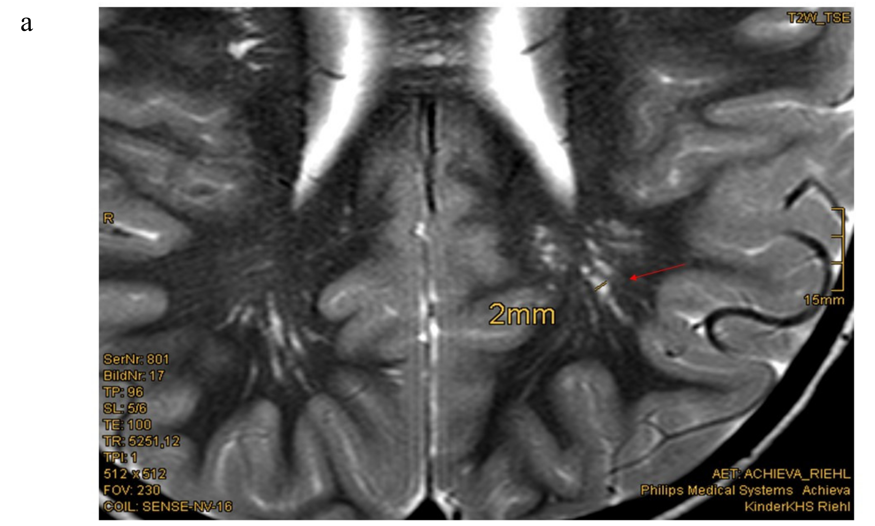
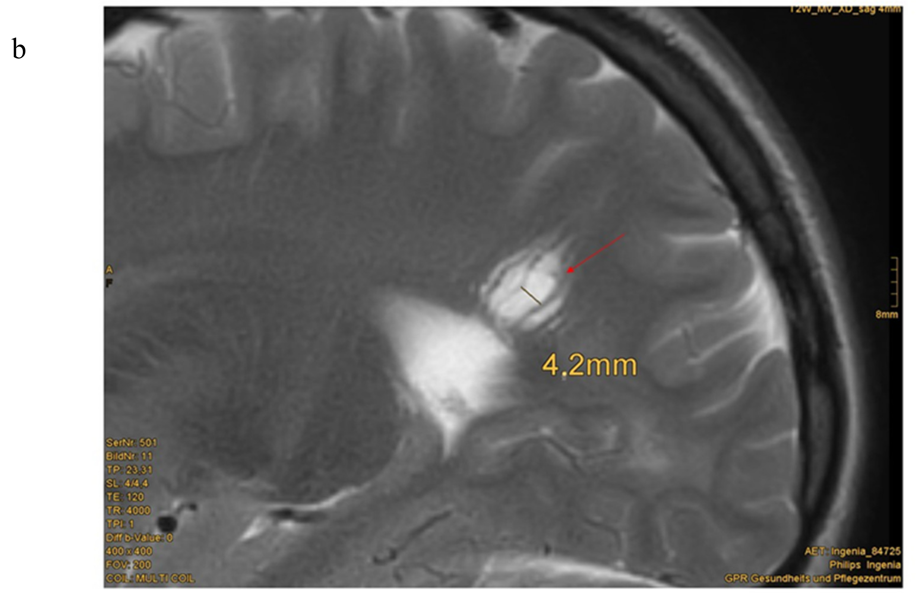
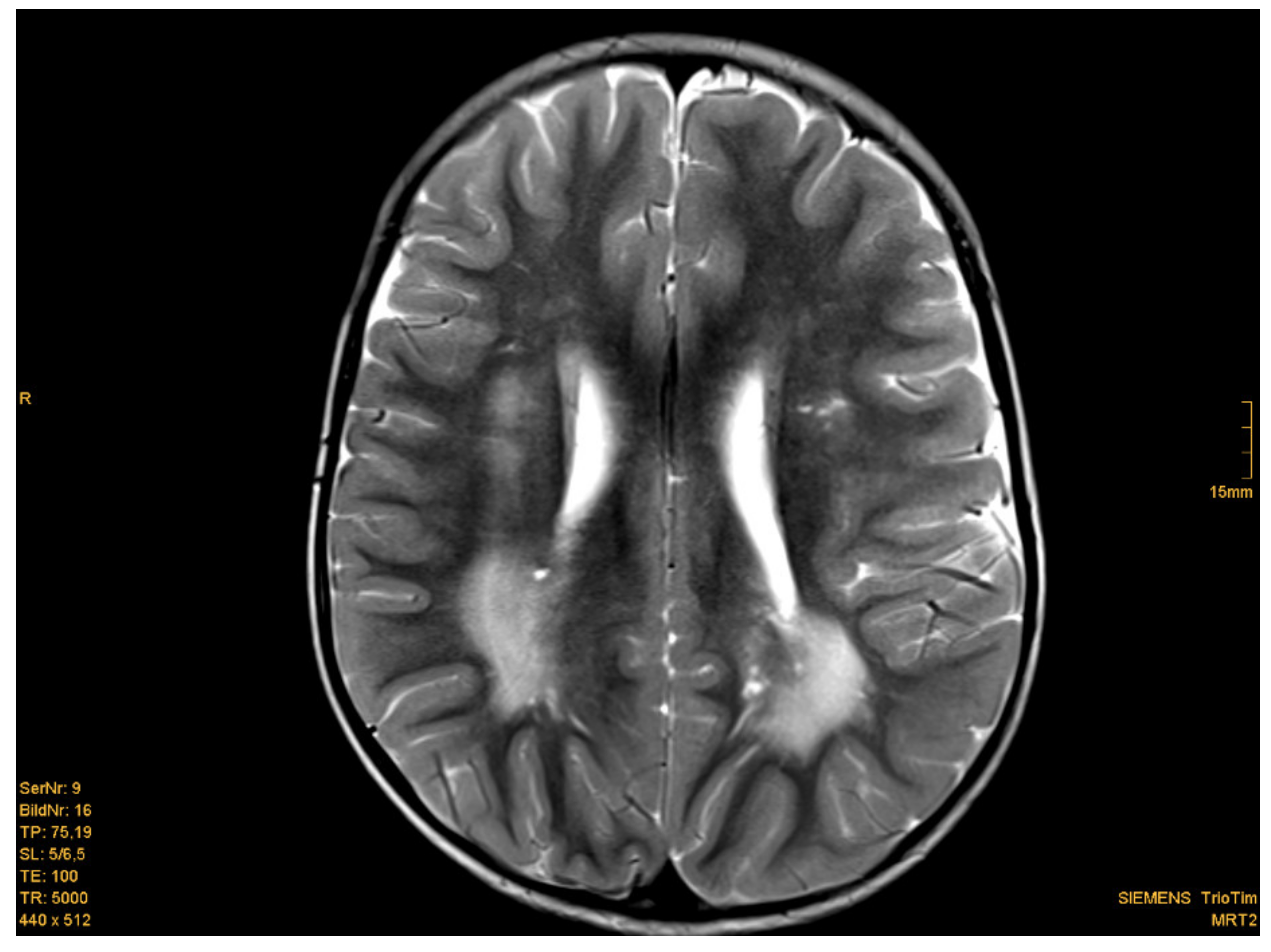
3.2. MRI
3.3. Psychomotor Development
3.4. Additional Phenotypical Features
4. Discussion
5. Conclusions
Author Contributions
Funding
Acknowledgments
Conflicts of Interest
References
- Tan, M.H.; Mester, J.L.; Ngeow, J.; Rybicki, L.A.; Orloff, M.S.; Eng, C. Lifetime cancer risks in individuals with germline PTEN mutations. Clin. Cancer Res. 2012, 18, 400–407. [Google Scholar] [CrossRef] [PubMed] [Green Version]
- Plamper, M.; Schreiner, F.; Gohlke, B.; Kionke, J.; Korsch, E.; Kirkpatrick, J.; Born, M.; Aretz, S.; Woelfle, J. Thyroid disease in children and adolescents with PTEN hamartoma tumor syndrome (PHTS). Eur. J. Pediatr. 2018, 177, 429–435. [Google Scholar] [CrossRef] [PubMed]
- Smith, J.R.; Marqusee, E.; Webb, S.; Nose, V.; Fishman, S.J.; Shamberger, R.C.; Frates, M.C.; Huang, S.A. Thyroid nodules and cancer in children with PTEN hamartoma tumor syndrome. J. Clin. Endocrinol. Metab. 2011, 96, 34–37. [Google Scholar] [CrossRef] [PubMed] [Green Version]
- Smpokou, P.; Fox, V.L.; Tan, W.H. PTEN hamartoma tumour syndrome: Early tumour development in children. Arch. Dis. Child. 2015, 100, 34–37. [Google Scholar] [CrossRef]
- Marsh, D.J.; Kum, J.B.; Lunetta, K.L.; Bennett, M.J.; Gorlin, R.J.; Ahmed, S.F.; Bodurtha, J.; Crowe, C.; Curtis, M.A.; Dasouki, M.; et al. PTEN mutation spectrum and genotype-phenotype correlations in Bannayan-Riley-Ruvalcaba syndrome suggest a single entity with Cowden syndrome. Hum. Mol. Genet. 1999, 8, 1461–1472. [Google Scholar] [CrossRef]
- Parisi, M.A.; Dinulos, M.B.; Leppig, K.A.; Sybert, V.P.; Eng, C.; Hudgins, L. The spectrum and evolution of phenotypic findings in PTEN mutation positive cases of Bannayan-Riley-Ruvalcaba syndrome. J. Med. Genet. 2001, 38, 52–58. [Google Scholar] [CrossRef]
- Tan, W.H.; Baris, H.N.; Burrows, P.E.; Robson, C.D.; Alomari, A.L.; Mulliken, J.B.; Fishman, S.J.; Irons, M.B. The spectrum of vascular anomalies in patients with PTEN mutations: Implications for diagnosis and management. J. Med. Genet. 2007, 44, 594–602. [Google Scholar] [CrossRef] [Green Version]
- Tan, M.H.; Mester, J.; Peterson, C.; Yang, Y.; Chen, J.L.; Rybicki, L.A.; Milas, K.; Pederson, H.; Remzi, B.; Orloff, M.S.; et al. A clinical scoring system for selection of patients for PTEN mutation testing is proposed on the basis of a prospective study of 3042 probands. Am. J. Hum. Genet. 2011, 88, 42–56. [Google Scholar] [CrossRef] [Green Version]
- Kato, K.; Mizuno, S.; Inaba, M.; Fukumura, S.; Kurahashi, N.; Maruyama, K.; Leda, D.; Ohashi, K.; Hori, I.; Negishi, Y.; et al. Distinctive facies, macrocephaly, and developmental delay are signs of PTEN mutation in childhood. Brain Dev. 2018, 40, 678–684. [Google Scholar] [CrossRef]
- Busa, T.; Milh, M.; Degardin, N.; Girard, N.; Sigaudy, S.; Longy, M.; Olshchwang, S.; Sobol, H.; Chabrol, B.; Philip, N. Clicical presentations of PTEN mutations in childhood in the absence of family history of Cowden syndrome. Eur. J. Paediatr. Neurol. 2015, 19, 188–192. [Google Scholar] [CrossRef]
- Plamper, M.; Gohlke, B.; Schreiner, F.; Woelfle, J. Phenotype-Driven Diagnostic of PTEN Hamartoma Tumor Syndrome: Macrocephaly, But Neither Height nor Weight Development, Is the Important Trait in Children. Cancers 2019, 11, 975. [Google Scholar] [CrossRef] [PubMed] [Green Version]
- Lok, C.; Viseux, V.; Avril, M.F.; Richard, M.A.; Gondry-Jouet, C.; Deramond, H.; Desfossez-Tribout, C.; Courtade, S.; Delaunay, M.; Piette, F.; et al. Cancerology Group of the French Society of Dermatology. Brain magnetic resonance imaging in patients with Cowden syndrome. Medicine (Baltimore) 2005, 84, 129–136. [Google Scholar] [CrossRef]
- Balci, T.B.; Davila, J.; Lewis, D.; Boafo, A.; Sell, E.; Richer, J.; Nikkel, S.M.; Armour, C.M.; Tomiak, E.; Lines, M.A.; et al. Broad spectrum of neuropsychiatric phenotypes associated with white matter disease in PTEN hamartoma tumor syndrome. Am. J. Med. Genet. Part B 2018, 177, 101–109. [Google Scholar] [CrossRef] [PubMed]
- Dhamija, R.; Weindling, S.M.; Porter, A.B.; Hu, L.S.; Wood, C.P.; Hoxworth, J.M. Neuroimaging abnormalities in patients with Cowden syndrome: Retrospective single-center study. Neurol. Clin. Pract. 2018, 8, 207–213. [Google Scholar] [CrossRef] [PubMed]
- Shiohama, T.; Levman, J.; Vasung, L.; Takahashi, E. Brain morphological analysis in PTEN hamartoma tumor syndrome. Am. J. Med. Genet. A 2020, 182, 1117–1129. [Google Scholar] [CrossRef]
- Farooq, A.; Walker, L.J.; Bowling, J.; Audisio, R.A. Cowden syndrome. Cancer Treat. Rev. 2010, 36, 577–583. [Google Scholar] [CrossRef]
- Nelen, M.R.; Kremer, H.; Konings, I.B.; Schoute, F.; van Essen, A.J.; Koch, R.; Woods, C.G.; Fryns, J.P.; Hamel, B.; Hoefsloot, L.H.; et al. Novel PTEN mutations in patients with Cowden disease: Absence of clear genotype-phenotype correlations. Eur. J. Hum. Genet. 1999, 7, 267–273. [Google Scholar] [CrossRef]
- Pilarski, R.; Burt, R.; Kohlman, W.; Pho, L.; Shannon, K.M.; Swisher, E. Cowden syndrome and the PTEN hamartoma tumor syndrome: Systematic review and revised diagnostic criteria. J. Natl. Cancer Inst. 2013, 105, 1607–1616. [Google Scholar] [CrossRef] [Green Version]
- Daly, M.B.; Pilarski, R.; Axilbund, J.E.; Buys, S.S.; Crawford, B.; Friedman, S.; Garber, J.E.; Horton, C.; Kaklamani, V.; Klein, C.; et al. Genetic/Familial High-Risk Assessment: Breast and Ovarien, Version 1. J. Natl. Compr. Cancer Netw. 2014, 12, 1326–1338. [Google Scholar] [CrossRef]
- Lachlan, K.L.; Lucassen, A.M.; Bunyan, D.; Temple, I.K. Cowden syndrome and Bannayan Riley Ruvalcaba syndrome represent one condition with variable expression and age-related penetrance: Results of a clinical study of PTEN mutation carriers. J. Med. Genet. 2007, 44, 579–585. [Google Scholar] [CrossRef] [Green Version]
- Pavone, P.; Pratico, A.D.; Rizzo, R.; Corsello, G.; Ruggieri, M.; Parano, E.; Falsaperla, R. A clinical review on megalencephaly: A large brain as a possible sign of cerebral impairment. Medicine 2017, 96, e6814. [Google Scholar] [CrossRef] [PubMed]
- Mirzaa, G.M.; Riviere, J.B.; Dobyns, W.B. Megalencephaly syndromes and activation mutatons in the PI3K-AKT pathway: MPPH and MCAP. Am. J. Med. Genet. C Semin. Med. Genet. 2013, 163, 122–130. [Google Scholar] [CrossRef] [PubMed]
- Mirzaa, G.M.; Pduri, A. Megalencephaly and hemimegalencephaly: Breaktroughs in molecular etiology. Am. J. Med. Genet. C Semin. Med. Genet. 2014, 166, 156–172. [Google Scholar] [CrossRef] [PubMed]
- Eichhorn, G.R.; Ammache, Z.; Bell, W.; Yuh, W.T. Unusually prominent perivascular spaces. Neurology 2011, 56, 1242. [Google Scholar] [CrossRef] [PubMed] [Green Version]
- Heier, L.A.; Bauer, C.J.; Schwartz, L.; Zimmerman, R.D.; Morgello, S.; Deck, M.D. Large Virchow-Robin spaces: MR-clinical correlation. AJNR Am. J. Neuroradiol. 1989, 10, 929–936. [Google Scholar] [PubMed]
- Rudie, J.D.; Rauschecker, A.M.; Nabavizadeh, S.A.; Mohan, S. Neuroimaging of Dilated Perivascular spaces: From Benign and Pathologic Causes to Mimics. J. Neuroimaging 2018, 28, 139–149. [Google Scholar] [CrossRef] [PubMed]
- MacLullich, A.M.; Wardlaw, J.M.; Ferguson, K.J.; Starr, J.M.; Seckl, J.R.; Deary, I.J. Enlarged perivascular spaces are associated with cognitive function in healthy elderly men. J. Neurol. Neurosurg. Psychiatry 2004, 75, 1519–1523. [Google Scholar] [CrossRef] [Green Version]
- Vanderver, A.; Tonduti, D.; Kahn, I.; Schmidt, J.; Medne, L.; Vento, J.; Van Der Knaap, M.S. Characteristic brain magnetic resonance imaging pattern in patients with macrocephaly and PTEN mutations. Am. J. Med. Genet. Part A 2014, 164, 627–633. [Google Scholar] [CrossRef] [Green Version]
- Busch, R.M.; Strivasta, S.; Hogue, O.; Frazier, T.W.; Klaas, P.; Hardan, A.; Martinez-Agosto, J.A.; Sahin, M.; Eng, C. Developmental Synaptopathies Consortium, Neurobehavioral phenotype of autism spectrum disorder associated with germline heterozygous mutations in PTEN. Transl. Psychiatry 2019, 9, 253. [Google Scholar] [CrossRef] [Green Version]
- Busch, R.M.; Chapin, J.S.; Mester, J.; Fergueson, L.; Haut, J.S.; Frazier, T.W.; Eng, C. The Cognitive Characteristics of PTEN Hamartoma Tumor Syndromes. Genet. Med. 2013, 15, 548–553. [Google Scholar] [CrossRef] [Green Version]
- Frazier, T.W.; Embacher, R.; Tilot, A.K.; Koenig, K.; Mester, J.; Eng, C. Molecular and phenotypic abnormalities in individuals with germline heterozygous PTEN mutations and autism. Mol. Psychiatry 2015, 20, 1132–1138. [Google Scholar] [CrossRef] [PubMed] [Green Version]
- AWMG Guidelins: S1-Leitlinie. Diagnostik und Management von Patientin mit PTEN Hamartom Tumor Syndrom (PHTS) im Kinder- und Jugendalter. Registernr.: 174-025. Available online: https://www.awmf.org/leitlinien/detail/ll/174-025.html (accessed on 30 October 2019).


| Pat.No | Sex | Mutation/Deletion in PTEN Gene (Localisation) | Age at DIAGNOSIS (Years) | Age at cMRI (Years) | Results of cMRI Scan | IQ > 85 | Delay in Motor Develop-ment | Muscle Hypotonia | Confirmed Autism | More Detailed Description of Neurological Features and Academic Performance | Age at Start of Walking Independently (Months) |
|---|---|---|---|---|---|---|---|---|---|---|---|
| 1 | male | c.389G > A; Arg130 Gln (exon 5) | 3.8 | n.d. | n.d. | + | + | - | - | moderate delay in motor development, normal intelligence, secondary school −>university | 22 |
| 2 | male | c.389G > A; Arg130 Gln (exon 5) | 4 | 0.25 2.5 | enlarged perivascular spaces | - | + | + | + | muscle hypotonia, autism, developmental delay in motor and language development, no expressive speech | 26 |
| 3 | male | c.540C > A; p.Y180X (exon 6) | 5.3 | 6 | enlarged perivascular spaces | + | + | - | - | delay of fine motor skills, normal intelligence, secondary school | 18 |
| 4 | male | c.737C > T.p.Pro246Leu (exon 7) | 1.3 | normal MRI scan (reported) | + | - | - | - | None, normal intelligence, secondary school | 17 | |
| 5 | male | c.209 + 5G > A (Intron 3) | 2 | 1.75 | white matter abnormalities, (periventricular posterior white matter), enlarged perivascular spaces | + | + | - | - | delay of gross and fine motor skills, normal intelligence, impulsivity, secondary school | 18 |
| 6 | male | c.445C > T; Gln149X (exon 5) | 3 | 2.0 | white matter abnormalities (posterior horn up to parietal white matter; smaller frontal and periventricular lesions), enlarged perivascular spaces | + | - | - | - | None, normal intelligence, elementary school | 18 |
| 7 | male | c.509G > A; pSer170Asn (exon 6) | 4 | n.d. | n.d. | + | + | - | - | moderat delay in fine motor skills, normal intelligence, elementary school | 14.5 |
| 8 | male | heterozygous deletion (exon 1–2) | 8 | 4.5 4.75 5.3 | Ventriculo-peritoneal shunt, enlarged perivascular spaces | - | + | - | (-) | social behaviour problems, impulsivity, developmental delay, pseudotumor cerebri, Difficulties in regular school | ? |
| 9 | male | partial deletion (exon 6) | 1.5 | 1 2.5 4.5 | Periventricular, occipital, parietal and and smaller frontal white matter abnormalities; enlarged perivascular spaces | + | + | + | - | muscle hypotonia, moderate delay in motor development, normal intelligence, secondary school diagnosis of PHTS because of MC and EPVS | 20 |
| 10 | male | c.697C > T;pArg233*(exon 7) | 11 | n.d. | n.d. | + | - | - | - | None, normal intelligence, secondary school | 18 |
| 11 | male | c.959T > G (p.Leu320*) | 7.5 | 0.3 7.8 8.3 9.3 | Cavernoma right side cerebellum, enlarged perivascular spaces; slight parieto-occipital white matter abnormalities | + IQ 91 | - | + | - | muscle hypotonia, difficulties in logical reasoning, impulsivity normal intelligence: HAWIK IV with 8 years: IQ 91 Special needs school | 14 |
| 12 | male | c.987dup T (p.Lys330*) (exon 8) | 6.5 | n.d. | n.d. | + IQ 84 | + | + | - | muscle hypotonia WPPSI-III, 2009–HAWIVA-III with 6 years: IQ 84, elementary school | 24 |
| 13 | male | c.(492 + 1_493–1)_(1026 + 1_1027–1)del | 0.9 | 0.6 | enlarged perivascular spaces | + | + | + | - | muscle hypotonia, delay in fine motor skills, normal intelligence, kindergarten | 19 |
| 14 | male | heterozygous deletion PTEN and BMPR1A Gene | 0.7 | 0.75 2 | arachnoid cysts left and right of the pineal region, enlarged perivascular spaces, parietal and temporal white matter abnormalities (left sided pronounced), parietal Pacchioni granulation | + | - | + | - | muscle hypotonia | 18 |
| 15 | male | c.800_801delAG (exon 7) | 10 | 4.5 | enlarged perivascular spaces, slight parietal white matter abnormalities | −/+ IQ with 4.5 years: 70; with 5.9 years: IQ 89 | + | - | - | developmental delay in speech, cognition and motor development, HAWIWA III with 4.5 years: 70; HAWIVA III with 5.9 years: IQ 89, special needs school | 14 |
| 16 | male | c.464a > G; p.Tyr155Cys | 12 | 11.75 | enlarged perivascular spaces | + IQ 85 | + | - | - | problems in sense of balance, dyslexia, panic attacks, diagnosis of PHTS because of MC and EPVS | ? |
| 17 | male | p.Arg130Ter*;c.388C > T | 4.5 | n.d. | n.d. | + | + | - | - | delay in motor development, normal intelligence | 30 |
| 18 | male | c.266C > G (p.Pro89Arg) | 9 | 0.75 2.25 8 | subependymal heterotopia at the top of the right lateral ventricle, enlarged perivascular spaces | + IQ 93 | + | + | + | delay in language and motor development. autism, ADHD, muscle hypotonia, obsessive-compulsive disorder, social behaviour problems, HAWIK: IQ 93, special needs school | 30 |
| 19 | female | c.741dupA; p.Pro248Thrfs*5 (exon 7) | 13.5 | n.d. | n.d. | + | - | - | - | None, secondary school −> university | 13 |
| 20 | female | c.302T > C; p.Ile101Thr (exon 5) | 5 | n.d. | n.d. | + IQ 89 | + | + | - | global developmental delay, muscle hypotonia, IQ testing: 89 | 24 |
| 21 | female | c.762dupA; p.Val255Serfs*43 (exon 7) | 5.5 | 6.2 | enlarged perivascular spaces | + IQ 96 | + | ++ | - | severe muscle hypotonia, difficulties in logical reasoning, HAWIK-IV/WISC-IV: IQ 96, special needs school | 24 |
| 22 | female | c.49C > T;p.Gln17* (exon1) | 6.8 | 8.5 | normal MRI scan, but enlarged perivascular spaces (reported) | + IQ 95 | + | + | - | ADHS, orofacial hypotonia, delay in motor development, normal intelligence, IQ with 6 years:95 Problems in elementary school | 18 |
| 23 | female | c.1008C > G;p.Tyr336* (exon 8) | 5.8 | 2.75 3 10.75 | extremely large perivascular spaces, arachnoidal cysts | - | + | - | - | problems in sense of balance, ataxia, global developmental delay, special needs school | 19 |
| 24 | female | c.492delG; p.Gly165Glufs*2 (exon 5) | 2.8 | 1.25 | normal MRI scan enlarged perivascular spaces | + | - | - | - | None, normal intelligence, secondary school | 17 |
| 25 | female | c.1133_1136del.pArg378ilefs*37 (exon 9) | 3.5 | 1 | Chiari malformation type I, enlarged perivascular spaces | ? | + | - | - | delay in cross motor skills, language developmental delay, kindergarten | 29 |
| 26 | female | c.389G > A; p.(Arg130 Gln) (exon 5) | 2.3 | 0.8 | Supraventricular white matter abnormalities, left-sided; enlarged perivascular spaces | ? | + | + | - | muscle hypotonia, delay in language and motor development, kindergarten | 28 |
| 27 | female | c.406T > C(p.Cys136Arg) | 3 | n.d. | n.d. | ? | + | + | - | autism, muscle hypotonia, delay in language development, kindergarten | 20 |
| (a) | |||
| Major Criteria | Minor Criteria | ||
| Macrocephaly | Autism spectrum disorder | ||
| Positive family history | Mental retardation (i.e., IQ of 75 and below) | ||
| Facial trichilemmomas (>/= 3) | Esophageal acanthosis | ||
| Oral papilloma | Lipoma | ||
| Macular pigmentation of glans penis | Renal cell carcinoma | ||
| Multiple GI hamartomas or ganglioneuroma | Testicular lipomatosis | ||
| Thyroid carcinoma/adenoma | Other thyroid lesions (e.g., adenoma, multinodular goiter) | ||
| Breast cancer | Vascular anomalies | ||
| Endometrial cancer | Enlarged perivascular spaces in cMRI | ||
| White matter abnormalities | |||
| (b) | |||
| Analysis of PTEN Gene, If | Macrocephaly Plus | No Macrocephaly/Positive Family History | Positive Family History (Positive PTEN Gene Mutation) |
| At least one of the following criteria: | 2 major criteria | Genetical testing without any other criteria, if a parent is positive for a PTEN gene mutation | |
| autism spectrum disorder or developmental delay | 1 major criteria +2 minor criteria | ||
| dermatologic features, including lipomas, trichilemmomas, oral papillomas, penile freckling | 3 minor criteria | ||
| vascular pathologies | |||
| multiple GI hamartomas or ganglioneuroma | |||
| thyroid lesions (especially adenoma and carcinoma) | |||
| enlarged perivascular spaces in cMRI | |||
© 2020 by the authors. Licensee MDPI, Basel, Switzerland. This article is an open access article distributed under the terms and conditions of the Creative Commons Attribution (CC BY) license (http://creativecommons.org/licenses/by/4.0/).
Share and Cite
Plamper, M.; Born, M.; Gohlke, B.; Schreiner, F.; Schulte, S.; Splittstößer, V.; Woelfle, J. Cerebral MRI and Clinical Findings in Children with PTEN Hamartoma Tumor Syndrome: Can Cerebral MRI Scan Help to Establish an Earlier Diagnosis of PHTS in Children? Cells 2020, 9, 1668. https://doi.org/10.3390/cells9071668
Plamper M, Born M, Gohlke B, Schreiner F, Schulte S, Splittstößer V, Woelfle J. Cerebral MRI and Clinical Findings in Children with PTEN Hamartoma Tumor Syndrome: Can Cerebral MRI Scan Help to Establish an Earlier Diagnosis of PHTS in Children? Cells. 2020; 9(7):1668. https://doi.org/10.3390/cells9071668
Chicago/Turabian StylePlamper, Michaela, Mark Born, Bettina Gohlke, Felix Schreiner, Sandra Schulte, Vera Splittstößer, and Joachim Woelfle. 2020. "Cerebral MRI and Clinical Findings in Children with PTEN Hamartoma Tumor Syndrome: Can Cerebral MRI Scan Help to Establish an Earlier Diagnosis of PHTS in Children?" Cells 9, no. 7: 1668. https://doi.org/10.3390/cells9071668