Issues of Acute Kidney Injury Staging and Management in Sepsis and Critical Illness: A Narrative Review

 ,
,
Abstract
:
1. Introduction
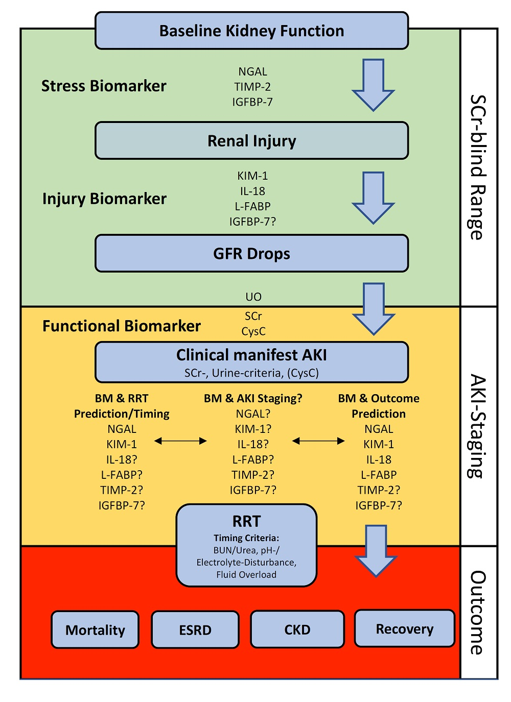
2. Staging of Acute Kidney Injury
Outcomes and Staging
3. Estimated Glomerular Filtration Rate and Its Limitations on Intensive Care Units
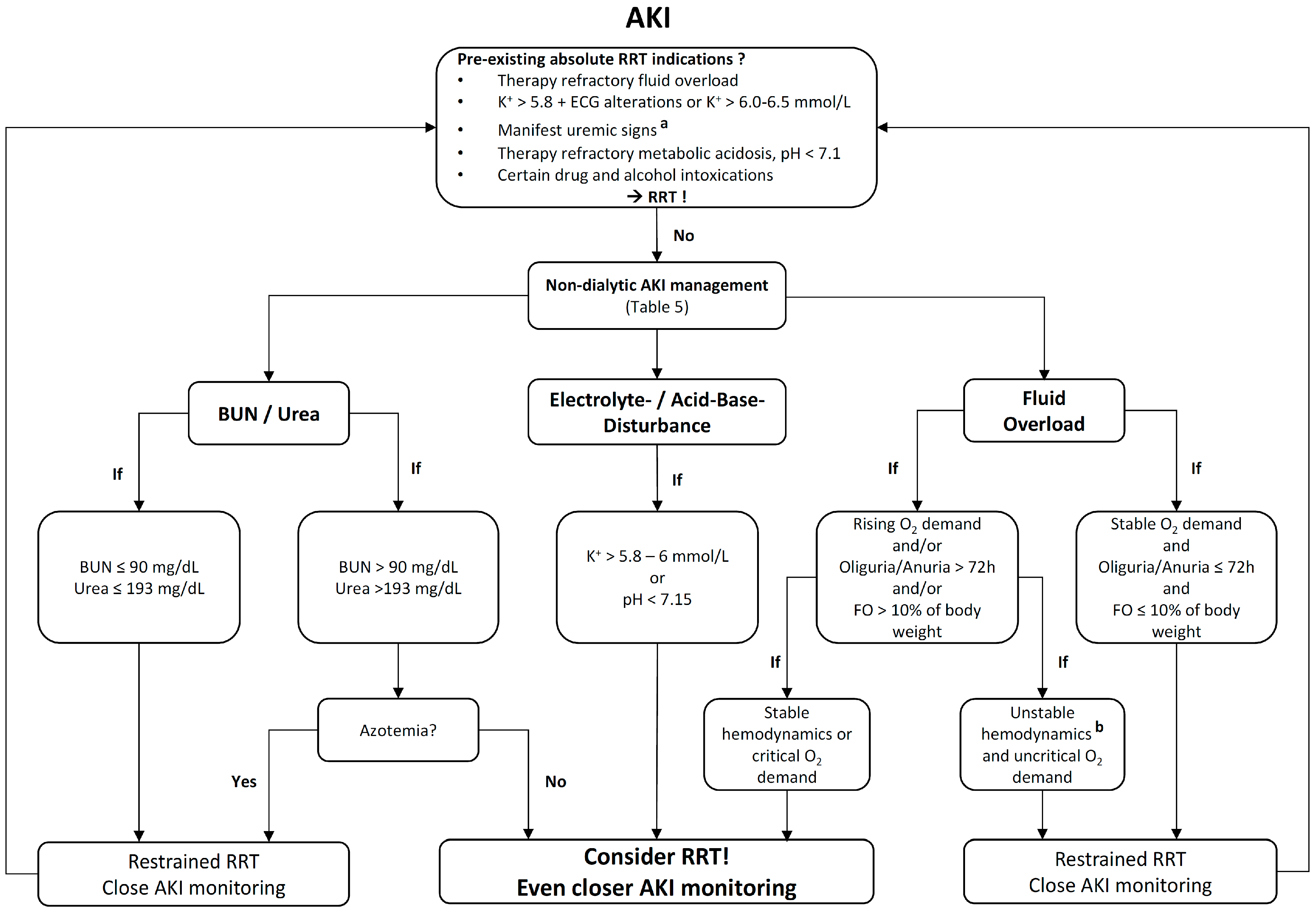
4. Renal Replacement Therapy—Pitfalls and Timing
4.1. Role of Serum Creatinine
4.2. Variation in Creatinine Production
4.3. Variations in Tubular Creatinine Secretion
4.4. Creatinine Measurements
4.5. Role of Blood Urea Nitrogen/Urea
4.6. Timing of Renal Replacement Therapy
5. Potential Harm of Renal Replacement Therapy
5.1. Decline in Urine Output
5.2. Renal Replacement Therapy-Induced Hypotension
5.3. Activation of Inflammatory Pathways
6. Conclusions
Acknowledgments
Authors Contributions
Conflicts of Interest
Abbreviations
| AKI | Acute kidney injury |
| AKIN | Acute Kidney Injury Network |
| BUN | Blood urea nitrogen |
| CKD | Chronic kidney disease |
| CKD-EPI | Chronic Kidney Disease Epidemiology Collaboration |
| COX-1 | Cyclooxygenase-1 |
| CrCl | Creatinine clearance |
| 51Cr-EDTA | Chromium-ethylenediaminetetraacetic acid |
| ESRD | End-stage renal disease |
| GFR | Glomerular filtration rate |
| eGFR | Estimated glomerular filtration rate |
| FO | Fluid overload |
| ICAM-1 | Intracellular adhesion molecule-1 |
| ICU | Intensive Care Unit |
| IGFBP-7 | Insulin like growth factor binding protein-7 |
| KDIGO | Kidney Disease Improving Global Outcomes |
| IL | Interleukin |
| MDRD | Modification of diet in renal disease |
| OAT | Organic anion transporter |
| PMMA | Polymethylmethacrylate |
| RBF | Renal blood flow |
| RRT | Renal replacement therapy |
| SCr | Serum creatinine |
| TIMP-2 | Tissue inhibitor of metalloproteinase-2 |
| TGF | Tubular glomerular feedback |
| TNFα | Tumor necrosis factor alpha |
| UO | Urine output |
| VOD | Volume of distribution |
References
- Uchino, S.; Kellum, J.A.; Bellomo, R.; Doig, G.S.; Morimatsu, H.; Morgera, S.; Schetz, M.; Tan, I.; Bouman, C.; Macedo, E.; et al. Acute renal failure in critically ill patients: A multinational, multicenter study. JAMA 2005, 294, 813–818. [Google Scholar] [CrossRef] [PubMed]
- Joannidis, M.; Metnitz, B.; Bauer, P.; Schusterschitz, N.; Moreno, R.; Druml, W.; Metnitz, P.G.H. Acute kidney injury in critically ill patients classified by AKIN versus RIFLE using the SAPS 3 database. Intensive Care Med. 2009, 35, 1692–1702. [Google Scholar] [CrossRef] [PubMed]
- Hoste, E.A.J.; Bagshaw, S.M.; Bellomo, R.; Cely, C.M.; Colman, R.; Cruz, D.N.; Edipidis, K.; Forni, L.G.; Gomersall, C.D.; Govil, D.; et al. Epidemiology of acute kidney injury in critically ill patients: The multinational AKI-EPI study. Intensive Care Med. 2015, 41, 1411–1423. [Google Scholar] [CrossRef] [PubMed]
- Ergin, B.; Kapucu, A.; Demirci-Tansel, C.; Ince, C. The renal microcirculation in sepsis. Nephrol. Dial. Transplant. 2015, 30, 169–177. [Google Scholar] [CrossRef] [PubMed]
- Bellomo, R.; Kellum, J.A.; Ronco, C.; Wald, R.; Mårtensson, J.; Maiden, M.; Bagshaw, S.M.; Glassford, N.J.; Lankadeva, Y.; Vaara, S.T.; et al. Acute kidney injury in sepsis. Intensive Care Med. 2017, 1–13. [Google Scholar] [CrossRef] [PubMed]
- Singer, M.; Deutschman, C.S.; Seymour, C.W.; Shankar-Hari, M.; Annane, D.; Bauer, M.; Bellomo, R.; Bernard, G.R.; Chiche, J.-D.; Coopersmith, C.M.; et al. The Third International Consensus Definitions for Sepsis and Septic Shock (Sepsis-3). JAMA 2016, 315, 801–810. [Google Scholar] [CrossRef] [PubMed]
- Kellum, J.A.; Lameire, N. KDIGO AKI Guideline Work Group Diagnosis, evaluation, and management of acute kidney injury: A KDIGO summary (Part 1). Crit. Care 2013, 17, 204. [Google Scholar] [CrossRef] [PubMed]
- Wald, R.; McArthur, E.; Adhikari, N.K.J.; Bagshaw, S.M.; Burns, K.E.A.; Garg, A.X.; Harel, Z.; Kitchlu, A.; Mazer, C.D.; Nash, D.M.; et al. Changing incidence and outcomes following dialysis-requiring acute kidney injury among critically ill adults: A population-based cohort study. Am. J. Kidney Dis. 2015, 65, 870–877. [Google Scholar] [CrossRef] [PubMed]
- Stads, S.; Fortrie, G.; van Bommel, J.; Zietse, R.; Betjes, M.G.H. Impaired kidney function at hospital discharge and long-term renal and overall survival in patients who received CRRT. Clin. J. Am. Soc. Nephrol. 2013, 8, 1284–1291. [Google Scholar] [CrossRef] [PubMed]
- Ishani, A.; Xue, J.L.; Himmelfarb, J.; Eggers, P.W.; Kimmel, P.L.; Molitoris, B.A.; Collins, A.J. Acute kidney injury increases risk of ESRD among elderly. J. Am. Soc. Nephrol. 2009, 20, 223–228. [Google Scholar] [CrossRef] [PubMed]
- Hoste, E.A.J.; Schurgers, M. Epidemiology of acute kidney injury: How big is the problem? Crit. Care Med. 2008, 36, S146–S151. [Google Scholar] [CrossRef] [PubMed]
- Srisawat, N.; Lawsin, L.; Uchino, S.; Bellomo, R.; Kellum, J.A. The BEST Kidney Investigators Cost of acute renal replacement therapy in the intensive care unit: Results from The Beginning and Ending Supportive Therapy for the Kidney (BEST Kidney) Study. Crit. Care 2010, 14, R46. [Google Scholar] [CrossRef] [PubMed]
- Farese, S.; Jakob, S.M.; Kalicki, R.; Frey, F.J.; Uehlinger, D.E. Treatment of acute renal failure in the intensive care unit: Lower costs by intermittent dialysis than continuous venovenous hemodiafiltration. Artif. Organs 2009, 33, 634–640. [Google Scholar] [CrossRef] [PubMed]
- Oeyen, S.; De Corte, W.; Benoit, D.; Annemans, L.; Dhondt, A.; Vanholder, R.; Decruyenaere, J.; Hoste, E. Long-term quality of life in critically ill patients with acute kidney injury treated with renal replacement therapy: A matched cohort study. Crit. Care 2015, 19, 1–11. [Google Scholar] [CrossRef] [PubMed]
- Orwelius, L.; Lobo, C.; Teixeira Pinto, A.; Carneiro, A.; Costa-Pereira, A.; Granja, C. Sepsis patients do not differ in health-related quality of life compared with other ICU patients. Acta Anaesthesiol. Scand. 2013, 57, 1201–1205. [Google Scholar] [CrossRef] [PubMed]
- Abelha, F.J.; Botelho, M.; Fernandes, V.; Barros, H. Outcome and quality of life of patients with acute kidney injury after major surgery. Nefrologia 2009, 29, 404–414. [Google Scholar] [PubMed]
- Bellomo, R.; Ronco, C.; Kellum, J.A.; Mehta, R.L.; Palevsky, P. Acute renal failure-definition, outcome measures, animal models, fluid therapy and information technology needs: The Second International Consensus Conference of the Acute Dialysis Quality Initiative (ADQI) Group. Crit. Care 2004, 8, R204–R212. [Google Scholar] [CrossRef] [PubMed]
- Mehta, R.L.; Kellum, J.A.; Shah, S.V.; Molitoris, B.A.; Ronco, C.; Warnock, D.G.; Levin, A. Acute kidney injury network: Report of an initiative to improve outcomes in acute kidney injury. Crit. Care 2007, 11, R31. [Google Scholar] [CrossRef] [PubMed]
- Luo, X.; Jiang, L.; Du, B.; Wen, Y.; Wang, M.; Xi, X. A comparison of different diagnostic criteria of acute kidney injury in critically ill patients. Crit. Care 2014, 18, R144. [Google Scholar] [CrossRef] [PubMed]
- Vincent, J.L.; Moreno, R.; Takala, J.; Willatts, S.; De Mendonça, A.; Bruining, H.; Reinhart, C.K.; Suter, P.M.; Thijs, L.G. The SOFA (Sepsis-related Organ Failure Assessment) score to describe organ dysfunction/failure. Intensive Care Med. 1996, 22, 707–710. [Google Scholar] [CrossRef] [PubMed]
- Siew, E.D.; Ikizler, T.A.; Matheny, M.E.; Shi, Y.; Schildcrout, J.S.; Danciu, I.; Dwyer, J.P.; Srichai, M.; Hung, A.M.; Smith, J.P.; et al. Estimating baseline kidney function in hospitalized patients with impaired kidney function. Clin. J. Am. Soc. Nephrol. 2012, 7, 712–719. [Google Scholar] [CrossRef] [PubMed]
- Siew, E.D.; Matheny, M.E.; Ikizler, T.A.; Lewis, J.B.; Miller, R.A.; Waitman, L.R.; Go, A.S.; Parikh, C.R.; Peterson, J.F. Commonly used surrogates for baseline renal function affect the classification and prognosis of acute kidney injury. Kidney Int. 2010, 77, 536–542. [Google Scholar] [CrossRef] [PubMed]
- Bagshaw, S.M.; Uchino, S.; Cruz, D.; Bellomo, R.; Morimatsu, H.; Morgera, S.; Schetz, M.; Tan, I.; Bouman, C.; Macedo, E.; et al. A comparison of observed versus estimated baseline creatinine for determination of RIFLE class in patients with acute kidney injury. Nephrol. Dial. Transplant. 2009, 24, 2739–2744. [Google Scholar] [CrossRef] [PubMed]
- Závada, J.; Hoste, E.; Cartin-Ceba, R.; Calzavacca, P.; Gajic, O.; Clermont, G.; Bellomo, R.; Kellum, J.A. A comparison of three methods to estimate baseline creatinine for RIFLE classification. Nephrol. Dial. Transplant. 2010, 25, 3911–3918. [Google Scholar] [CrossRef] [PubMed]
- Pickering, J.W.; Endre, Z.H. Back-calculating baseline creatinine with MDRD misclassifies acute kidney injury in the intensive care unit. Clin. J. Am. Soc. Nephrol. 2010, 5, 1165–1173. [Google Scholar] [CrossRef] [PubMed]
- Siew, E.D.; Peterson, J.F.; Eden, S.K.; Moons, K.G.; Ikizler, T.A.; Matheny, M.E. Use of multiple imputation method to improve estimation of missing baseline serum creatinine in acute kidney injury research. Clin. J. Am. Soc. Nephrol. 2013, 8, 10–18. [Google Scholar] [CrossRef] [PubMed]
- Meola, M.; Nalesso, F.; Petrucci, I.; Samoni, S.; Ronco, C. Ultrasound in Acute Kidney Disease. Contrib. Nephrol. 2016, 188, 11–20. [Google Scholar] [PubMed]
- Hedgeman, E.; Lipworth, L.; Lowe, K.; Saran, R.; Do, T.; Fryzek, J. International burden of chronic kidney disease and secondary hyperparathyroidism: A systematic review of the literature and available data. Int. J. Nephrol. 2015, 2015, 1–15. [Google Scholar] [CrossRef] [PubMed]
- Izawa, J.; Uchino, S.; Takinami, M. A detailed evaluation of the new acute kidney injury criteria by KDIGO in critically ill patients. J Anesth 2016, 30, 215–222. [Google Scholar] [CrossRef] [PubMed]
- Kellum, J.A.; Sileanu, F.E.; Murugan, R.; Lucko, N.; Shaw, A.D.; Clermont, G. Classifying AKI by urine output versus serum creatinine level. J. Am. Soc. Nephrol. 2015, 26, 2231–2238. [Google Scholar] [CrossRef] [PubMed]
- Ricci, Z.; Cruz, D.; Ronco, C. The RIFLE criteria and mortality in acute kidney injury: A systematic review. Kidney Int. 2008, 73, 538–546. [Google Scholar] [CrossRef] [PubMed]
- Bagshaw, S.M.; George, C.; Bellomo, R. A comparison of the RIFLE and AKIN criteria for acute kidney injury in critically ill patients. Nephrol. Dial. Transplant. 2008, 23, 1569–1574. [Google Scholar] [CrossRef] [PubMed]
- Lopes, J.A.; Fernandes, P.; Jorge, S.; Gonçalves, S.; Alvarez, A.; Costa e Silva, Z.; França, C.; Prata, M.M. Acute kidney injury in intensive care unit patients: A comparison between the RIFLE and the Acute Kidney Injury Network Classifications. Crit. Care 2008, 12, R110. [Google Scholar] [CrossRef] [PubMed]
- Macedo, E.; Malhotra, R.; Bouchard, J.; Wynn, S.K.; Mehta, R.L. Oliguria is an early predictor of higher mortality in critically ill patients. Kidney Int. 2011, 80, 760–767. [Google Scholar] [CrossRef] [PubMed]
- Vaara, S.T.; Parviainen, I.; Pettilä, V.; Nisula, S.; Inkinen, O.; Uusaro, A. Association of oliguria with the development of acute kidney injury in the critically ill. Kidney Int. 2015, 89, 200–208. [Google Scholar] [CrossRef] [PubMed]
- Murray, P.T.; Mehta, R.L.; Shaw, A.; Ronco, C.; Endre, Z.; Kellum, J.A.; Chawla, L.S.; Cruz, D.; Ince, C.; Okusa, M.D. Potential use of biomarkers in acute kidney injury: Report and summary of recommendations from the 10th Acute Dialysis Quality Initiative Consensus Conference. Kidney Int. 2014, 85, 513–521. [Google Scholar] [CrossRef] [PubMed]
- Endre, Z.H.; Kellum, J.A.; Di Somma, S.; Doi, K.; Goldstein, S.L.; Koyner, J.L.; Macedo, E.; Mehta, R.L.; Murray, P.T. Differential diagnosis of AKI in clinical practice by functional and damage biomarkers: Workgroup statements from the Tenth Acute Dialysis Quality Initiative Consensus Conference. Contrib. Nephrol. 2013, 182, 30–44. [Google Scholar] [PubMed]
- Dewitte, A.; Joannes-Boyau, O.; Sidobre, C.; Fleureau, C.; Bats, M.L.; Derache, P.; Leuillet, S.; Ripoche, J.; Combe, C.; Ouattara, A. kinetic eGFR and novel AKI biomarkers to predict renal recovery. Clin. J. Am. Soc. Nephrol. 2015, 10, 1900–1910. [Google Scholar] [CrossRef] [PubMed]
- Alge, J.L.; Arthur, J.M. Biomarkers of AKI: A review of mechanistic relevance and potential therapeutic implications. Clin. J. Am. Soc. Nephrol. 2015, 10, 147–155. [Google Scholar] [CrossRef] [PubMed]
- Emlet, D.R.; Pastor-Soler, N.; Marciszyn, A.; Wen, X.; Gomez, H.; Humphries, W.H., IV; Morrisroe, S.; Volpe, J.K.; Kellum, J.A. Insulin-like growth factor binding protein 7 and tissue inhibitor of metalloproteinases-2: Differential expression and secretion in human kidney tubule cells. AJP Renal Physiol. 2017, 312, F284–F296. [Google Scholar] [CrossRef] [PubMed]
- Bihorac, A.; Chawla, L.S.; Shaw, A.D.; Al-Khafaji, A.; Davison, D.L.; DeMuth, G.E.; Fitzgerald, R.; Gong, M.N.; Graham, D.D.; Gunnerson, K.; et al. Validation of cell-cycle arrest biomarkers for acute kidney injury using clinical adjudication. Am. J. Respir. Crit. Care. Med. 2014, 189, 932–939. [Google Scholar] [CrossRef] [PubMed]
- Hoste, E.A.J.; McCullough, P.A.; Kashani, K.; Chawla, L.S.; Joannidis, M.; Shaw, A.D.; Feldkamp, T.; Uettwiller-Geiger, D.L.; McCarthy, P.; Shi, J.; et al. Derivation and validation of cutoffs for clinical use of cell cycle arrest biomarkers. Nephrol. Dial. Transplant. 2014, 29, 2054–2061. [Google Scholar] [CrossRef] [PubMed]
- Kashani, K.; Cheungpasitporn, W.; Ronco, C. Biomarkers of acute kidney injury: The pathway from discovery to clinical adoption. Clin. Chem. Lab. Med. 2017. [Google Scholar] [CrossRef] [PubMed]
- Gist, K.M.; Goldstein, S.L.; Wrona, J.; Alten, J.A.; Basu, R.K.; Cooper, D.S.; Soranno, D.E.; Duplantis, J.; Altmann, C.; Gao, Z.; et al. Kinetics of the cell cycle arrest biomarkers (TIMP-2*IGFBP-7) for prediction of acute kidney injury in infants after cardiac surgery. Pediatr. Nephrol. 2017, 368–369. [Google Scholar] [CrossRef] [PubMed]
- Sime, F.B.; Roberts, J.A. Antibiotic dosing in critically ill patients receiving renal replacement therapy. Expert Rev. Clin. Pharmacol. 2015, 1–3. [Google Scholar] [CrossRef] [PubMed]
- Moran, S.M.; Myers, B.D. Course of acute renal failure studied by a model of creatinine kinetics. Kidney Int. 1985, 27, 928–937. [Google Scholar] [CrossRef] [PubMed]
- Endre, Z.H.; Pickering, J.W. Acute kidney injury: Cell cycle arrest biomarkers win race for AKI diagnosis. Nat. Rev. Nephrol. 2014, 10, 683–685. [Google Scholar] [CrossRef] [PubMed]
- Macedo, E.; Bouchard, J.; Soroko, S.H.; Chertow, G.M.; Himmelfarb, J.; Ikizler, T.A.; Paganini, E.P.; Mehta, R.L. Fluid accumulation, recognition and staging of acute kidney injury in critically-ill patients. Crit. Care 2010, 14, R82. [Google Scholar] [CrossRef] [PubMed]
- Bragadottir, G.; Redfors, B.; Ricksten, S.E. Assessing glomerular filtration rate (GFR) in critically ill patients with acute kidney injury-true GFR versus urinary creatinine clearance and estimating equations. Crit. Care 2013, 17, R108. [Google Scholar] [CrossRef] [PubMed]
- Levey, A.S.; Bosch, J.P.; Lewis, J.B.; Greene, T.; Rogers, N.; Roth, D. A more accurate method to estimate glomerular filtration rate from serum creatinine: A new prediction equation. Ann. Intern. Med. 1999, 130, 461–470. [Google Scholar] [CrossRef] [PubMed]
- Levey, A.S. A New equation to estimate glomerular filtration rate. Ann. Intern. Med. 2009, 150, 604–612. [Google Scholar] [CrossRef] [PubMed]
- Macedo, E.; Mehta, R.L. Measuring renal function in critically ill patients: Tools and strategies for assessing glomerular filtration rate. Curr. Opin. Crit. Care 2013, 19, 560–566. [Google Scholar] [CrossRef] [PubMed]
- Pickering, J.W.; Frampton, C.M.; Walker, R.J.; Shaw, G.M.; Endre, Z.H. Four hour creatinine clearance is better than plasma creatinine for monitoring renal function in critically ill patients. Crit. Care 2012, 16, R107. [Google Scholar] [CrossRef] [PubMed]
- Hoste, E.A.J.; Damen, J.; Vanholder, R.C.; Lameire, N.H.; Delanghe, J.R.; van den Hauwe, K.; Colardyn, F.A. Assessment of renal function in recently admitted critically ill patients with normal serum creatinine. Nephrol. Dial. Transplant. 2005, 20, 747–753. [Google Scholar] [CrossRef] [PubMed]
- Herrera-Gutiérrez, M.E.; Seller-Pérez, G.; Banderas-Bravo, E.; Muñoz-Bono, J.; Lebrón-Gallardo, M.; Fernandez-Ortega, J.F. Replacement of 24-h creatinine clearance by 2-h creatinine clearance in intensive care unit patients: A single-center study. Intensive Care Med. 2007, 33, 1900–1906. [Google Scholar] [CrossRef] [PubMed]
- Herget-Rosenthal, S.; Kribben, A.; Pietruck, F.; Ross, B.; Philipp, T. Two by two hour creatinine clearance—Repeatable and valid. Clin. Nephrol. 1999, 51, 348–354. [Google Scholar] [PubMed]
- Coresh, J.; Astor, B.C.; McQuillan, G.; Kusek, J.; Greene, T.; Van Lente, F.; Levey, A.S. Calibration and random variation of the serum creatinine assay as critical elements of using equations to estimate glomerular filtration rate. Am. J. Kidney Dis. 2002, 39, 920–929. [Google Scholar] [CrossRef] [PubMed]
- Barrett, A.J.; Davies, M.E.; Grubb, A. The place of human γ-trace (cystatin C) amongst the cysteine proteinase inhibitors. Biochem. Biophys. Res. Commun. 1984, 120, 631–636. [Google Scholar] [CrossRef]
- Madero, M.; Sarnak, M.J.; Stevens, L.A. Serum cystatin C as a marker of glomerular filtration rate. Curr. Opin. Nephrol. Hypertens. 2006, 15, 610–616. [Google Scholar] [CrossRef] [PubMed]
- Nejat, M.; Pickering, J.W.; Walker, R.J.; Endre, Z.H. Rapid detection of acute kidney injury by plasma cystatin C in the intensive care unit. Nephrol. Dial. Transplant. 2010, 25, 3283–3289. [Google Scholar] [CrossRef] [PubMed]
- Zhang, Z.; Lu, B.; Sheng, X.; Jin, N. Cystatin C in prediction of acute kidney injury: A systemic review and meta-analysis. Am. J. Kidney Dis. 2011, 58, 356–365. [Google Scholar] [CrossRef] [PubMed]
- Herget-Rosenthal, S.; Bökenkamp, A.; Hofmann, W. How to estimate GFR-serum creatinine, serum cystatin C or equations? Clin. Biochem. 2007, 40, 153–161. [Google Scholar] [CrossRef] [PubMed]
- Dai, X.; Zeng, Z.; Fu, C.; Zhang, S.; Cai, Y.; Chen, Z. Diagnostic value of neutrophil gelatinase-associated lipocalin, cystatin C, and soluble triggering receptor expressed on myeloid cells-1 in critically ill patients with sepsis-associated acute kidney injury. Crit. Care 2015, 19, 223. [Google Scholar] [CrossRef] [PubMed]
- Carlier, M.; Dumoulin, A.; Janssen, A.; Picavet, S.; Vanthuyne, S.; Van Eynde, R.; Vanholder, R.; Delanghe, J.; De Schoenmakere, G.; De Waele, J.J.; et al. Comparison of different equations to assess glomerular filtration in critically ill patients. Intensive Care Med. 2015, 41, 427–435. [Google Scholar] [CrossRef] [PubMed]
- Pottel, H.; Delanaye, P.; Schaeffner, E.; Dubourg, L.; Eriksen, B.O.; Melsom, T.; Lamb, E.J.; Rule, A.D.; Turner, S.T.; Glassock, R.J.; et al. Estimating glomerular filtration rate for the full age spectrum from serum creatinine and cystatin C. Nephrol. Dial. Transplant. 2017, 32, 497–507. [Google Scholar] [CrossRef] [PubMed]
- Delanaye, P.; Cavalier, E.; Morel, J.; Mehdi, M.; Maillard, N.; Claisse, G.; Lambermont, B.; Dubois, B.E.; Damas, P.; Krzesinski, J.M.; et al. Detection of decreased glomerular filtration rate in intensive care units: Serum cystatin C versusserum creatinine. BMC Nephrol. 2014, 15, 877. [Google Scholar] [CrossRef] [PubMed]
- Diego, E.; Castro, P.; Soy, D.; Poch, E.; Nicolás, J.M. Predictive performance of glomerular filtration rate estimation equations based on cystatin C versus serum creatinine values in critically ill patients. Am. J. Health Syst. Pharm. 2016, 73, 206–215. [Google Scholar] [CrossRef] [PubMed]
- Liu, X.; Foster, M.C.; Tighiouart, H.; Anderson, A.H.; Beck, G.J.; Contreras, G.; Coresh, J.; Eckfeldt, J.H.; Feldman, H.I.; Greene, T.; et al. Non-GFR determinants of low-molecular-weight serum protein filtration markers in CKD. Am. J. Kidney Dis. 2016, 68, 892–900. [Google Scholar] [CrossRef] [PubMed]
- Stevens, L.A.; Schmid, C.H.; Greene, T.; Li, L.; Beck, G.J.; Joffe, M.M.; Froissart, M.; Kusek, J.W.; Zhang, Y.L.; Coresh, J.; et al. Factors other than glomerular filtration rate affect serum cystatin C levels. Kidney Int. 2009, 75, 652–660. [Google Scholar] [CrossRef] [PubMed]
- Mårtensson, J.; Martling, C.R.; Oldner, A.; Bell, M. Impact of sepsis on levels of plasma cystatin C in AKI and non-AKI patients. Nephrol. Dial. Transplant. 2012, 27, 576–581. [Google Scholar] [CrossRef] [PubMed]
- Filler, G.; Bökenkamp, A.; Hofmann, W.; Le Bricon, T.; Martínez-Brú, C.; Grubb, A. Cystatin C as a marker of GFR-history, indications, and future research. Clin. Biochem. 2005, 38, 1–8. [Google Scholar] [CrossRef] [PubMed]
- Leelahavanichkul, A.; Souza, A.C.P.; Street, J.M.; Hsu, V.; Tsuji, T.; Doi, K.; Li, L.; Hu, X.; Zhou, H.; Kumar, P.; et al. Comparison of serum creatinine and serum cystatin C as biomarkers to detect sepsis-induced acute kidney injury and to predict mortality in CD-1 mice. Am. J. Physiol. Renal. Physiol. 2014, 307, F939–F948. [Google Scholar] [CrossRef] [PubMed]
- Di Nardo, M.; Ficarella, A.; Ricci, Z.; Luciano, R.; Stoppa, F.; Picardo, S.; Picca, S.; Muraca, M.; Cogo, P. Impact of severe sepsis on serum and urinary biomarkers of acute kidney injury in critically ill children: An observational study. Blood Purif. 2013, 35, 172–176. [Google Scholar] [CrossRef] [PubMed]
- Gaudry, S.; Hajage, D.; Schortgen, F.; Martin-Lefevre, L.; Pons, B.; Boulet, E.; Boyer, A.; Chevrel, G.; Lerolle, N.; Carpentier, D.; et al. Initiation strategies for renal-replacement therapy in the intensive care unit. N. Engl. J. Med. 2016, 375, 122–133. [Google Scholar] [CrossRef] [PubMed]
- Zhang, L.; Chen, Z.; Diao, Y.; Yang, Y.; Fu, P. Associations of fluid overload with mortality and kidney recovery in patients with acute kidney injury: A systematic review and meta-analysis. J. Crit. Care 2015, 30, 860. [Google Scholar] [CrossRef] [PubMed]
- Jones, C.A.; McQuillan, G.M.; Kusek, J.W.; Eberhardt, M.S.; Herman, W.H.; Coresh, J.; Salive, M.; Jones, C.P.; Agodoa, L.Y. Serum creatinine levels in the US population: Third National Health And Nutrition Examination Survey. Am. J. Kidney Dis. 1998, 32, 992–999. [Google Scholar] [CrossRef]
- Thomas, M.E.; Blaine, C.; Dawnay, A.; Devonald, M.A.J.; Ftouh, S.; Laing, C.; Latchem, S.; Lewington, A.; Milford, D.V.; Ostermann, M. The definition of acute kidney injury and its use in practice. Kidney Int. 2014, 87, 62–73. [Google Scholar] [CrossRef] [PubMed]
- Clark, W.R.; Mueller, B.A.; Kraus, M.A.; Macias, W.L. Quantification of creatinine kinetic parameters in patients with acute renal failure. Kidney Int. 1998, 54, 554–560. [Google Scholar] [CrossRef] [PubMed]
- Oshima, T.; Deutz, N.E.; Doig, G.; Wischmeyer, P.E.; Pichard, C. Protein-energy nutrition in the ICU is the power couple: A hypothesis forming analysis. Clin. Nutr. 2016, 35, 968–974. [Google Scholar] [CrossRef] [PubMed]
- Sherman, D.S.; Fish, D.N.; Teitelbaum, I. Assessing renal function in cirrhotic patients: Problems and pitfalls. Am. J. Kidney Dis. 2003, 41, 269–278. [Google Scholar] [CrossRef] [PubMed]
- Doi, K.; Yuen, P.S.T.; Eisner, C.; Hu, X.; Leelahavanichkul, A.; Schnermann, J.; Star, R.A. Reduced production of creatinine limits its use as marker of kidney injury in sepsis. J. Am. Soc. Nephrol. 2009, 20, 1217–1221. [Google Scholar] [CrossRef] [PubMed]
- Liu, K.D.; Thompson, B.T.; Ancukiewicz, M.; Steingrub, J.S.; Douglas, I.S.; Matthay, M.A.; Wright, P.; Peterson, M.W.; Rock, P.; Hyzy, R.C.; et al. Acute kidney injury in patients with acute lung injury: Impact of fluid accumulation on classification of acute kidney injury and associated outcomes. Crit. Care Med. 2011, 39, 2665–2671. [Google Scholar] [CrossRef] [PubMed]
- Endre, Z.H.; Pickering, J.W. Biomarkers and creatinine in AKI: The trough of disillusionment or the slope of enlightenment? Kidney Int. 2013, 84, 644–647. [Google Scholar] [CrossRef] [PubMed]
- Perrone, R.D.; Madias, N.E.; Levey, A.S. Serum creatinine as an index of renal function: New insights into old concepts. Clin. Chem. 1992, 38, 1933–1953. [Google Scholar] [PubMed]
- Chen, S. Retooling the creatinine clearance equation to estimate kinetic GFR when the plasma creatinine is changing acutely. J. Am. Soc. Nephrol. 2013, 24, 877–888. [Google Scholar] [CrossRef] [PubMed]
- Bosch, X.; Poch, E.; Grau, J.M. Rhabdomyolysis and acute kidney injury. N. Engl. J. Med. 2009, 361, 62–72. [Google Scholar] [CrossRef] [PubMed]
- Woodrow, G.; Brownjohn, A.M.; Turney, J.H. The clinical and biochemical features of acute renal failure due to rhabdomyolysis. Ren. Fail. 1995, 17, 467–474. [Google Scholar] [CrossRef] [PubMed]
- Levey, A.S. Measurement of renal function in chronic renal disease. Kidney Int. 1990, 38, 167–184. [Google Scholar] [CrossRef] [PubMed]
- Perrone, R.D.; Steinman, T.I.; Beck, G.J.; Skibinski, C.I.; Royal, H.D.; Lawlor, M.; Hunsicker, L.G. Utility of radioisotopic filtration markers in chronic renal insufficiency: Simultaneous comparison of 125I-iothalamate, 169Yb-DTPA, 99mTc-DTPA, and inulin. Am. J. Kidney Dis. 1990, 16, 224–235. [Google Scholar] [CrossRef]
- Bostom, A.G.; Kronenberg, F.; Ritz, E. Predictive performance of renal function equations for patients with chronic kidney disease and normal serum creatinine levels. J. Am. Soc. Nephrol. 2002, 13, 2140–2144. [Google Scholar] [CrossRef] [PubMed]
- Shemesh, O.; Golbetz, H.; Kriss, J.P.; Myers, B.D. Limitations of creatinine as a filtration marker in glomerulopathic patients. Kidney Int. 1985, 28, 830–838. [Google Scholar] [CrossRef] [PubMed]
- Berg, K.J.; Gjellestad, A.; Nordby, G.; Rootwelt, K.; Djøseland, O.; Fauchald, P.; Mehl, A.; Narverud, J.; Talseth, T. Renal effects of trimethoprim in ciclosporin- and azathioprine-treated kidney-allografted patients. Nephron 1989, 53, 218–222. [Google Scholar] [CrossRef] [PubMed]
- Duncker, D.; Oswald, H.; Gardiwal, A.; Lüsebrink, U.; König, T.; Schreyer, H.; Klein, G. Stable cystatin C serum levels confirm normal renal function in patients with dronedarone-associated increase in serum creatinine. J. Cardiovasc. Pharmacol. Ther. 2013, 18, 109–112. [Google Scholar] [CrossRef] [PubMed]
- Hilbrands, L.B.; Artz, M.A.; Wetzels, J.F.; Koene, R.A. Cimetidine improves the reliability of creatinine as a marker of glomerular filtration. Kidney Int. 1991, 40, 1171–1176. [Google Scholar] [CrossRef] [PubMed]
- Rocci, M.L.; Vlasses, P.H.; Ferguson, R.K. Creatinine serum concentrations and H2-receptor antagonists. Clin. Nephrol. 1984, 22, 214–215. [Google Scholar] [PubMed]
- Mitchell, E.K. Flucytosine and false elevation of serum creatinine level. Ann. Intern. Med. 1984, 101, 278. [Google Scholar] [CrossRef] [PubMed]
- Saah, A.J.; Koch, T.R.; Drusano, G.L. Cefoxitin falsely elevates creatinine levels. JAMA 1982, 247, 205–206. [Google Scholar] [CrossRef] [PubMed]
- Molitch, M.E.; Rodman, E.; Hirsch, C.A.; Dubinsky, E. Spurious serum creatinine elevations in ketoacidosis. Ann. Intern. Med. 1980, 93, 280–281. [Google Scholar] [CrossRef] [PubMed]
- Miller, W.G.; Myers, G.L.; Ashwood, E.R.; Killeen, A.A.; Wang, E.; Thienpont, L.M.; Siekmann, L. Creatinine measurement: State of the art in accuracy and interlaboratory harmonization. Arch. Pathol. Lab. Med. 2005, 129, 297–304. [Google Scholar] [PubMed]
- Soldin, S.J.; Henderson, L.; Hill, J.G. The effect of bilirubin and ketones on reaction rate methods for the measurement of creatinine. Clin. Biochem. 1978, 11, 82–86. [Google Scholar] [CrossRef]
- Myers, G.L.; Miller, W.G.; Coresh, J.; Fleming, J.; Greenberg, N.; Greene, T.; Hostetter, T.; Levey, A.S.; Panteghini, M.; Welch, M.; et al. Recommendations for improving serum creatinine measurement: A report from the Laboratory Working Group of the National Kidney Disease Education Program. Clin. Chem. 2006, 52, 5–18. [Google Scholar] [CrossRef] [PubMed]
- Peake, M.; Whiting, M. Measurement of serum creatinine-current status and future goals. Clin. Biochem. Rev. 2006, 27, 173–184. [Google Scholar] [PubMed]
- Davenport, A.; Cholongitas, E.; Xirouchakis, E.; Burroughs, A.K. Pitfalls in assessing renal function in patients with cirrhosis-potential inequity for access to treatment of hepatorenal failure and liver transplantation. Nephrol. Dial. Transplant. 2011, 26, 2735–2742. [Google Scholar] [CrossRef] [PubMed]
- Delanghe, J.R.; Cobbaert, C.; Harmoinen, A.; Jansen, R.; Laitinen, P.; Panteghini, M. Focusing on the clinical impact of standardization of creatinine measurements: A report by the EFCC working group on creatinine standardization. Clin. Chem. Lab. Med. 2011, 49, 977–982. [Google Scholar] [CrossRef] [PubMed]
- Preiss, D.J.; Godber, I.M.; Lamb, E.J.; Dalton, R.N.; Gunn, I.R. The influence of a cooked-meat meal on estimated glomerular filtration rate. Ann. Clin. Biochem. 2007, 44, 35–42. [Google Scholar] [CrossRef] [PubMed]
- Pickering, J.W.; Ralib, A.M.; Endre, Z.H. Combining creatinine and volume kinetics identifies missed cases of acute kidney injury following cardiac arrest. Crit. Care 2013, 17, R7. [Google Scholar] [CrossRef] [PubMed]
- Conte, G.; Dal Canton, A.; Terribile, M.; Cianciaruso, B.; Di Minno, G.; Pannain, M.; Russo, D.; Andreucci, V.E. Renal handling of urea in subjects with persistent azotemia and normal renal function. Kidney Int. 1987, 32, 721–727. [Google Scholar] [CrossRef] [PubMed]
- Dossetor, J.B. Creatininemia versus uremia. Ann. Intern. Med. 1966, 65, 1287. [Google Scholar] [CrossRef] [PubMed]
- Uchino, S.; Bellomo, R.; Goldsmith, D. The meaning of the blood urea nitrogen/creatinine ratio in acute kidney injury. Clin. Kidney. J. 2012, 5, 187–191. [Google Scholar] [CrossRef]
- Möller, E.; McIntosh, J.F.; Van Slyke, D.D. Studies of urea excretion. II: Relationship between urine volume and the rate of urea excretion by normal adults. J. Clin. Investig. 1928, 6, 427–465. [Google Scholar] [CrossRef] [PubMed]
- Papadakis, M.A.; Arieff, A.I. Unpredictability of clinical evaluation of renal function in cirrhosis. Prospective study. Am. J. Med. 1987, 82, 945–952. [Google Scholar] [CrossRef]
- Macias, W.L.; Alaka, K.J.; Murphy, M.H.; Miller, M.E.; Clark, W.R.; Mueller, B.A. Impact of the nutritional regimen on protein catabolism and nitrogen balance in patients with acute renal failure. J. Parenter. Enteral Nutr. 1996, 20, 56–62. [Google Scholar] [CrossRef] [PubMed]
- Casaer, M.P.; Mesotten, D.; Schetz, M.R.C. Bench-to-bedside review: Metabolism and nutrition. Crit. Care 2008, 12, 222. [Google Scholar] [CrossRef] [PubMed]
- Keiserman, W. Effects of urea loading in patients with far advanced renal failure. Urology 1973, 1, 272. [Google Scholar] [CrossRef]
- Seabra, V.F.; Balk, E.M.; Liangos, O.; Sosa, M.A.; Cendoroglo, M.; Jaber, B.L. Timing of renal replacement therapy initiation in acute renal failure: A meta-analysis. Am. J. Kidney Dis. 2008, 52, 272–284. [Google Scholar] [CrossRef] [PubMed]
- Jamale, T.E.; Hase, N.K.; Kulkarni, M.; Pradeep, K.J.; Keskar, V.; Jawale, S.; Mahajan, D. Earlier-start versus usual-start dialysis in patients with community-acquired acute kidney injury: A randomized controlled trial. Am. J. Kidney Dis. 2013, 62, 1116–1121. [Google Scholar] [CrossRef] [PubMed]
- Liu, Y.; Davari-Farid, S.; Arora, P.; Porhomayon, J.; Nader, N.D. Early versus late initiation of renal replacement therapy in critically ill patients with acute kidney injury after cardiac surgery: A systematic review and meta-analysis. J. Cardiothorac. Vasc. Anestha 2014, 28, 557–563. [Google Scholar] [CrossRef] [PubMed]
- Wald, R.; Bagshaw, S.M. The timing of renal replacement therapy initiation in acute kidney injury. Crit. Care Med. 2014, 42, 1933–1934. [Google Scholar] [CrossRef] [PubMed]
- Karvellas, C.J.; Farhat, M.R.; Sajjad, I.; Mogensen, S.S.; Leung, A.A.; Wald, R.; Bagshaw, S.M. A comparison of early versus late initiation of renal replacement therapy in critically ill patients with acute kidney injury: A systematic review and meta-analysis. Crit. Care 2011, 15, R72. [Google Scholar] [CrossRef] [PubMed]
- Gaudry, S.; Hajage, D.; Schortgen, F.; Martin-Lefevre, L.; Ricard, J.; Dreyfuss, D. Comparison of two strategies for initiating renal replacement therapy in the ICU: Study protocol plan for a multicenter, randomized, controlled trial. Crit. Care 2015, 19, P299. [Google Scholar] [CrossRef]
- Jun, M.; Bellomo, R.; Cass, A.; Gallagher, M.; Lo, S.; Lee, J. Timing of renal replacement therapy and patient outcomes in the randomized evaluation of normal versus augmented level of replacement therapy study. Crit Care Med. 2014, 42, 1756–1765. [Google Scholar] [CrossRef] [PubMed]
- García-Fernández, N.; Pérez-Valdivieso, J.R.; Bes-Rastrollo, M.; Vives, M.; Lavilla, J.; Herreros, J.; Monedero, P.; GEDRCC. Timing of renal replacement therapy after cardiac surgery: A retrospective multicenter spanish cohort study. Blood Purif. 2011, 32, 104–111. [Google Scholar]
- Vaara, S.T.; Reinikainen, M.; Wald, R.; Bagshaw, S.M.; Pettila, V. Timing of RRT based on the presence of conventional indications. Clin. J. Am. Soc. Nephrol. 2014, 9, 1577–1585. [Google Scholar] [CrossRef] [PubMed]
- Leite, T.T.; Macedo, E.; Pereira, S.M.; Bandeira, S.R.; Pontes, P.H.; Garcia, A.S.; Militão, F.R.; Sobrinho, I.M.; Assunção, L.M.; Libório, A.B. Timing of renal replacement therapy initiation by AKIN classification system. Crit. Care 2013, 17, R62. [Google Scholar] [CrossRef] [PubMed]
- Wald, R.; Adhikari, N.K.J.; Smith, O.M.; Weir, M.A.; Pope, K.; Cohen, A.; Thorpe, K.; McIntyre, L.; Lamontagne, F.; Soth, M.; et al. Comparison of standard and accelerated initiation of renal replacement therapy in acute kidney injury. Kidney Int. 2015, 88, 1–8. [Google Scholar] [CrossRef] [PubMed]
- Zarbock, A.; Kellum, J.A. Timing of initiation of renal replacement therapy in critically ill patients with acute kidney injury-reply. JAMA 2016, 316, 1214. [Google Scholar] [CrossRef] [PubMed]
- Kashani, K.; Al-Khafaji, A.; Ardiles, T.; Artigas, A.; Bagshaw, S.M.; Bell, M.; Bihorac, A.; Birkhahn, R.; Cely, C.M.; Chawla, L.S.; et al. Discovery and validation of cell cycle arrest biomarkers in human acute kidney injury. Crit. Care 2013, 17, R25. [Google Scholar] [CrossRef] [PubMed]
- Bonventre, J.V.; Yang, L. Cellular pathophysiology of ischemic acute kidney injury. J. Clin. Investig. 2011, 121, 4210–4221. [Google Scholar] [CrossRef] [PubMed]
- Price, P.M.; Safirstein, R.L.; Megyesi, J. The cell cycle and acute kidney injury. Kidney Int. 2009, 76, 604–613. [Google Scholar] [CrossRef] [PubMed]
- Parikh, C.R.; Mansour, S.G. Perspective on clinical application of biomarkers in AKI. J. Am. Soc. Nephrol. 2017, 28, 1677–1685. [Google Scholar] [CrossRef] [PubMed]
- Sugahara, S.; Suzuki, H. Early start on continuous hemodialysis therapy improves survival rate in patients with acute renal failure following coronary bypass surgery. Hemodial. Int. 2004, 8, 320–325. [Google Scholar] [CrossRef] [PubMed]
- Durmaz, I.; Yagdi, T.; Calkavur, T.; Mahmudov, R.; Apaydin, A.Z.; Posacioglu, H.; Atay, Y.; Engin, C. Prophylactic dialysis in patients with renal dysfunction undergoing on-pump coronary artery bypass surgery. Ann. Thorac. Surg. 2003, 75, 859–864. [Google Scholar] [CrossRef]
- Bouman, C.S.C.; Oudemans-van Straaten, H.M.; Tijssen, J.G.P.; Zandstra, D.F.; Kesecioglu, J. Effects of early high-volume continuous venovenous hemofiltration on survival and recovery of renal function in intensive care patients with acute renal failure: A prospective, randomized trial. Crit. Care Med. 2002, 30, 2205–2211. [Google Scholar] [CrossRef] [PubMed]
- Bagshaw, S.M.; Uchino, S.; Bellomo, R.; Morimatsu, H.; Morgera, S.; Schetz, M.; Tan, I.; Bouman, C.; Macedo, E.; Gibney, N.; et al. Beginning and ending supportive therapy for the kidney (BEST Kidney) investigators timing of renal replacement therapy and clinical outcomes in critically ill patients with severe acute kidney injury. J. Crit Care 2009, 24, 129–140. [Google Scholar] [CrossRef] [PubMed]
- Liu, K.D. Timing of initiation of dialysis in critically ill patients with acute kidney injury. Clin. J. Am. Soc. Nephrol. 2006, 1, 915–919. [Google Scholar] [CrossRef] [PubMed]
- Ji, Q.; Mei, Y.; Wang, X.; Feng, J.; Cai, J.; Zhou, Y.; Sun, Y.; Xie, S.; Hu, D. Timing of continuous veno-venous hemodialysis in the treatment of acute renal failure following cardiac surgery. Heart Vessels 2011, 26, 183–189. [Google Scholar] [CrossRef] [PubMed]
- Carl, D.E.; Grossman, C.; Behnke, M.; Sessler, C.N.; Gehr, T.W.B. Effect of timing of dialysis on mortality in critically ill, septic patients with acute renal failure. Hemodial. Int. 2010, 14, 11–17. [Google Scholar] [CrossRef] [PubMed]
- Iyem, H.; Tavli, M.; Akcicek, F.; Büket, S. Importance of early dialysis for acute renal failure after an open-heart surgery. Hemodial. Int. 2009, 13, 55–61. [Google Scholar] [CrossRef] [PubMed]
- Shiao, C.C.; Wu, V.C.; Li, W.Y.; Lin, Y.F.; Hu, F.C.; Young, G.H.; Kuo, CC.; Kao, T.W.; Huang, D.M.; Chen, Y.M.; et al. Late initiation of renal replacement therapy is associated with worse outcomes in acute kidney injury after major abdominal surgery. Crit. Care 2009, 13, R171. [Google Scholar] [CrossRef] [PubMed]
- Manché, A.; Casha, A.; Rychter, J.; Farrugia, E.; Debono, M. Early dialysis in acute kidney injury after cardiac surgery. Interact. Cardiovasc. Thorac. Surg. 2008, 7, 829–832. [Google Scholar] [CrossRef] [PubMed]
- Andrade, L.; Cleto, S.; Seguro, A.C. Door-to-dialysis time and daily hemodialysis in patients with leptospirosis: Impact on mortality. Clin. J. Am. Soc. Nephrol. 2007, 2, 739–744. [Google Scholar] [CrossRef] [PubMed]
- Wu, V.C.; Ko, W.J.; Chang, H.W.; Chen, Y.S.; Chen, Y.W.; Chen, Y.M.; Hu, F.C.; Lin, Y.H.; Tsai, P.R.; Wu, K.D. Early renal replacement therapy in patients with postoperative acute liver failure associated with acute renal failure: Effect on postoperative outcomes. J. Am. Coll. Surg. 2007, 205, 266–276. [Google Scholar] [CrossRef] [PubMed]
- Piccinni, P.; Dan, M.; Barbacini, S.; Carraro, R.; Lieta, E.; Marafon, S.; Zamperetti, N.; Brendolan, A.; D’Intini, V.; Tetta, C.; et al. Early isovolaemic haemofiltration in oliguric patients with septic shock. Intensive Care Med. 2005, 32, 80–86. [Google Scholar] [CrossRef] [PubMed]
- Demirkilic, U.; Kuralay, E.; Yenicesu, M.; Caglar, K.; Oz, B.S.; Cingoz, F.; Gunay, C.; Yildirim, V.; Ceylan, S.; Arslan, M.; et al. Timing of replacement therapy for acute renal failure after cardiac surgery. J. Card. Surg. 2004, 19, 17–20. [Google Scholar] [CrossRef] [PubMed]
- Elahi, M.M.; Lim, M.Y.; Joseph, R.N.; Dhannapuneni, R.R.V.; Spyt, T.J. Early hemofiltration improves survival in post-cardiotomy patients with acute renal failure. Eur. J. Cardiothorac. Surg. 2004, 26, 1027–1031. [Google Scholar] [CrossRef] [PubMed]
- Gettings, L.G.; Reynolds, H.N.; Scalea, T. Outcome in post-traumatic acute renal failure when continuous renal replacement therapy is applied early vs. late. Intensive Care Med. 1999, 25, 805–813. [Google Scholar] [CrossRef] [PubMed]
- Cooper, B.A.; Branley, P.; Bulfone, L.; Collins, J.F.; Craig, J.C.; Fraenkel, M.B.; Harris, A.; Johnson, D.W.; Kesselhut, J.; Li, J.J.; et al. A randomized, controlled trial of early versus late initiation of dialysis. N. Engl. J. Med. 2010, 363, 609–619. [Google Scholar] [CrossRef] [PubMed]
- Porter, C.J.; Juurlink, I.; Bisset, L.H.; Bavakunji, R.; Mehta, R.L.; Devonald, M.A.J. A real-time electronic alert to improve detection of acute kidney injury in a large teaching hospital. Nephrol. Dial. Transplant. 2014, 29, 1888–1893. [Google Scholar] [CrossRef] [PubMed]
- Handler, S.M.; Kane-Gill, S.L.; Kellum, J.A. Optimal and early detection of acute kidney injury requires effective clinical decision support systems. Nephrol. Dial. Transplant. 2014, 29, 1802–1803. [Google Scholar] [CrossRef] [PubMed]
- Kirkendall, E.S.; Spires, W.L.; Mottes, T.A.; Schaffzin, J.K.; Barclay, C.; Goldstein, S.L. Development and performance of electronic acute kidney injury triggers to identify pediatric patients at risk for nephrotoxic medication-associated harm. Appl. Clin. Inform. 2014, 5, 313–333. [Google Scholar] [CrossRef] [PubMed]
- Perazella, M.A.; Coca, S.G. Three feasible strategies to minimize kidney injury in “incipient AKI”. Nat. Rev. Nephrol. 2013, 9, 484–490. [Google Scholar] [CrossRef] [PubMed]
- Mutter, T.C.; Ruth, C.A.; Dart, A.B. Hydroxyethyl starch (HES) versus other fluid therapies: Effects on kidney function. Cochrane Database Syst. Rev. 2013, CD007594. [Google Scholar] [CrossRef] [PubMed]
- Heung, M.; Wolfgram, D.F.; Kommareddi, M.; Hu, Y.; Song, P.X.; Ojo, A.O. Fluid overload at initiation of renal replacement therapy is associated with lack of renal recovery in patients with acute kidney injury. Nephrol. Dial. Transplant. 2012, 27, 956–961. [Google Scholar] [CrossRef] [PubMed]
- Ho, K.M.; Sheridan, D.J. Meta-analysis of frusemide to prevent or treat acute renal failure. BMJ 2006, 333, 420. [Google Scholar] [CrossRef] [PubMed]
- Brunkhorst, F.M.; Engel, C.; Bloos, F.; Meier-Hellmann, A.; Ragaller, M.; Weiler, N.; Moerer, O.; Gruendling, M.; Oppert, M.; Grond, S.; et al. Intensive insulin therapy and pentastarch resuscitation in severe sepsis. N. Engl. J. Med. 2008, 358, 125–139. [Google Scholar] [CrossRef] [PubMed]
- Kohli, H.S.; Bhaskaran, M.C.; Muthukumar, T.; Thennarasu, K.; Sud, K.; Jha, V.; Gupta, K.L.; Sakhuja, V. Treatment-related acute renal failure in the elderly: A hospital-based prospective study. Nephrol. Dial. Transplant. 2000, 15, 212–217. [Google Scholar] [CrossRef] [PubMed]
- Asfar, P.; Meziani, F.; Hamel, J.F.; Grelon, F.; Megarbane, B.; Anguel, N.; Mira, J.P.; Dequin, P.F.; Gergaud, S.; Weiss, N.; et al. High versus low blood-pressure target in patients with septic shock. N. Engl. J. Med. 2014, 370, 1583–1593. [Google Scholar] [CrossRef] [PubMed]
- Yeh, B.P.; Tomko, D.J.; Stacy, W.K.; Bear, E.S.; Haden, H.T.; Falls, W.F. Factors influencing sodium and water excretion in uremic man. Kidney Int. 1975, 7, 103–110. [Google Scholar] [CrossRef] [PubMed]
- Finn, W.F. Nephron heterogeneity in polyuric acute renal failure. J. Lab. Clin. Med. 1981, 98, 21–29. [Google Scholar] [PubMed]
- Bagshaw, S.M.; Delaney, A.; Haase, M.; Ghali, W.A.; Bellomo, R. Loop diuretics in the management of acute renal failure: A systematic review and meta-analysis. Crit. Care Resusc. 2007, 9, 60–68. [Google Scholar] [PubMed]
- Bagshaw, S.M.; Bellomo, R.; Kellum, J.A. Oliguria, volume overload, and loop diuretics. Crit. Care Med. 2008, 36, S172–S178. [Google Scholar] [CrossRef] [PubMed]
- Ejaz, A.A.; Mohandas, R. Are diuretics harmful in the management of acute kidney injury? Curr. Opin. Nephrol. Hypertens. 2014, 23, 155–160. [Google Scholar] [CrossRef] [PubMed]
- Van der Voort, P.H.J.; Boerma, E.C.; Koopmans, M.; Zandberg, M.; de Ruiter, J.; Gerritsen, R.T.; Egbers, P.H.M.; Kingma, W.P.; Kuiper, M.A. Furosemide does not improve renal recovery after hemofiltration for acute renal failure in critically ill patients: A double blind randomized controlled trial. Crit. Care Med. 2009, 37, 533–538. [Google Scholar] [CrossRef] [PubMed]
- Mehta, R.L. Diuretics, mortality, and nonrecovery of renal function in acute renal failure. JAMA 2002, 288, 2547–2553. [Google Scholar] [CrossRef] [PubMed]
- Wu, V.C.; Lai, C.F.; Shiao, C.C.; Lin, Y.F.; Wu, P.C.; Chao, C.T.; Hu, F.C.; Huang, T.M.; Yeh, Y.C.; Tsai, I.J.; et al. Effect of diuretic use on 30-day postdialysis mortality in critically ill patients receiving acute dialysis. PLoS ONE 2012, 7, e30836. [Google Scholar] [CrossRef] [PubMed]
- Wang, N.; Jiang, L.; Zhu, B.; Wen, Y.; Xi, X.M. Fluid balance and mortality in critically ill patients with acute kidney injury: A multicenter prospective epidemiological study. Crit. Care 2015, 19, 1–11. [Google Scholar] [CrossRef] [PubMed]
- Conger, J.D. Does hemodialysis delay recovery from acute renal failure? Semin. Dial. 1990, 3, 146–148. [Google Scholar] [CrossRef]
- Palevsky, P.M.; Baldwin, I.; Davenport, A.; Goldstein, S.; Paganini, E. Renal replacement therapy and the kidney: Minimizing the impact of renal replacement therapy on recovery of acute renal failure. Curr. Opin. Crit. Care 2005, 11, 548–554. [Google Scholar] [CrossRef] [PubMed]
- Adams, P.L.; Adams, F.F.; Bell, P.D.; Navar, L.G. Impaired renal blood flow autoregulation in ischemic acute renal failure. Kidney Int. 1980, 18, 68–76. [Google Scholar] [CrossRef] [PubMed]
- Malek, M.; Nematbakhsh, M. Renal ischemia/reperfusion injury; from pathophysiology to treatment. J. Renal Inj. Prev. 2014, 4, 1–8. [Google Scholar]
- Morrell, E.D.; Kellum, J.A.; Hallows, K.R.; Pastor-Soler, N.M. Epithelial transport during septic acute kidney injury. Nephrol. Dial. Transplant. 2014, 29, 1312–1319. [Google Scholar] [CrossRef] [PubMed]
- Singh, P.; Okusa, M.D. The role of tubuloglomerular feedback in the pathogenesis of acute kidney injury. Contrib. Nephrol. 2011, 174, 12–21. [Google Scholar] [PubMed]
- Preising, C.; Schneider, R.; Bucher, M.; Gekle, M.; Sauvant, C. Regulation of expression of renal organic anion transporters OAT1 and OAT3 in a model of ischemia/reperfusion injury. Cell Physiol. Biochem. 2015, 37, 1–13. [Google Scholar] [CrossRef] [PubMed]
- Schneider, R.; Meusel, M.; Betz, B.; Held, C.; Möller-Ehrlich, K.; Büttner-Herold, M.; Wanner, C.; Gekle, M.; Sauvant, C. Oat1/3 restoration protects against renal damage after ischemic AKI. Am. J. Physiol. Renal Physiol. 2015, 308, F198–F208. [Google Scholar] [CrossRef] [PubMed]
- Bergström, J.; Asaba, H.; Fürst, P.; Oulès, R. Dialysis, Ultrafiltration, and blood pressure. Blood Purif. 2006, 24, 222–229. [Google Scholar] [CrossRef] [PubMed]
- Henrich, W.L.; Woodard, T.D.; Blachley, J.D.; Gomez-Sanchez, C.; Pettinger, W.; Cronin, R.E. Role of osmolality in blood pressure stability after dialysis and ultrafiltration. Kidney Int. 1980, 18, 480–488. [Google Scholar] [CrossRef] [PubMed]
- Swartz, R.D.; Somermeyer, M.G.; Hsu, C.H. Preservation of plasma volume during hemodialysis depends on dialysate osmolality. Am. J. Nephrol. 1982, 2, 189–194. [Google Scholar] [CrossRef] [PubMed]
- Kimura, G.; Irie, A.; Kuroda, K.; Kojima, S.; Satani, M. Absence of transcellular fluid shift during haemofiltration. Proc. Eur. Dial. Transplant. Assoc. 1980, 17, 192–196. [Google Scholar] [PubMed]
- Schuenemann, B.; Borghardt, J.; Falda, Z.; Jacob, I.; Kramer, P.; Kraft, B.; Quellhorst, E. Reactions of blood pressure and body spaces to hemofiltration treatment. Trans. Am. Soc. Artif. Intern. Organs 1978, 24, 687–689. [Google Scholar] [PubMed]
- Kovacs, B.; Sullivan, K.J.; Hiremath, S.; Patel, R.V. The effect of sustained low efficient dialysis versus continuous renal replacement therapy on renal recovery after acute kidney injury in the intensive care unit: A systematic review and meta-analysis. Nephrology 2017, 22, 343–353. [Google Scholar] [CrossRef] [PubMed]
- Truche, A.S.; Darmon, M.; Bailly, S.; Clec’h, C.; Dupuis, C.; Misset, B.; Azoulay, E.; Schwebel, C.; Bouadma, L.; Kallel, H.; et al. Continuous renal replacement therapy versus intermittent hemodialysis in intensive care patients: Impact on mortality and renal recovery. Intensive Care Med. 2016, 42, 1408–1417. [Google Scholar] [CrossRef] [PubMed]
- Zhang, L.; Yang, J.; Eastwood, G.M.; Zhu, G.; Tanaka, A.; Bellomo, R. Extended daily dialysis versus continuous renal replacement therapy for acute kidney injury: A meta-analysis. Am. J. Kidney Dis. 2015, 66, 322–330. [Google Scholar] [CrossRef] [PubMed]
- Schwenger, V.; Weigand, M.A.; Hoffmann, O.; Dikow, R.; Kihm, L.P.; Seckinger, J.; Miftari, N.; Schaier, M.; Hofer, S.; Haar, C.; et al. Sustained low efficiency dialysis using a single-pass batch system in acute kidney injury—A randomized interventional trial: The renal replacement therapy study in intensive care unit patients. Crit. Care 2012, 16, R140. [Google Scholar] [CrossRef] [PubMed]
- Sun, Z.; Ye, H.; Shen, X.; Chao, H.; Wu, X.; Yang, J. Continuous venovenous hemofiltration versus extended daily hemofiltration in patients with septic acute kidney injury: A retrospective cohort study. Crit. Care 2014, 18, 1–9. [Google Scholar] [CrossRef] [PubMed]
- Wald, R.; Shariff, S.Z.; Adhikari, N.K.J.; Bagshaw, S.M.; Burns, K.E.A.; Friedrich, J.O.; Garg, A.X.; Harel, Z.; Kitchlu, A.; Ray, J.G. The association between renal replacement therapy modality and long-term outcomes among critically ill adults with acute kidney injury: A retrospective cohort study. Crit. Care Med. 2014, 42, 868–877. [Google Scholar] [CrossRef] [PubMed]
- Schneider, A.G.; Bellomo, R.; Bagshaw, S.M.; Glassford, N.J.; Lo, S.; Jun, M.; Cass, A.; Gallagher, M. Choice of renal replacement therapy modality and dialysis dependence after acute kidney injury: A systematic review and meta-analysis. Intensive Care Med. 2013, 39, 987–997. [Google Scholar] [CrossRef] [PubMed]
- Schefold, J.C.; von Haehling, S.; Pschowski, R.; Bender, T.O.; Berkmann, C.; Briegel, S.; Hasper, D.; Jörres, A. The effect of continuous versus intermittent renal replacement therapy on the outcome of critically ill patients with acute renal failure (CONVINT): A prospective randomized controlled trial. Crit. Care 2014, 18, 1–11. [Google Scholar] [CrossRef] [PubMed]
- Friedrich, J.O.; Wald, R.; Bagshaw, S.M.; Burns, K.E.; Adhikari, N.K. Hemofiltration compared to hemodialysis for acute kidney injury: Systematic review and meta-analysis. Crit. Care 2012, 16, R146. [Google Scholar] [CrossRef] [PubMed]
- Nash, D.M.; Przech, S.; Wald, R.; O'Reilly, D. Systematic review and meta-analysis of renal replacement therapy modalities for acute kidney injury in the intensive care unit. J. Crit. Care 2017, 41, 138–144. [Google Scholar] [CrossRef] [PubMed]
- Schulman, G.; Fogo, A.; Gung, A.; Badr, K.; Hakim, R. Complement activation retards resolution of acute ischemic renal failure in the rat. Kidney Int. 1991, 40, 1069–1074. [Google Scholar] [CrossRef] [PubMed]
- Cheung, A.K. Biocompatibility of hemodialysis membranes. J. Am. Soc. Nephrol. 1990, 1, 150–161. [Google Scholar] [PubMed]
- Hakim, R.M. Clinical implications of hemodialysis membrane biocompatibility. Kidney Int. 1993, 44, 484–494. [Google Scholar] [CrossRef] [PubMed]
- Hakim, R.M.; Wingard, R.L.; Parker, R.A. Effect of the dialysis membrane in the treatment of patients with acute renal failure. N. Engl. J. Med. 1994, 331, 1338–1342. [Google Scholar] [CrossRef] [PubMed]
- Schiffl, H.; Lang, S.M.; König, A.; Strasser, T.; Haider, M.C.; Held, E. Biocompatible membranes in acute renal failure: Prospective case-controlled study. Lancet 1994, 344, 570–572. [Google Scholar] [CrossRef]
- Himmelfarb, J.; Tolkoff Rubin, N.; Chandran, P.; Parker, R.A.; Wingard, R.L.; Hakim, R. A multicenter comparison of dialysis membranes in the treatment of acute renal failure requiring dialysis. J. Am. Soc. Nephrol. 1998, 9, 257–266. [Google Scholar] [PubMed]
- Subramanian, S.; Venkataraman, R.; Kellum, J.A. Influence of dialysis membranes on outcomes in acute renal failure: A meta-analysis. Kidney Int. 2002, 62, 1819–1823. [Google Scholar] [CrossRef] [PubMed]
- Bozza, F.A.; Salluh, J.I.; Japiassu, A.M.; Soares, M.; Assis, E.F.; Gomes, R.N.; Bozza, M.T.; Castro-Faria-Neto, H.C.; Bozza, P.T. Cytokine profiles as markers of disease severity in sepsis: A multiplex analysis. Crit. Care 2007, 11, R49. [Google Scholar] [CrossRef] [PubMed]
- Clermont, G.; Acker, C.G.; Angus, D.C.; Sirio, C.A.; Pinsky, M.R.; Johnson, J.P. Renal failure in the ICU: Comparison of the impact of acute renal failure and end-stage renal disease on ICU outcomes. Kidney Int. 2002, 62, 986–996. [Google Scholar] [CrossRef] [PubMed]
- Simmons, E.M.; Himmelfarb, J.; Sezer, M.T.; Chertow, G.M.; Mehta, R.L.; Paganini, E.P.; Soroko, S.; Freedman, S.; Becker, K.; Spratt, D.; et al. Plasma cytokine levels predict mortality in patients with acute renal failure. Kidney Int. 2004, 65, 1357–1365. [Google Scholar] [CrossRef] [PubMed]
- Kellum, J.A.; Kong, L.; Fink, M.P.; Weissfeld, L.A.; Yealy, D.M.; Pinsky, M.R.; Fine, J.; Krichevsky, A.; Delude, R.L.; Angus, D.C. Understanding the inflammatory cytokine response in pneumonia and sepsis: Results of the genetic and inflammatory markers of sepsis (GenIMS) study. Arch. Intern. Med. 2007, 167, 1655–1663. [Google Scholar] [CrossRef] [PubMed]
- Vandissel, J.; Vanlangevelde, P.; Westendorp, R.; Kwappenberg, K.; Frölich, M. Anti-inflammatory cytokine profile and mortality in febrile patients. Lancet 1998, 351, 950–953. [Google Scholar] [CrossRef]
- Nakamura, M.; Oda, S.; Sadahiro, T.; Hirayama, Y.; Watanabe, E.; Tateishi, Y.; Nakada, T.A.; Hirasawa, H. Treatment of severe sepsis and septic shock by CHDF using a PMMA membrane hemofilter as a cytokine modulator. Contrib. Nephrol. 2010, 166, 73–82. [Google Scholar] [PubMed]
- Galli, F.; Benedetti, S.; Floridi, A.; Canestrari, F.; Piroddi, M.; Buoncristiani, E.; Buoncristiani, U. Glycoxidation and inflammatory markers in patients on treatment with PMMA-based protein-leaking dialyzers. Kidney Int. 2005, 67, 750–759. [Google Scholar] [CrossRef] [PubMed]
- Mayeur, N.; Rostaing, L.; Nogier, M.B.; Jaafar, A.; Cointault, O.; Kamar, N.; Conil, J.M.; Fourcade, O.; Lavayssiere, L. Kinetics of plasmatic cytokines and cystatin C during and after hemodialysis in septic shock-related acute renal failure. Crit. Care 2010, 14, R115–R119. [Google Scholar] [CrossRef] [PubMed]
- Nakada, T.A.; Oda, S.; Matsuda, K.I.; Sadahiro, T.; Nakamura, M.; Abe, R.; Hirasawa, H. Continuous hemodiafiltration with PMMA hemofilter in the treatment of patients with septic shock. Mol. Med. 2008, 14, 257–263. [Google Scholar] [CrossRef] [PubMed]
- Atan, R.; Crosbie, D.; Bellomo, R. Techniques of extracorporeal cytokine removal: A systematic review of the literature. Blood Purif. 2012, 33, 88–100. [Google Scholar] [CrossRef] [PubMed]
- Weidenbusch, M.; Rodler, S.; Anders, H.J. Interleukin-22 in kidney injury and regeneration. Am. J. Physiol. Ren. Physiol. 2015, 308, F1041–F1046. [Google Scholar] [CrossRef] [PubMed]
- Xu, M.J.; Feng, D.; Wang, H.; Guan, Y.; Yan, X.; Gao, B. IL-22 ameliorates renal ischemia-reperfusion injury by targeting proximal tubule epithelium. J. Am. Soc. Nephrol. 2014, 25, 967–977. [Google Scholar] [CrossRef] [PubMed]
- Haase, M.; Bellomo, R.; Baldwin, I.; Haase-Fielitz, A.; Fealy, N.; Davenport, P.; Morgera, S.; Goehl, H.; Storr, M.; Boyce, N.; et al. Hemodialysis membrane with a high-molecular-weight cutoff and cytokine levels in sepsis complicated by acute renal failure: A phase 1 randomized trial. Am. J. Kidney Dis. 2007, 50, 296–304. [Google Scholar] [CrossRef] [PubMed]
- Honore, P.M.; Jacobs, R.; Joannes-Boyau, O.; De Regt, J.; De Waele, E.; Van Gorp, V.; Boer, W.; Verfaillie, L.; Spapen, H.D. Newly designed CRRT membranes for sepsis and SIRS-a pragmatic approach for bedside intensivists summarizing the more recent advances: A systematic structured review. ASAIO J. 2013, 59, 99–106. [Google Scholar] [CrossRef] [PubMed]
- Kogelmann, K.; Jarczak, D.; Scheller, M.; Drüner, M. Hemoadsorption by CytoSorb in septic patients: A case series. Crit. Care 2017, 21, 74. [Google Scholar] [CrossRef] [PubMed]


| AKI | Stage 1 | Stage 2 | Stage 3 |
|---|---|---|---|
| Serum creatinine | I: 1.5–1.9 times baseline a II: ≥0.3 mg/dL increase within 48 h | I: 2.0–2.9 times baseline a | I: ≥3 times baseline a II: Increase ≥4 mg/dL III: Initiation of RRT |
| Urine output | III: <0.5 mL/kg/h for 6–12 h | II: <0.5 mL/kg/h for ≥12 h | IV: <0.3 mL/kg/h for ≥24 h V: Anuria ≥12 h |
| “Renal Damage” Biomarkers | |||
|---|---|---|---|
| “Indicate renal damage at a certain time point and give insights in heterogeneous AKI pathophysiology” | |||
| Molecular Weight | Origin | Kinetics | Evidence |
| NGAL | |||
| 25 kDa | thick ascending limb and collecting ducts | Peak approximately 6 h after injury Detectable 3 h after injury Sustained elevation up to 5 days | Reno-protective and anti-apoptotic Bind to iron-siderophore complexes Bacteriostatic agent Iron-trafficking in developing renal epithelium Induces tubulogenesis in nephrogenesis |
| KIM-1 | |||
| 38.7 kDa | Proliferating, dedifferentiated epithelial cells in proximal tubule | Peak 48 h (2–3 days) after injury | Reno-protective Confers kidney cells to phagocytic cell type Promote phagocytosis, role in tubular regeneration and renal recovery Distinguish period of injury and recovery |
| IL-18 | |||
| 22 kDa | Predominantly immune cells | Peak 12–18 h after injury First detection within 6 h after injury | Promotes renal injury Promotes inflammation via NF-κB, release of TNFα, iNOS and chemokines |
| L-FABP | |||
| 14 kDa | Predominantly proximal tubule | Peak within 6 h after injury | Reno-protective Antioxidant Gene induction by hypoxia = marker for renal hypoxia protection from reactive oxygen species promotes metabolism of fatty acids |
| TIMP-2 | |||
| 24 kDa | Proximal and distal tubule, predominantly distal | Peak probably within 12–24 h after injury | Reno-protective and pro-recovery role Predicts AKI KDIGO stage 2–3 within 12 h Induces G1 cell cycle arrest prevents cell death role in nephrogenesis special role not fully understood |
| IGFBP-7 | |||
| 29 kDa | In AKI proximal and distal tubule, predominantly proximal Ubiquitously expressed | Peak probably within 12–24 h after injury | Reno-protective by inducing G1-cell cycle arrest Potentially impairs renal perfusion and recovery Predicts AKI KDIGO stage 2–3 within 12 h G1-cell cycle arrest marker Known as tumor suppressor and regulator of cellular senescence Potential deleterious effect as IGF-1 receptor antagonist |
| Rise in Creatinine | Fall in Creatinine |
|---|---|
| Acute rise in creatinine: | Acute fall or attenuated rise in creatinine: |
| |
| Chronic creatinine elevation: | Chronic reduction in creatinine: |
|
|
| False creatinine elevation: | False reduction in creatinine: |
|
|
| Author | Study Design | Patients Early/Late | Purposed Early Criteria | Purposed Late Criteria | Cutoff before Early RRT | Cutoff before Late RRT | Mortality Early/Late |
|---|---|---|---|---|---|---|---|
| Gaudry et al., 2016 [74] | RCT | 311/308 | After randomization + AKI Stage III | Hyperkalemia > 6 mmol/L, metabolic acidosis pH < 7.15, pulmonary edema > 5 L O2, BUN > 112 mg/dL, Oliguria >72 h | BUN 52 mg/dL SCr 3.27 mg/dL | BUN 90 mg/dL SCr 5.33 mg/dL | 49%/50% (p = 0.790) |
| Zarbock et al., 2016 [126] | RCT | 112/119 | Within 8 h of KDIGO stage 2 diagnosis | Within 12 h of stage 3 diagnosis | BUN 38.5 mg/dL SCr 1.5 mg/dL | BUN 47.5 mg/dL 2.4 mg/dL | 39%/55% (p = 0.030) |
| Wald et al., 2015 [125] | RCT | 48/33 | Within 12 h after fulfilling study criteria | Potassium > 6 mmol/L, bicarbonate < 10 mmol/L, Horowitz < 200+ infiltrates X-ray | Urea 115.9 mg/dL SCr 3.68 mg/dL | Urea 161.6 mg/dL SCr 4.57 mg/dL | 38%/37% (p = 0.920) |
| Jamale et al., 2013 [116] | RCT | 102/106 | BUN > 70 mg/dL or SCr > 7 mg/dL | Clinically indicated or jugged by nephrologist | BUN > 71.7 mg/dL SCr > 7.4 mg/dL | BUN 100.9 mg/dL SCr 10.4 mg/dL | 21%/12% (p = 0.200) |
| Sugahara et al., 2004 [131] | RCT | 14/14 | 3h after UO < 30 mL/h | 2 h after UO < 20 mL/h | SCr 2.9 mg/dL | SCr 3.0 mg/dL | Survival 86%/14%, (p = 0.010) |
| Durmaz et al., 2003 [132] | RCT | 21/23 | 10% increase of SCr after surgery | 50% increase or UO < 400 mL/24 h | BUN 53.7 mg/dL SCr 3.1 mg/dL | BUN 65.0 mg/dL SCr 4.3 mg/dL | 4.7%/ 0% (p = 0.048) |
| Bouman et al., 2002 [133] | RCT | 35/36 | within 12 h: UO < 30 mL/h and 3 h CrCl < 20 mL/min | Urea > 40 mmol/L or K > 6.5 mmol/L or severe pulmonary edema | Urea 17.1 mmol/L | Urea 37.4 mmol/L | Survival 67%/75% (p = 0.800) |
| Vaara et al., 2014 [123] | Prospective cohort | 105/134 | RRT without classic indications = pre-emptive | Classic RRT indications | Urea 19.1 mmol/L SCr 2.6 mg/dL | Urea 23.2 mmol/L SCr 3.7 mg/dL | 27%/49% (p = 0.010) |
| Leite et al., 2013 [124] | Prospective cohort | 64/86 | <24 h after AKIN 3 | ≥24 h after AKIN 3 | Urea 100.1 mg/dL SCr 2.7 mg/dL | Urea 108.2 mg/dL SCr 2.8 mg/dL | 51%/82% (p = 0.002) |
| Bagshaw et al., 2009 [134] | Prospective cohort | 618/619 618/618 | Median Urea of all patients Median SCr of all patients | Urea < 24.2 mmol/L SCr < 3.5 mg/dL | Urea >24.2 mmol/L SCr >3.5 mg/dL | 62%/59% (p < 0.001) | |
| Liu et al., 2006 [135] | Prospective cohort | 122/121 | BUN ≤ 76 mg/dL | BUN > 76 mg/dL | BUN 47 mg/dL SCr 3.4 mg/dL | BUN 115 mg/dL SCr 4.7 mg/dL | Survival 65%/59% (p = 0.090) |
| Gaudry et al., 2015 [120] | Retrospective cohort | 34/27 | UO < 100 mL/8 h and no response to 50 mg furosemide | SCr > 5 mg/dL or K >5.5 mEq/L irrespective of UO | NR | NR | 24%/56% (p = 0.016) |
| Jun et al., 2014 [121] | Retrospective cohort | I: 109 II: 110 III: 109 IV: 111 | Time between meeting Rifle I and Randomization (=CRRT) I: <7.1 h II: ≥7.1 to 17.6 h III: ≥17.6 to 46 h IV: ≥46 h | I: urea 103.3 mg/dL, SCr 2.97 mg/dL (reference group) II: urea 102.7 mg/dL, SCr 2.81 mg/dL III: urea 124.7 mg/dL, SCr 3.59 mg/dL IV: urea 173 mg/dL, SCr 3.75 mg/dL | I: 36% II: 39% III: 37% IV: 40% (p = 0.923) | ||
| Fernandez et al., 2011 [9] | Retrospective cohort | 101/102 | Within first 3 days after surgery | After the third day | NR | NR | 53%/80% (p < 0.001) |
| Ji et al., 2011 [136] | Retrospective cohort | 34/24 | Within 12 h UO ≤ 0.5 mg/kg/h after surgery + 50% increase in baseline of crea and urea | 12 h after the onset of early criteria | BUN 60.8 mg/dL SCr 2.8 mg/dL | BUN 93.6 mg/dL SCr 4.5 mg/dL | 9%/38% (p = 0.020) |
| Carl et al., 2010 [137] | Retrospective cohort | 85/62 | BUN < 100 mg/dL | BUN ≥ 100 mg/dL | BUN 66 mg/dL SCr 5 mg/dL | BUN 137 mg/dL SCr 5.8 mg/dL | 52%/68% (p = NR) |
| Iyem et al., 2009 [138] | Retrospective cohort | 95/90 | UO ≤ 0.5 mL/kg/h after surgery and 50% increase of baseline crea and urea | 48 h after the onset of early criteria | BUN 54.6 mg/dL SCr 2.1 mg/dL | BUN 68.2 mg/dL SCr 2.9 mg/dL | 5%/7% (p = NR, reported as not significant) |
| Shiao et al., 2009 [139] | Retrospective cohort | 51/47 | RIFLE Risk | RIFLE Injury/Failure | BUN 68.8 mg/dL SCr 3.3 mg/dL | BUN 81.9 mg/dL SCr 3.8 mg/dL | 43%/75% (p = 0.002) |
| Manche et al., 2008 [140] | Retrospective cohort | 56/15 | Hyperkaliemia | UO <0.5 mL/kg/h | Urea 14.4 mmol/L SCr 2.64 mg/dL | Urea 35.2 mmol/L SCr 4.56 mg/dL | Survival 75%/13% (p < 0.001) |
| Andrade et al., 2007 [141] | Retrospective cohort | 18/15 | On admission | 24 h | Urea 107 mg/dL | Urea 153 mg/dL | 17%/67% (p = 0.010) |
| Wu et al., 2007 [142] | Retrospective cohort | 54/26 | BUN < 80 mg/dL | BUN > 80 mg/dL | BUN 46.2 mg/dL SCr 2.9 mg/dL | BUN 118.8 mg/dL Scr 4.7 mg/dL | 63%/85% (p = 0.040) |
| Piccinni et al., 2005 [143] | Retrospective cohort | 40/40 | Within 12 h after admission and diagnosis of septic shock | Classic RRT indications | BUN 120 mg/dL SCr 1.8 mg/dL | BUN 110 mg/dL SCr 1.7 mg/dL | Survival 55%/27% (p = 0.005) |
| Demirkilic et al., 2004 [144] | Retrospective cohort | 27/34 | UO < 100 mL/8 h despite 50 mg furosemide | SCr > 5 mg/dL or K > 5.5 mmol/L | NR | NR | 24%/56% (p = 0.016) |
| Elahi et al., 2004 [145] | Retrospective cohort | 28/36 | UO < 100 mL/8 h despite furosemide | Urea ≥ 30 mmol/L, Crea ≥ 250 mmol/L or K ≥ 6.5 mmol/L | Urea 23.9 mg/dL SCr 3.7 mg/dL | Urea 26.8 mg/dL SCr 4.3 mg/dL | 43%/22% (p = < 0.050) |
| Gettings et al. 1999 [146] | Retrospective cohort | 51/49 | BUN < 60 mg/dL | BUN ≥ 60 mg/dL | BUN 43 mg/dL SCr 2.69 mg/dL SCr 5.73 mg/dL a | BUN 94 mg/dL SCr 3.59 mg/dL SCr 6.5mg/dL a | Survival 39%/20% (p = 0.041) |
| Strategy | Therapeutic Measures |
|---|---|
| Early AKI recognition [148,149,150] | Remove obstruction in post-renal AKI |
| Appropriate fluid removal in pre-renal AKI | |
| Avoidance of contrast agents and nephrotoxins | |
| Enhanced AKI monitoring [148,149,150] | Monitoring renal function parameter, electrolytes and urine output |
| Avoidance of nephrotoxines [148,149,150] | Check medication |
| Discontinue nephrotoxic drugs | |
| Appropriate intravenous fluid administration [151,152] | Avoid sodium- and chloride-rich solution |
| Avoid hydroxyethyl starches | |
| Use balanced electrolyte solutions | |
| Preventing fluid overload/hyperkaliemia [75,151,153,154] | Appropriate use of diuretics |
| Use of cation exchanger and loop diuretics | |
| Cautious fluid administration | |
| Low salt diet | |
| Limiting potassium and phosphate intake | Low potassium and phosphate diet |
| Appropriate parenteral nutrition | |
| Avoid potassium and phosphate-rich intravenous solutions or oral supplements | |
| Optimization of renal perfusion pressure [155,156,157] | Mean arterial pressure ≥ 65 or ≥ 80–85 mmHg in patients with chronic hypertension |
| Fluid administration | |
| Use of catecholamines | |
| Recognize and avoid azotemia | Check for BUN/urea to SCr dissociation |
| Exclude low SCr production (muscle wasting, etc.) | |
| Avoid excessive, nutritional protein supply | |
| Evaluate metabolic status (catabolic vs. anabolic) | |
| Check for gastrointestinal bleeding or steroid therapy |
© 2017 by the authors. Licensee MDPI, Basel, Switzerland. This article is an open access article distributed under the terms and conditions of the Creative Commons Attribution (CC BY) license (http://creativecommons.org/licenses/by/4.0/).
Share and Cite
Nusshag, C.; Weigand, M.A.; Zeier, M.; Morath, C.; Brenner, T. Issues of Acute Kidney Injury Staging and Management in Sepsis and Critical Illness: A Narrative Review. Int. J. Mol. Sci. 2017, 18, 1387. https://doi.org/10.3390/ijms18071387
Nusshag C, Weigand MA, Zeier M, Morath C, Brenner T. Issues of Acute Kidney Injury Staging and Management in Sepsis and Critical Illness: A Narrative Review. International Journal of Molecular Sciences. 2017; 18(7):1387. https://doi.org/10.3390/ijms18071387
Chicago/Turabian StyleNusshag, Christian, Markus A. Weigand, Martin Zeier, Christian Morath, and Thorsten Brenner. 2017. "Issues of Acute Kidney Injury Staging and Management in Sepsis and Critical Illness: A Narrative Review" International Journal of Molecular Sciences 18, no. 7: 1387. https://doi.org/10.3390/ijms18071387