Review

Hereditary Diffuse Gastric Cancer: Molecular Genetics, Biological Mechanisms and Current Therapeutic Approaches

Department and Outpatients Department of Internal Medicine I, University Hospital Regensburg, 93053 Regensburg, Germany
*
Author to whom correspondence should be addressed.
Int. J. Mol. Sci. 2022, 23(14), 7821; https://doi.org/10.3390/ijms23147821
Submission received: 24 May 2022 / Revised: 1 July 2022 / Accepted: 6 July 2022 / Published: 15 July 2022

Abstract

:
Hereditary diffuse gastric cancer is an autosomal dominant syndrome characterized by a high prevalence of diffuse gastric cancer and lobular breast cancer. It is caused by inactivating mutations in the tumor suppressor gene CDH1. Genetic testing technologies have become more efficient over the years, also enabling the discovery of other susceptibility genes for gastric cancer, such as CTNNA1 among the most important genes. The diagnosis of pathogenic variant carriers with an increased risk of developing gastric cancer is a selection process involving a multidisciplinary team. To achieve optimal long-term results, it requires shared decision-making in risk management. In this review, we present a synopsis of the molecular changes and current therapeutic approaches in HDGC based on the current literature.

1. Introduction

Hereditary diffuse gastric cancer (HDGC) is a cancer syndrome characterized by a high prevalence of diffuse gastric cancer (DGC) and lobular breast cancer (LBC). It was first described in a New Zealand Māori family in 1998 [1].
Gastric cancer ranks as the fifth cause of cancer mortality worldwide after lung, colorectal, breast, and prostate cancer [2]. Approximately 1–3% of gastric cancers are hereditary [1,3]. Worldwide, a population incidence of approximately 5–10/100.000 births is estimated for HDGC [4]. Both the overall incidence and mortality of non-cardia gastric cancer have declined over the past four decades. However, the incidence is increasing among persons younger than 50 years [5]. Among the young, gastric cancer is associated with a high incidence of poor differentiation and signet ring cell morphology. In this patient cohort, gastric cancer presents in advanced stages at first diagnosis with poor survival rates even with surgical resection [6].
Approximately 40% of HDGC families exhibit germline mutations within the CDH1 gene (cadherin 1) [4]. CDH1 encodes for E-cadherin, a homophilic transmembrane protein with a tumor suppressor function that is localized to the adherens junctions in epithelial tissue. In normal cells, E-cadherin expression is crucial for cell–cell adhesion, cell mechanosensitivity, epithelial-mesenchymal transition and signaling for contact inhibition of cell proliferation [7].
Blair et al. published an update of the International Gastric Cancer Linkage Consortium (IGCLC) clinical management guidelines [8]. According to these 2020 IGCLC guidelines, HDGC is defined either by the presence of a CDH1 or CTNNA1 (catenin alpha 1) pathogenic variant in an isolated case of diffuse gastric cancer (DGC) or a family with two or more cases of DGC in first- or second-degree relatives [8]. With regard to HDGC screening, it remains clinically challenging to identify patients with a high pretest probability of HDGC and to screen for a CDH1 or CTNNA1 mutation. A CDH1 test is recommended if defined criteria of the IGCLC are met. Individuals who fulfil criteria for genetic testing but are found to be negative for a CDH1 variant should be subsequently tested for CTNNA1.
The clinical phenotype of HDGC shows considerable heterogeneity regarding the histomorphological type of cancer and age of onset [4,9]. The histopathology of advanced HDGC is comparable to sporadic DGC, although the presence of typical precursor lesions, in situ or pagetoid signet-ring cells, are very specific for CDH1-mutation related HDGC [10]. Early HDGC has an indolent phenotype, while advanced HDGC displays an aggressive phenotype with a mixture of pleomorphic cells, increased proliferation, and aberrant p53 expression [10,11].
If CDH1 mutation is detected without the phenotypical manifestation of gastric cancer, it is an ongoing debate whether to recommend surgical resection or endoscopic control. CDH1 mutation carriers harbor an approximately 70% lifetime risk to develop gastric cancer for male gender and 56% in women [4]. Therefore, CDH1 variant carriers from confirmed HDGC families should be advised to consider prophylactic total gastrectomy (PTG), regardless of endoscopic findings [8]. Surgery is recommended in early adulthood, generally between 20 and 30 years of age [10]. PTG is not recommended for patients over 70 years unless there are significant mitigating circumstances. For those declining or wishing to postpone PTG, annual endoscopy carried out by experienced endoscopists with knowledge of HDGC is recommended [8,10]. The decision to perform PTG or continue with surveillance (endoscopic screening and biopsy) is influenced by a number of different and interrelated factors from the patients like objective risk confirmation, perceived familial cancer burden, subjective risk perceptions, experiences, and perceptions of the different risk management options and life stage [12].

2. Molecular Genetics and Histopathological Alterations in HDGC

2.1. Driving Mutations and Cancer-Predisposing Genes for HDGC

As introduced, several driver mutations and mutations of cancer-predisposing genes are known in HDGC. CDH1 mutations are the most common germline mutations detected in gastric cancer and underlie HDGC syndrome. On the global level, approximately 30–40% of cases that fit the clinical criteria for HDGC carry a pathogenic variant in the germline CDH1 gene [3,4,10]. CDH1 encodes for the cell-to-cell adhesion protein E-cadherin [13]. Moreover, several CDH1 germline mutations have been identified in different ethnic groups [9,14].
In patients with a strong HDGC family history but without CDH1 mutations, other genetic causes have been described. The rarity of patients with HDGC without pathogenic CDH1 variants makes the collection of large datasets challenging. Germline mutations of some related genes, such as CTNNA1, MAP3K6, INSR, FBXO24, DOT1L, CD44, PALB2, MSH2, BRCA1, RAD51C, and MET are susceptible to specific HDGC families (see Table 1) [15,16,17].
CTNNA1 encodes for Catenin alpha-1, a CDH1-binding partner. The first HDGC family carrying a CTNNA1 germline mutation was reported in 2013 [15]. Since then, four additional families have been reported, establishing CTNNA1 as a second HDGC susceptibility gene in addition to CDH1 [18]. However, little is known about the penetrance of pathogenic CTNNA1 variants [19]. Nevertheless, the 2020 IGCLC clinical criteria apply and advocate the indication for genetic testing for CTNNA1 in addition to genetic testing for CDH1 [8].
Mutations in homologous recombination (HR) repair genes, such as PALB2, likely explain a significant fraction of inherited gastric cancer (GC) [4,20]. Fewings et al. found that PALB2 loss of Function (LOF) variants are >7.5-fold more common in HDGC families than in the general population [17]. Its risk can be affected by mutation hotspots, modifier genes, and other unaccounted environmental factors [17,21]. There is limited evidence for a modifier role of Helicobacter pylori in PALB2 mutation carriers.
There are also limited data that patients with PALB2-associated non-HDGC may potentially benefit from PARP inhibitor therapies [22]. Therefore, the emergence of PALB2 as a new familial GC gene may offer double significance for both prevention and targeted and personalized treatment of GC.
Although whole-genome sequencing might identify some additional candidates in regulatory elements or structural variants for many families with HDGC without pathogenic CDH1 or CTNNA1 variants, the underlying cause often remains unexplained even after whole-exome sequencing [17]. Future studies of genes associated with disease predisposition could focus on cancer genes with mild to moderate impact, such as PALB2, to expand options for personalized therapy.

2.2. Spectrum of CDH1 Germline Variants

The gene CDH1 is located on chromosome 16q22.1 and consists of 16 exons [23]. Germline variants are scattered across the entire gene [24,25]. In the HDGC clinical setting, CDH1 germline abnormalities are distributed evenly throughout the gene with no apparent genotype–phenotype correlation [26]. While the first germline mutation of CDH1 was reported in families with HDGC in 1998 [1], more than 100 CDH1 other germline mutations are described in HDGC at present. The known variants are mainly truncating mutations, usually caused by frameshift, single nucleotide variants, or exon/intron splice site mutations [4]. The missense fraction accounts for 16% of the mutations described so far [24]. The clinical and functional impact of missense mutations still raises controversy among specialists because a full-length protein is preserved in most cases, and regular levels of E-cadherin are usually expressed [24,27].
CDH1 mutations are inherited in only one allele presenting an autosomal-dominant familial disorder. For initiation of the neoplastic process, downregulation of the second copy of the CDH1 gene or somatic inactivation has to be induced. Epigenetic alterations are suggested to play an essential role in this process. The most common established mechanism leading to biallelic CDH1 inactivation is promoter hypermethylation, while mutation or deletion of the second CDH1 allele is less frequently described [28,29,30,31]. Approximately 27% (41 of 155) of total reported CDH1 pathogenic mutations have been reported in multiple families, suggesting that germline mutations can either arise from a common ancestor or be the result of novel events at mutational hotspots [4,9,32].
Hansford et al. describe the cumulative risks of GC and breast cancer (BC) for CDH1 mutation carriers. The cumulative incidence of GC by 80 years was 70% for male participants and 56% for female participants. The risk of BC for female participants was 42% [4]. However, CDH1 germline variants are associated with multiple disorders beyond elevated susceptibility for diffuse type gastric cancer and LBC. For example, it currently remains undetermined whether colorectal cancer is also a potential manifestation of HDGC [33,34].

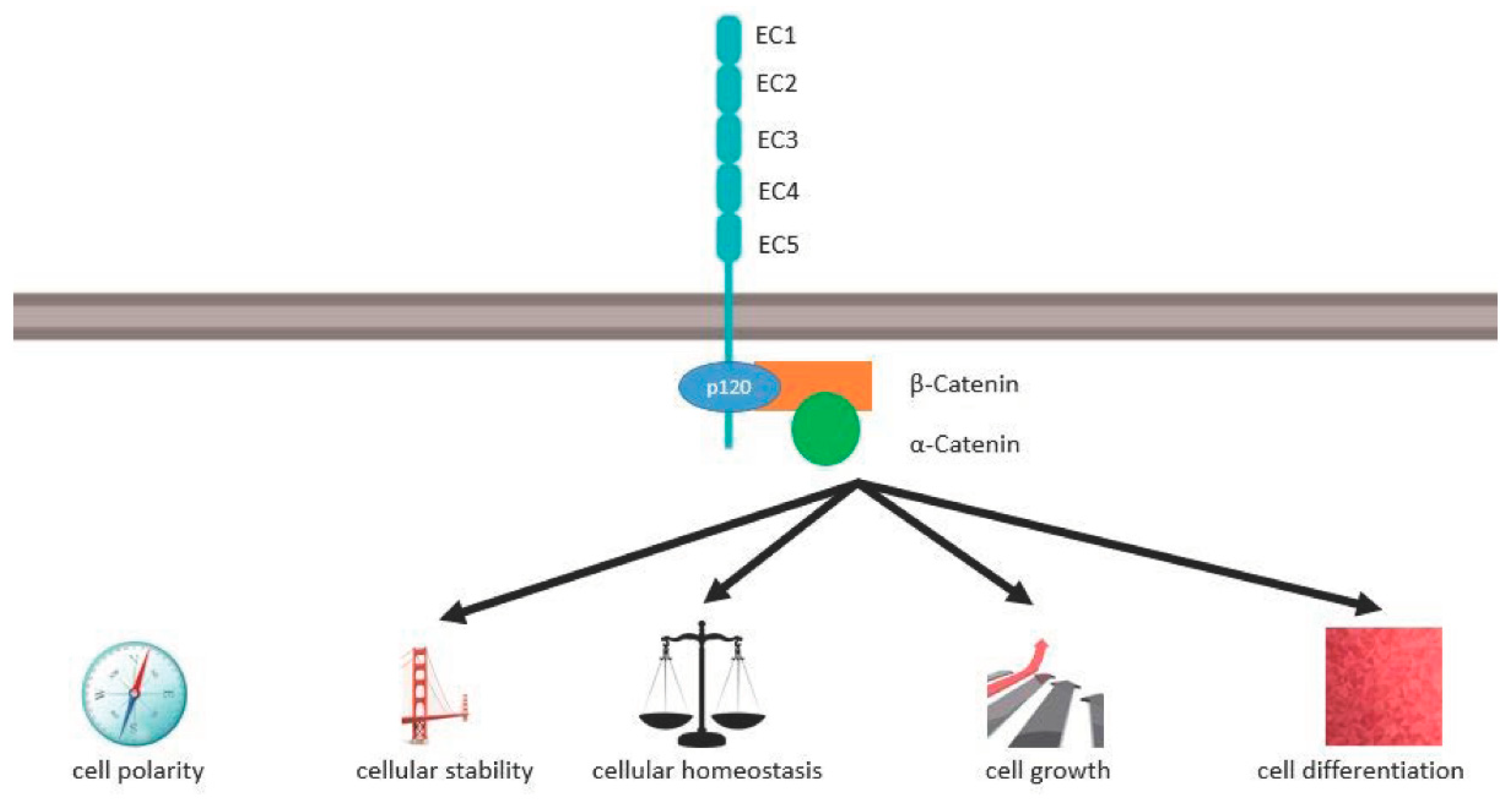
2.3. E-Cadherin Structure, Molecular Function, and Signal Pathways in Cancer

CDH1 encodes the 120-kDa transmembrane glycoprotein E-cadherin. E-cadherin is a classical Type I-cadherin that is detected in the adherens junctions of all epithelial tissues [35]. Its structure is highly conserved and consists of an extracellular domain, a transmembrane domain, and an intracellular domain (see Figure 1).
The extracellular domain has five extracellular cadherin repeats (EC1–EC5) with about 100 amino acids each. There are four negatively poled calcium ion binding sides between the five EC-domains each binding three Calcium ions. The EC1s of neighbouring cells interact calcium-dependently in a homophilic manner and mediate rigid cell–cell adhesion [36,37]. Cis interactions of the EC1 domain with the EC2 domain lead to further clustering [38].
The single transmembrane domain bridges the extracellular domain to the intracellular domain. The intracellular domain of E-cadherin has a highly phosphorylated region that binds p120 catenin as well as α catenin via β catenin. α catenin links the actin cytoskeleton to the plasma membrane which is vital for the stability of the epithelial architecture and cellular mechanical signaling.
Truncating and missense variants occur in every domain, truncating domains most frequently in EC2, EC3, EC5 and IC [34]. Missense mutations of the extracellular region of E-cadherin are more likely to result in increased motility of the affected cell than missense mutations of the intracellular region [24].
As shown in Figure 1, via the cadherin–catenin complex, E-cadherin controls cell polarity, cellular stability, cellular homeostasis, cell growth and differentiation. α-E-catenin, the protein of CTNNA1, interacts with E-cadherin via β-catenin. Lobo et al. describe mutations in α-E-catenin, which occur in areas of the gene that code for the amino acids 97–148 [39]. These amino acids are forming the N-terminal region of α-E-catenin which binds β-catenin. β-catenin finally binds to the C-terminal region of E-cadherin.
E-cadherin interacts directly or indirectly with many other membrane components (e.g., tight junctions, gap junctions, desmosomes, integrin-based adhesions, and pathogenetic proteins) [36]. The extracellular fragment of E-cadherin can be cleaved by membrane-bound metalloproteinases from the membrane after proteolysis of E-cadherin [40]. Soluble E-cadherin controls multiple signaling pathways via paracrine and autocrine signaling. Its function is modulated by various direct and indirect interactions (e.g., mechanical forces at the junctions, direct phosphorylation, binding and phosphorylation of its catenins, endocytosis, recycling and degradation, multiple signaling pathways, developmental processes) [41].
E-cadherin is a crucial part of signaling pathways like cyclin kinase inhibitor p27-mediated signaling, activation of mitogen-activated kinase (MAPK), rat sarcoma viral oncogene (Ras) and ras-related C3 botulinum toxin substrate (Rac1) signaling, phosphatidylinositol-3-kinase (PI3K)/AKT signaling, HIPPO signaling and epithelial mesenchymal transition [41,42,43].
Loss of E-cadherin weakens the adherens junctions in epithelial tissues and intercellular stability by wnt, the HIPPO pathway, growth factor receptor tyrosine kinases (RTKs) and GTPases [43]. This is the first crucial step towards metastasis, angiogenesis, adhesion and invasion. Uncontrolled activation of intracellular signaling pathways results in uncontrolled proliferation [44].
Table 1. Molecular profiling in HDGC.
GenesCorresponding ProteinsCancers in Which the Related Genes ExpressEncoded FunctionsReference
CDH1E-cadherin Gastric cancer (diffuse type including HDGC), lobular breast cancer, colorectal cancer, hepatocellular carcinoma, squamous cell carcinomas of the skin, neck, and head, esophageal carcinoma, pancreatic ductal adenocarcinomaTumor suppressor and adhesion, adhesion in cell-cell contact [4,10,11,13,14,15,18,24,25,26,27,28,29,30,31,32,33,34,35,38,39,40,41,42,43,44,45,46,47,48]
CTNNA1Alpha E-cadherinHDGC (HDGC without CDH1-mutation, as well), diffuse type GC and colorectal cancerTumor suppressor and adhesion, adhesion in cell-cell contact[16,19,20]
MAP3K6A serine/threonine protein kinaseHDGC,
diffuse and intestinal type GC
Tumor suppressor[24,25]
BRCA1, PALB2, RAD51CCorresponding Proteins on their ownHDGC, breast cancer, pancreatic cancer, pancreatic ductal adenocarcinomaRegulate homologous DNA recombination[16,17,18]
DOT1LHistone MethyltransferaseDGCEffect on DNA Repair[17,24]
MSH2Codes for a DNA mismatch repair (MMR) proteinHereditary nonpolyposis colorectal cancer (HNPCC)
HDGC
diffuse and intestinal type gastric cancer
Component of the post-replicative DNA mismatch repair system (MMR)[16,17,18]
META protein with an extracellular, transmembrane and a tyrosine kinase domainGastric cancer (intestinal and diffuse type, including HDGC, breast-, prostate-, ovarian- cancer, hereditary papillary renal carcinoma Functions in cellular survival, embryogenesis, cellular migration, and invasion [26]
CD44A cell-surface glycoproteinHyperplastic polyps, intestinal metaplasia, gastric cancer (intestinal and diffuse type, including HDGC) Cell surface glycoprotein[27]
INSRReceptor tyrosine kinaseHDGCEffect on tumor cell invasion[17]
FBXO24F-box proteinDGCTumor driver[17]

2.4. Other Contributing Risk Factors for HDGC

Despite a considerable decline in mortality over the last century, GC still remains the fifth cause of cancer-related death worldwide, with an incidence of over 1.08 million new cases [2]. To study its etiology and risk factors remains an important issue as it may allow to identify major targets for primary prevention.
A consortium of epidemiological investigations initiated in 2012 the “Stomach cancer Pooling (StoP) Project” which included over twenty studies in order to analyze the role of lifestyle and genetic determinants in the etiology of GC in general [45]. Patients with gastric cancer from different parts of the world were included (40% from Asia, 43% from Europe, 17% from North America), regardless of the histological subtype; intestinal type as well as diffuse type GC. Among the identified risk factors, Helicobacter pylori is still the most prominent, with a significantly higher prevalence in men than women (in men: OR 1.33, 95% confidence interval 1.04–1.70) [45]. Other identified risk factors were cigarette smoking (40% increased risk in smokers versus non-smokers), heavy alcohol consumption (50% increased risk for heavy drinkers compared to never drinkers), meat consumption (especially red, processed, and total meat, with the highest OR observed for an intake of 150 g/day of red meat) and low socioeconomic status [45,46,47].

2.5. Histological Alterations in CDH1 Gene Mutation

HDGC has no predilection to occur in a specific area of the stomach. Therefore, total gastric mucosa embedding and mapping should be performed to detect HDGC in prophylactic gastrectomies. This is time consuming and might contribute to a lack of expertise to assess HDGC appropriately. Furthermore, background alterations in CDH1-carriers make it even more difficult to discover cancer lesions: Chronic gastritis, foveolar hyperplasia, cystic gland dilatation and epithelial tufting, as well as globoid change, have been described in CDH1-carriers [48]. For its histological detection, standardized protocols, WHO and Laurén classification should be used.
Two precursor lesions for HDGC have been described in CDH1-carriers—first, “in situ signet-ring cell carcinoma” (SRCC), which is characterized by signet-ring cells (SRC) with hyperchromatic and depolarized nuclei located in glandular basal membranes; second, rows of SRCs with pagetoid growth patterns inside the glandular basal membrane and below the non-neoplastic glandular epithelium and foveola.
In advanced stages of HDGC, SRC and pleomorphic poorly differentiated non-SRCs infiltrate the gastric wall, diffusely in a scattered manner. They are poorly cohesive. The gastric wall appears thickened and rigid. The cells increase in size from the neck gland zone to the surface [8]. Glandular and tubular structures, mucinous areas and rosettes are especially found in areas of lymphovascular invasion and lymph node metastasis [49]. A high KI-67 proliferation index and overexpression of p53 indicate a highly aggressive phenotype of HDGC cancers [49].

3. Implications of CDH1 Gene Alterations in the Diagnosis and Management of HDGC

3.1. IGCLC Diagnostic Criteria for HDGC Syndrome

Due to the relatively low incidence of HDGC, randomized clinical trial data specific to HDGC are lacking. Consequently, recommendations concerning genetic testing criteria rely on consensus expert opinion and evidence as well as observational studies. Over the years, multiple criteria have been developed to facilitate the screening of CDH1 mutation carriers. The updated guidelines for HDGC of the IGCLC provide recommendations for medical care givers and patients at a risk for the HDGC syndrome. To restore the balance between the accessibility, costs and acceptance of genetic testing and greater identification of pathogenic variant carriers, the HDGC genetic testing criteria have been broadened, mainly through less restrictive age limits [8]. The recommendations of CDH1 testing according to the IGCLC guidelines are summarized in Table 2.
Furthermore, the IGCLC guidelines intend a better understanding of the nature of the disease and associated risk factors, facilitating decision-making and providing guidance for lifetime management of HDGC families [8].
Due to the association of CTNNA1 mutations and HDGC [4,15,16,18], IGCLC guidelines recommend that individuals who fulfil criteria for genetic testing but are found to be negative for a CDH1 variant should be subsequently tested for CTNNA1 mutation [4,8]. In a study by Clark et al., CTNNA1 loss-of-function variants were detected on multiplex genetic panel testing (MGPT) with a majority of the individuals diagnosed with gastric or breast cancer [19]. In another study, in CDH1 mutation-negative index cases, candidate mutations within genes of high and moderate penetrance including CTNNA1 and BRCA2 were identified [4].
Although there is only limited evidence that PALB2 pathogenic gene variants might be associated with an increased risk for GC in CDH1-negative patients [50], IGCLC recommends considering PALB2 testing in the case of unexplained gastric cancer in some families, alongside other genes associated with an increased risk of gastric cancer (eg. ATM, BRCA2, Lynch syndrome genes, APC and TP53) [8].

3.2. Genetic Counseling and Genetic Testing for HDGC Syndrome

The clinical phenotype of HDGC shows considerable heterogeneity concerning the type of cancer and age of onset [4,51]. In some families, GC onset has been described in a very young patient’s age [10]. In other families, the main cancer phenotype is LBC and either no other family members or only older family members are affected by HDGC. Some reports also suggest an association with colorectal cancer [34].
The age of testing at-risk relatives of index patients should take into consideration the earliest age of cancer onset in that specific family. Testing from the late teens or early 20ies is favored in families with early-onset DGC. Because of the autosomal-dominant inheritance and high penetrance of CDH1 germline mutations [52,53], genetic testing guided by personal history, family history, detailed three-generation pedigree analysis, and other risk models are currently used to identify the individuals at risk for HDGC syndrome [8].
Retrospective data provide evidence that early genetic counseling and detection of CDH1 mutation in asymptomatic carriers improves survival in HDGC [54,55]. However, a study by Xicola et al., conducted with 113 CDH1 pathogenic variant probands and 476 relatives, demonstrated that, in unselected CDH1 pathogenic variant carrier families who do not fulfil the criteria of HDGC, the GC risk is low [52]. In addition, the age at first diagnosis was higher than previously reported in families pre-selected for HDGC criteria. A substantial proportion of families with cancer limited to breast do not present with any GC [52].
Genetic testing includes two main clinical approaches: single-gene testing or multiplex genetic panel testing (MGPT). Both tests can be performed on DNA extracted from blood or buccal samples except for patients who have received allogenic bone marrow transplantation or a recent diagnosis of hematologic malignancy. In these patients, DNA from a fibroblast culture is the preferable tissue sample.
The spread of MGPT has led to the identification of CDH1 and CTNNA1 germline pathogenic and likely pathogenic variants in individuals without a personal and family history suggestive of HDGC. This issue poses challenges regarding the clinical management [19,56].
The CDH1 gene analysis includes the examination of the entire coding sequence by PCR amplification and DNA sequencing, including intron-exon boundaries [57,58,59]. The sequence analysis by PCR based Sanger sequencing and direct sequencing allows for detecting small intragenic deletions/insertions and missense, nonsense, as well as splice variants of CDH1 and other germline mutations. Exonic or whole-gene deletions and duplications are detected by quantitative PCR, long-range PCR, multiplex ligation-dependent probe amplification (MPLA) and chromosomal microarray analysis (CMA) [60].
A study reported by Molinaro et al. supports a multimethod approach for CDH1 testing using a series of molecular methods, including DNA sequencing, MPLA, single-nucleotide primer extension, bisulfite sequencing, reverse-transcription PCR and bioinformatics tools [61]. It demonstrates that both DNA and RNA analysis are required to increase the detection rate of pathogenic mutation. The number of patients without a clear molecular diagnosis could be reduced by this approach [61]. Another study suggests that a multigene analysis is important to detect germline mutations and genetic variants in patients with cancer at multiple sites in the family [62].
Bio-imaging analysis of in situ fluorescence microscopy has been described to quantify missense CDH1 variants [63]. The study illustrated that this method could be applied to evaluate the pathogenicity of the CDH1 mutant variant as a complementary approach and to detect a wide range of proteins and diseases characterized by aberrant protein expression or trafficking deregulation.
The updated variant curation guidelines published in 2018 by the American College of Medical Genetics and Genomics and the Association for Molecular Pathology (ACMG/AMP) provide a framework to assess variant pathogenicity of the germline CDH1 sequence variants. They facilitate a better detection and classification in CDH1 variants and thus identification of individuals and families that will benefit from cancer surveillance and risk-reducing surgery [64].
With the help of the rapid development of genetic testing technologies and data analysis tools over the last few years, genetic testing has become more efficient, enabling the discovery of susceptibility genes for HDGC.
However, in the assessment of the appropriate clinical management and decision-making, a multidisciplinary team consisting of gastroenterologists, genetic counselors, medical geneticists, pathologists and oncological psychologists is of key importance [65]. Due to the variable penetrance of the CDH1 or CTNNA1 mutation, unaffected carriers from HDGC families face difficult decisions and can be assisted best through education and interactions with counseling by an informed interdisciplinary team [66].
Genetic counseling should include the initial evaluation of personal history, family history and a three-generation family pedigree as well as any history of cleft lip or cleft palate and histopathological confirmation of cancer diagnosis or precursor lesions [8].
Due to the high psychosocial impact on quality of life and long-time sequelae, a discussion on lifetime risks of HDGC or LBC, as well as the possibility of undergoing PTG or other options of surveillance such as endoscopy, should be provided [55,67,68]. The counselee should also be informed about the potential significance of the test results for the family, and about reproductive options, such as the availability of genetic testing through prenatal and preimplantation genetic diagnosis [12].

3.3. Management of CDH1 Mutation Gene Carriers

The heterogeneity of the clinical phenotype in HDGC makes a balanced screening strategy necessary. For risk stratification, it may help to identify different CDH1 mutation types. Lo et al. describe associations of CDH1 germline variant location and cancer phenotype in families with HDGC [34]. The frequency of truncating germline CDH1 variants varied across functional domains of the E-cadherin receptor gene and was highest in linker and PRE regions. Families with truncating CDH1 germline variants located in the PRE-PRO region were six times more likely to have family members affected by colorectal cancer (OR 6.2) compared with germline variants in other regions. Variants in the intracellular E-cadherin region were protective for cancer at a young age (OR 0.2) and in the linker regions for breast cancer (OR 0.35). Different CDH1 genotypes were associated with different intracellular signaling activation levels including different p-ERK, p-mTOR, and β-catenin levels in early submucosal T1a lesions of HDGC families with different CDH1 variants [34].
These data suggest that the location and type of CDH1 germline variants may help to identify families at increased risk for concomitant cancers that might benefit from individualized surveillance and intervention strategies. However, there is no strong evidence that the risk of other cancers is significantly increased in individuals with a particular CDH1 pathogenic variant [4,61,69].
For female CDH1 mutation gene carriers, breast surveillance with annual MRI should start at the age of 30 years [70]. The benefit of adding mammography to MRI in young women remains uncertain [71]. A woman with a CDH1 pathogenic variant may choose breast-conserving surgery depending on the individual risk profile [72].
Current data suggest that a germline mutation in the CDH1 gene is present in a proportion of signet-ring carcinomas in the colorectum. A subset of these tumors could be familial. However, the loss of E-cadherin staining in the absence of CDH1 mutations also suggests a role for environmental factors in a subset of these tumors [73,74,75]. Interestingly, the colorectal adenocarcinoma associated with HDGC syndrome is not necessarily signet ring cell carcinoma [76]. However, there is currently no evidence to suggest that the risk of colorectal cancer in CDH1 mutation carriers is significantly elevated and up to now there are insufficient data to give recommendations on colorectal cancer screening [69].
In CDH1 and CTNNA1 gene mutation carriers, the question arises as to whether endoscopic surveillance for HDGC might be sufficient or surgery be indicated. A significant challenge in HDGC syndrome is balancing the high risk of GC against the morbidity of total gastrectomy that includes dumping syndrome, weight loss, malnutrition and depression.
Castro et al. describe that, in their cohort of patients with CDH1 mutation gene carriers, gastroscopy with targeted and random biopsies had low sensitivity. In that cohort, 332 random and 22 targeted biopsies were needed to identify a single SRCC focus. Total gastrectomy was performed in 13 patients and SRCC foci were identified in 12 surgical specimens [77]. In contrast, Dieren et al. describe a higher sensitivity of gastroscopy regarding SRCC in CDH1 mutation gene carriers. Out of 30 patients who underwent gastrectomy, at least one SRCC lesion was found in 26 of 30 gastrectomy specimens. In 18 of these 26 surgical specimens (69%), SRCC had been identified by endoscopic biopsies. The low number of SRCC detected through random sampling stresses the importance of targeted biopsies [78].
Another study by Mi et al. reports on endoscopic surveillance for HDGC according to CDH1 mutation status. Eighty-five individuals fulfilling HDGC criteria were included and underwent 201 endoscopies. Fifty-four patients (63.5%) tested positive for CDH1 mutation. SRCC yield was 61.1% in CDH1 mutation carriers compared with 9.7% in non-carriers, and mutation-positive patients had a 10-fold risk of SRCC on endoscopy compared with those with no mutation detected (p < 0.0005) [79].
The reported studies show that a combination of random and targeted therapies increases the sensitivity of gastroscopy in CDH1 mutation gene carriers and that endoscopic surveillance is an option especially with high resolution and high-definition endoscopes with advanced endoscopic imaging [80]. As CDH1 mutation carriers harbor an approximately 70% lifetime risk of gastric cancer in men and 56% in women [4], the critical question remains as to whether endoscopic surveillance only unnecessarily delays a gastrectomy. An ongoing study (NCT04253106) examines the role of liquid biopsies in blood and gastric fluid in asymptomatic carriers who decline gastrectomy. This might be a complementary approach to endoscopic surveillance for these patients in the near future.
These studies further stress that one of the major problems is the variable penetrance of CDH1 gene mutation carriers and other distinct gene mutations as well to a clinical phenotype and HDGC. This issue might be addressed in a pooled data analysis of the data that are already available including all patients that underwent PTG. Furthermore, this aspect might be addressed in prospective, multicentric register studies. As we are dealing with a rare disease but tremendous impact on patients and families, all patients with manifestation of HDGC, LBC but also CDH1 gene mutation carriers without penetrance of the malignant phenotype should be prospectively included in such registers.

3.4. Therapy Strategies for HDGC

Endoscopic surveillance and/or gastrectomy are the pillars of management for patients with HDGC. In case of locally restricted expansion, surgical resection is indicated. In stage IB-III, resection is combined with perioperative or adjuvant chemotherapy in accordance with sporadic, non-HDGC [81]. Local progressed, non-resectable or metastatic disease should be treated with chemotherapy. The FDA approved Pembrolizumab combined with Trastuzumab and Chemotherapy as first-line treatment in locally advanced unresectable or metastatic HER2-positive gastric or gastroesophageal junction adenocarcinoma and for recurrent locally advanced or metastatic gastric or gastroesophageal junction cancer with PD-L1-expression. For patients without HER2-overexpression but with high PD-L1 expression on the tumor cells and infiltrating immune cells (PD-L1 CPS > 5%), Nivolumab in combination with chemotherapy is recommended and approved by FDA and EMA.
Unfortunately, there is currently no specific molecular genetic therapy available for HDGC. Due to the rarity of the tumor disease, there are currently no studies investigating specific molecular treatment strategies specific for HDGC. Drug screening studies in isogenic CDH1−/−-mutant mammary epithelial MCF10A and c.1380delA CDH1-mutant gastric cancer cells raise the hope that HDGC might respond well to PI3K, mTOR, or ALK/ROS-1 like tyrosine kinase inhibition [25]. These inhibitors are already widely used in breast cancer and non-small cell lung cancer (NSCLC), among others. New trials on gastric cancer focus on microRNAs, suppressor-tRNA, antibodies targeting tyrosine kinase receptors, immune checkpoints (PDL-1, CTLA-4, TIM-3, LAG-3), and cytokines (TGF-β, Il-6), cell therapy, and virotherapy [82].

4. Conclusions

The clinical phenotype of HDGC shows considerable heterogeneity with regard to the type of cancer and age of onset, rendering the identification of patients with a high pretest probability of HDGC challenging. Due to the rapid development of genetic testing technologies and data analysis tools over the last few years, genetic testing has become more efficient, enabling the discovery of susceptibility genes for HDGC. Screening for CDH1 and CTNNA1 mutation (alongside other genes associated with increased risk of gastric cancer in the case of unexplained gastric cancer in some families) should be performed in individuals who fulfil criteria for genetic testing, followed by a thorough evaluation and interdisciplinary approach to establish the best therapeutic approach (endoscopic surveillance versus prophylactic gastrectomy). In the assessment of the appropriate management and decision-making, a multidisciplinary team consisting of gastroenterologists, surgeons, genetic counselors, medical geneticists, pathologists, and oncological psychologists is therefore of key importance.

Author Contributions

A.K., M.M. and L.-S.C. conceived the study concept. L.-S.C., S.S. and H.C.T. reviewed the relevant literature. L.-S.C. designed the structure of the manuscript. L.-S.C., S.S. and H.C.T. wrote separate chapters (L.-S.C.: Section 2.4, Section 3.1, Section 3.2 and Section 4, Abstract; S.S.: Section 2.3, Section 2.5 and Section 3.4; H.C.T.: Section 1, Section 2.1, Section 2.2 and Section 3.3). A.K. and M.M. revised the manuscript. All authors have read and agreed to the published version of the manuscript.

Funding

This research received no external funding.

Institutional Review Board Statement

Not applicable.

Informed Consent Statement

Not applicable.

Data Availability Statement

Not applicable.

Conflicts of Interest

The authors declare no conflict of interest.

Abbreviations

ACMGAmerican College of Medical Genetics and Genomics
AMPAssociation for Molecular Pathology
APCadenomatous polyposis coli
ATMataxia telangiectasia mutated gene
BRCA2breast cancer gene 2
CDH1cadherin-1 or E-cadherin
CMAchromosomal microarray analysis
CTNNA1catenin alpha-1
DNAdeoxyribonucleic acid
DGCdiffuse gastric cancer
HDGChereditary diffuse gastric cancer
HNPCChereditary nonpolyposis colorectal cancer
IGCLCinternational gastric cancer linkage consortium
LBClobular breast cancer
MGPTmultiplex genetic panel testing
MPLAmultiplex ligation-dependent probe amplification
ORodds ratio
PALB2partner and localizer of BRCA2
PCRpolymerase chain reaction
PTGprophylactic total gastrectomy
RNAribonucleic acid
SRCsignet-ring cells
StoPstomach cancer pooling project
TP53tumor protein 53

References

  1. Guilford, P.; Hopkins, J.; Harraway, J.; McLeod, M.; McLeod, N.; Harawira, P.; Taite, H.; Scoular, R.; Miller, A.; Reeve, A.E. E-Cadherin Germline Mutations in Familial Gastric Cancer. Nature 1998, 392, 402–405. [Google Scholar] [CrossRef] [PubMed]
  2. Sung, H.; Ferlay, J.; Siegel, R.L.; Laversanne, M.; Soerjomataram, I.; Jemal, A.; Bray, F. Global Cancer Statistics 2020: Globocan Estimates of Incidence and Mortality Worldwide for 36 Cancers in 185 Countries. CA A Cancer J. Clin. 2021, 71, 209–249. [Google Scholar] [CrossRef] [PubMed]
  3. Fitzgerald, R.C.; Hardwick, R.; Huntsman, D.; Carneiro, F.; Guilford, P.; Blair, V.; Chung, D.C.; Norton, J.; Ragunath, K.; Van Krieken, J.H.; et al. Hereditary Diffuse Gastric Cancer: Updated Consensus Guidelines for Clinical Management and Directions for Future Research. J. Med. Genet. 2010, 47, 436–444. [Google Scholar] [CrossRef] [PubMed] [Green Version]
  4. Hansford, S.; Kaurah, P.; Li-Chang, H.; Woo, M.; Senz, J.; Pinheiro, H.; Schrader, K.A.; Schaeffer, D.F.; Shumansky, K.; Zogopoulos, G.; et al. Hereditary Diffuse Gastric Cancer Syndrome: CDH1 Mutations and Beyond. JAMA Oncol. 2015, 1, 23–32. [Google Scholar] [CrossRef] [Green Version]
  5. Wang, Z.; Graham, D.Y.; Khan, A.; Balakrishnan, M.; Abrams, H.R.; El-Serag, H.B.; Thrift, A.P. Incidence of Gastric Cancer in the USA during 1999 to 2013: A 50-State Analysis. Int. J. Epidemiol. 2018, 47, 966–975. [Google Scholar] [CrossRef]
  6. Rona, K.A.; Schwameis, K.; Zehetner, J.; Samakar, K.; Green, K.; Samaan, J.; Sandhu, K.; Bildzukewicz, N.; Katkhouda, N.; Lipham, J.C. Gastric Cancer in the Young: An Advanced Disease with Poor Prognostic Features. J. Surg. Oncol. 2017, 115, 371–375. [Google Scholar] [CrossRef]
  7. Lecuit, T.; Yap, A.S. E-Cadherin Junctions as Active Mechanical Integrators in Tissue Dynamics. Nat. Cell Biol. 2015, 17, 533–539. [Google Scholar] [CrossRef]
  8. Blair, V.R.; McLeod, M.; Carneiro, F.; Coit, D.G.; D’Addario, J.L.; van Dieren, J.M.; Harris, K.L.; Hoogerbrugge, N.; Oliveira, C.; van der Post, R.S.; et al. Hereditary Diffuse Gastric Cancer: Updated Clinical Practice Guidelines. Lancet Oncol. 2020, 21, e386–e397. [Google Scholar] [CrossRef]
  9. Kaurah, P.; MacMillan, A.; Boyd, N.; Senz, J.; De Luca, A.; Chun, N.; Suriano, G.; Zaor, S.; Van Manen, L.; Gilpin, C.; et al. Founder and Recurrent CDH1 Mutations in Families with Hereditary Diffuse Gastric Cancer. JAMA 2007, 297, 2360–2372. [Google Scholar] [CrossRef] [Green Version]
  10. van der Post, R.S.; Vogelaar, I.P.; Carneiro, F.; Guilford, P.; Huntsman, D.; Hoogerbrugge, N.; Caldas, C.; Schreiber, K.E.C.; Hardwick, R.H.; Ausems, M.G.E.M.; et al. Hereditary Diffuse Gastric Cancer: Updated Clinical Guidelines with an Emphasis on Germline CDH1 Mutation Carriers. J. Med. Genet. 2015, 52, 361–374. [Google Scholar] [CrossRef] [Green Version]
  11. van der Post, R.S.; Gullo, I.; Oliveira, C.; Tang, L.H.; Grabsch, H.I.; O’Donovan, M.; Fitzgerald, R.C.; van Krieken, H.; Carneiro, F. Histopathological, Molecular, and Genetic Profile of Hereditary Diffuse Gastric Cancer: Current Knowledge and Challenges for the Future. Adv. Exp. Med. Biol. 2016, 908, 371–391. [Google Scholar] [CrossRef] [PubMed]
  12. Hallowell, N.; Badger, S.; Richardson, S.; Caldas, C.; Hardwick, R.H.; Fitzgerald, R.C.; Lawton, J. High-Risk Individuals’ Perceptions of Reproductive Genetic Testing for CDH1 Mutations. Fam. Cancer 2017, 16, 531–535. [Google Scholar] [CrossRef] [PubMed]
  13. Luo, W.; Fedda, F.; Lynch, P.; Tan, D. CDH1 Gene and Hereditary Diffuse Gastric Cancer Syndrome: Molecular and Histological Alterations and Implications for Diagnosis And Treatment. Front. Pharmacol. 2018, 9, 1421. [Google Scholar] [CrossRef] [PubMed] [Green Version]
  14. Benusiglio, P.R.; Malka, D.; Rouleau, E.; De Pauw, A.; Buecher, B.; Noguès, C.; Fourme, E.; Colas, C.; Coulet, F.; Warcoin, M.; et al. CDH1 Germline Mutations and the Hereditary Diffuse Gastric and Lobular Breast Cancer Syndrome: A Multicentre Study. J. Med. Genet. 2013, 50, 486–489. [Google Scholar] [CrossRef]
  15. Majewski, I.J.; Kluijt, I.; Cats, A.; Scerri, T.S.; de Jong, D.; Kluin, R.J.C.; Hansford, S.; Hogervorst, F.B.L.; Bosma, A.J.; Hofland, I.; et al. An α-E-Catenin (CTNNA1) Mutation in Hereditary Diffuse Gastric Cancer. J. Pathol. 2013, 229, 621–629. [Google Scholar] [CrossRef]
  16. Donner, I.; Kiviluoto, T.; Ristimäki, A.; Aaltonen, L.A.; Vahteristo, P. Exome Sequencing Reveals Three Novel Candidate Predisposition Genes for Diffuse Gastric Cancer. Fam. Cancer 2015, 14, 241–246. [Google Scholar] [CrossRef]
  17. Fewings, E.; Larionov, A.; Redman, J.; Goldgraben, M.A.; Scarth, J.; Richardson, S.; Brewer, C.; Davidson, R.; Ellis, I.; Evans, D.G.; et al. Germline Pathogenic Variants in PALB2 and Other Cancer-Predisposing Genes in Families with Hereditary Diffuse Gastric Cancer without CDH1 Mutation: A Whole-Exome Sequencing Study. Lancet Gastroenterol. Hepatol. 2018, 3, 489–498. [Google Scholar] [CrossRef] [Green Version]
  18. Benusiglio, P.R.; Colas, C.; Guillerm, E.; Canard, A.; Delhomelle, H.; Warcoin, M.; Bellanger, J.; Eyries, M.; Zizi, M.; Netter, J.; et al. Clinical Implications of CTNNA1 Germline Mutations in Asymptomatic Carriers. Gastric Cancer 2019, 22, 899–903. [Google Scholar] [CrossRef] [Green Version]
  19. Clark, D.F.; Michalski, S.T.; Tondon, R.; Nehoray, B.; Ebrahimzadeh, J.; Hughes, S.K.; Soper, E.R.; Domchek, S.M.; Rustgi, A.K.; Pineda-Alvarez, D.; et al. Loss-of-Function Variants in CTNNA1 Detected on Multigene Panel Testing in Individuals with Gastric or Breast Cancer. Genet. Med. 2020, 22, 840–846. [Google Scholar] [CrossRef] [Green Version]
  20. Sahasrabudhe, R.; Lott, P.; Bohorquez, M.; Toal, T.; Estrada, A.P.; Suarez, J.J.; Brea-Fernández, A.; Cameselle-Teijeiro, J.; Pinto, C.; Ramos, I.; et al. Germline Mutations in PALB2, BRCA1, and RAD51C, Which Regulate DNA Recombination Repair, in Patients With Gastric Cancer. Gastroenterology 2017, 152, 983–986. [Google Scholar] [CrossRef] [Green Version]
  21. Carvajal-Carmona, L.G. PALB2 as a Familial Gastric Cancer Gene: Is the Wait Over? Lancet Gastroenterol. Hepatol. 2018, 3, 451–452. [Google Scholar] [CrossRef] [Green Version]
  22. Alexandrov, L.B.; Nik-Zainal, S.; Siu, H.C.; Leung, S.Y.; Stratton, M.R. A Mutational Signature in Gastric Cancer Suggests Therapeutic Strategies. Nat. Commun. 2015, 6, 8683. [Google Scholar] [CrossRef] [Green Version]
  23. Berx, G.; Becker, K.F.; Höfler, H.; van Roy, F. Mutations of the Human E-Cadherin (CDH1) Gene. Hum. Mutat. 1998, 12, 226–237. [Google Scholar] [CrossRef]
  24. Melo, S.; Figueiredo, J.; Fernandes, M.S.; Gonçalves, M.; Morais-de-Sá, E.; Sanches, J.M.; Seruca, R. Predicting the Functional Impact of CDH1 Missense Mutations in Hereditary Diffuse Gastric Cancer. Int. J. Mol. Sci. 2017, 18, 2687. [Google Scholar] [CrossRef] [PubMed] [Green Version]
  25. Li, D.; Lo, W.; Rudloff, U. Merging Perspectives: Genotype-Directed Molecular Therapy for Hereditary Diffuse Gastric Cancer (HDGC) and E-Cadherin-EGFR Crosstalk. Clin. Transl. Med. 2018, 7, 7. [Google Scholar] [CrossRef] [PubMed] [Green Version]
  26. Oliveira, C.; Pinheiro, H.; Figueiredo, J.; Seruca, R.; Carneiro, F. E-Cadherin Alterations in Hereditary Disorders with Emphasis on Hereditary Diffuse Gastric Cancer. Prog. Mol. Biol. Transl. Sci. 2013, 116, 337–359. [Google Scholar] [CrossRef] [PubMed]
  27. Figueiredo, J.; Söderberg, O.; Simões-Correia, J.; Grannas, K.; Suriano, G.; Seruca, R. The Importance of E-Cadherin Binding Partners to Evaluate the Pathogenicity of E-Cadherin Missense Mutations Associated to HDGC. Eur. J. Hum. Genet. 2013, 21, 301–309. [Google Scholar] [CrossRef] [Green Version]
  28. Oliveira, C.; Sousa, S.; Pinheiro, H.; Karam, R.; Bordeira-Carriço, R.; Senz, J.; Kaurah, P.; Carvalho, J.; Pereira, R.; Gusmão, L.; et al. Quantification of Epigenetic and Genetic 2nd Hits in CDH1 during Hereditary Diffuse Gastric Cancer Syndrome Progression. Gastroenterology 2009, 136, 2137–2148. [Google Scholar] [CrossRef] [PubMed]
  29. Grady, W.M.; Willis, J.; Guilford, P.J.; Dunbier, A.K.; Toro, T.T.; Lynch, H.; Wiesner, G.; Ferguson, K.; Eng, C.; Park, J.G.; et al. Methylation of the CDH1 Promoter as the Second Genetic Hit in Hereditary Diffuse Gastric Cancer. Nat. Genet. 2000, 26, 16–17. [Google Scholar] [CrossRef]
  30. Oliveira, C.; de Bruin, J.; Nabais, S.; Ligtenberg, M.; Moutinho, C.; Nagengast, F.M.; Seruca, R.; van Krieken, H.; Carneiro, F. Intragenic Deletion of CDH1 as the Inactivating Mechanism of the Wild-Type Allele in an HDGC Tumour. Oncogene 2004, 23, 2236–2240. [Google Scholar] [CrossRef] [Green Version]
  31. Figueiredo, J.; Melo, S.; Carneiro, P.; Moreira, A.M.; Fernandes, M.S.; Ribeiro, A.S.; Guilford, P.; Paredes, J.; Seruca, R. Clinical Spectrum and Pleiotropic Nature of CDH1 Germline Mutations. J. Med. Genet. 2019, 56, 199–208. [Google Scholar] [CrossRef] [PubMed] [Green Version]
  32. Oliveira, C.; Senz, J.; Kaurah, P.; Pinheiro, H.; Sanges, R.; Haegert, A.; Corso, G.; Schouten, J.; Fitzgerald, R.; Vogelsang, H.; et al. Germline CDH1 Deletions in Hereditary Diffuse Gastric Cancer Families. Hum. Mol. Genet. 2009, 18, 1545–1555. [Google Scholar] [CrossRef] [PubMed]
  33. Kluijt, I.; Siemerink, E.J.M.; Ausems, M.G.E.M.; van Os, T.A.M.; de Jong, D.; Simões-Correia, J.; van Krieken, J.H.; Ligtenberg, M.J.; Figueiredo, J.; van Riel, E.; et al. CDH1-Related Hereditary Diffuse Gastric Cancer Syndrome: Clinical Variations and Implications for Counseling. Int. J. Cancer 2012, 131, 367–376. [Google Scholar] [CrossRef] [PubMed] [Green Version]
  34. Lo, W.; Zhu, B.; Sabesan, A.; Wu, H.-H.; Powers, A.; Sorber, R.A.; Ravichandran, S.; Chen, I.; McDuffie, L.A.; Quadri, H.S.; et al. Associations of CDH1 Germline Variant Location and Cancer Phenotype in Families with Hereditary Diffuse Gastric Cancer (HDGC). J. Med. Genet. 2019, 56, 370–379. [Google Scholar] [CrossRef]
  35. Yu, W.; Yang, L.; Li, T.; Zhang, Y. Cadherin Signaling in Cancer: Its Functions and Role as a Therapeutic Target. Front. Oncol. 2019, 9, 989. [Google Scholar] [CrossRef] [PubMed]
  36. Biswas, K.H. Molecular Mobility-Mediated Regulation of E-Cadherin Adhesion. Trends Biochem. Sci. 2020, 45, 163–173. [Google Scholar] [CrossRef] [PubMed]
  37. Rasool, S.; Geethakumari, A.M.; Biswas, K.H. Role of Actin Cytoskeleton in E-Cadherin-Based Cell–Cell Adhesion Assembly and Maintenance. J. Indian Inst. Sci. 2021, 101, 51–62. [Google Scholar] [CrossRef]
  38. Gloushankova, N.A.; Rubtsova, S.N.; Zhitnyak, I.Y. Cadherin-Mediated Cell-Cell Interactions in Normal and Cancer Cells. Tissue Barriers 2017, 5, e1356900. [Google Scholar] [CrossRef] [Green Version]
  39. Lobo, S.; Benusiglio, P.R.; Coulet, F.; Boussemart, L.; Golmard, L.; Spier, I.; Hüneburg, R.; Aretz, S.; Colas, C.; Oliveira, C. Cancer Predisposition and Germline CTNNA1 Variants. Eur. J. Med. Genet. 2021, 64, 104316. [Google Scholar] [CrossRef]
  40. Bernegger, S.; Jarzab, M.; Wessler, S.; Posselt, G. Proteolytic Landscapes in Gastric Pathology and Cancerogenesis. Int. J. Mol. Sci. 2022, 23, 2419. [Google Scholar] [CrossRef]
  41. Kourtidis, A.; Lu, R.; Pence, L.J.; Anastasiadis, P.Z. A Central Role for Cadherin Signaling in Cancer. Exp. Cell Res. 2017, 358, 78–85. [Google Scholar] [CrossRef] [PubMed]
  42. Kaszak, I.; Witkowska-Piłaszewicz, O.; Niewiadomska, Z.; Dworecka-Kaszak, B.; Ngosa Toka, F.; Jurka, P. Role of Cadherins in Cancer—A Review. Int. J. Mol. Sci. 2020, 21, 7624. [Google Scholar] [CrossRef] [PubMed]
  43. Mendonsa, A.M.; Na, T.-Y.; Gumbiner, B.M. E-Cadherin in Contact Inhibition and Cancer. Oncogene 2018, 37, 4769–4780. [Google Scholar] [CrossRef] [PubMed]
  44. Cureus | Significance of E-Cadherin Gene Mutations in Patients With Hereditary Diffuse Gastric Cancer Syndrome: A Systematic Review. Available online: https://www.cureus.com/articles/39243-significance-of-e-cadherin-gene-mutations-in-patients-with-hereditary-diffuse-gastric-cancer-syndrome-a-systematic-review (accessed on 23 August 2021).
  45. Pelucchi, C.; Lunet, N.; Boccia, S.; Zhang, Z.-F.; Praud, D.; Boffetta, P.; Levi, F.; Matsuo, K.; Ito, H.; Hu, J.; et al. The Stomach Cancer Pooling (StoP) Project: Study Design and Presentation. Eur. J. Cancer Prev. 2015, 24, 16–23. [Google Scholar] [CrossRef]
  46. Ferro, A.; Rosato, V.; Rota, M.; Costa, A.R.; Morais, S.; Pelucchi, C.; Johnson, K.C.; Hu, J.; Palli, D.; Ferraroni, M.; et al. Meat Intake and Risk of Gastric Cancer in the Stomach Cancer Pooling (StoP) Project. Int. J. Cancer 2020, 147, 45–55. [Google Scholar] [CrossRef]
  47. Corso, G.; Montagna, G.; Figueiredo, J.; La Vecchia, C.; Fumagalli Romario, U.; Fernandes, M.S.; Seixas, S.; Roviello, F.; Trovato, C.; Guerini-Rocco, E.; et al. Hereditary Gastric and Breast Cancer Syndromes Related to CDH1 Germline Mutation: A Multidisciplinary Clinical Review. Cancers 2020, 12, 1598. [Google Scholar] [CrossRef]
  48. Rocha, J.P.; Gullo, I.; Wen, X.; Devezas, V.; Baptista, M.; Oliveira, C.; Carneiro, F. Pathological Features of Total Gastrectomy Specimens from Asymptomatic Hereditary Diffuse Gastric Cancer Patients and Implications for Clinical Management. Histopathology 2018, 73, 878–886. [Google Scholar] [CrossRef]
  49. Spoto, C.P.E.; Gullo, I.; Carneiro, F.; Montgomery, E.A.; Brosens, L.A.A. Hereditary Gastrointestinal Carcinomas and Their Precursors: An Algorithm for Genetic Testing. Semin. Diagn. Pathol. 2018, 35, 170–183. [Google Scholar] [CrossRef]
  50. Carreño, M.; Pena-Couso, L.; Mercadillo, F.; Perea, J.; Urioste, M. Investigation on the Role of PALB2 Gene in CDH1-Negative Patients With Hereditary Diffuse Gastric Cancer. Clin. Transl. Gastroenterol. 2020, 11, e00280. [Google Scholar] [CrossRef]
  51. Pharoah, P.D.; Guilford, P.; Caldas, C. Incidence of Gastric Cancer and Breast Cancer in CDH1 (E-Cadherin) Mutation Carriers from Hereditary Diffuse Gastric Cancer Families. Gastroenterology 2001, 121, 1348–1353. [Google Scholar] [CrossRef]
  52. Xicola, R.M.; Li, S.; Rodriguez, N.; Reinecke, P.; Karam, R.; Speare, V.; Black, M.H.; LaDuca, H.; Llor, X. Clinical Features and Cancer Risk in Families with Pathogenic CDH1 Variants Irrespective of Clinical Criteria. J. Med. Genet. 2019, 56, 838–843. [Google Scholar] [CrossRef] [PubMed] [Green Version]
  53. Huynh, J.M.; Laukaitis, C.M. Panel Testing Reveals Nonsense and Missense CDH1 Mutations in Families without Hereditary Diffuse Gastric Cancer. Mol. Genet. Genomic. Med. 2016, 4, 232–236. [Google Scholar] [CrossRef] [PubMed]
  54. Moslim, M.A.; Heald, B.; Tu, C.; Burke, C.A.; Walsh, R.M. Early Genetic Counseling and Detection of CDH1 Mutation in Asymptomatic Carriers Improves Survival in Hereditary Diffuse Gastric Cancer. Surgery 2018, 164, 754–759. [Google Scholar] [CrossRef] [PubMed]
  55. Choi, H.-J.; Ki, C.-S.; Suh, S.-P.; Kim, J.-W. Presymptomatic Identification of CDH1 Germline Mutation in a Healthy Korean Individual with Family History of Gastric Cancer. Ann. Lab. Med. 2014, 34, 386–389. [Google Scholar] [CrossRef] [Green Version]
  56. Katona, B.W.; Clark, D.F.; Domchek, S.M. CDH1 on Multigene Panel Testing: Look Before You Leap. JNCI J. Natl. Cancer Inst. 2020, 112, 330–334. [Google Scholar] [CrossRef]
  57. López, M.; Cervera-Acedo, C.; Santibáñez, P.; Salazar, R.; Sola, J.-J.; Domínguez-Garrido, E. A Novel Mutation in the CDH1 Gene in a Spanish Family with Hereditary Diffuse Gastric Cancer. Springerplus 2016, 5, 1181. [Google Scholar] [CrossRef] [Green Version]
  58. Suriano, G.; Yew, S.; Ferreira, P.; Senz, J.; Kaurah, P.; Ford, J.M.; Longacre, T.A.; Norton, J.A.; Chun, N.; Young, S.; et al. Characterization of a Recurrent Germ Line Mutation of the E-Cadherin Gene: Implications for Genetic Testing and Clinical Management. Clin. Cancer Res. 2005, 11, 5401–5409. [Google Scholar] [CrossRef] [Green Version]
  59. Hakkaart, C.; Ellison-Loschmann, L.; Day, R.; Sporle, A.; Koea, J.; Harawira, P.; Cheng, S.; Gray, M.; Whaanga, T.; Pearce, N.; et al. Germline CDH1 Mutations Are a Significant Contributor to the High Frequency of Early-Onset Diffuse Gastric Cancer Cases in New Zealand Māori. Fam. Cancer 2019, 18, 83–90. [Google Scholar] [CrossRef] [Green Version]
  60. Zhang, H.; Feng, M.; Feng, Y.; Bu, Z.; Li, Z.; Jia, S.; Ji, J. Germline Mutations in Hereditary Diffuse Gastric Cancer. Chin. J. Cancer Res. 2018, 30, 122–130. [Google Scholar] [CrossRef]
  61. Molinaro, V.; Pensotti, V.; Marabelli, M.; Feroce, I.; Barile, M.; Pozzi, S.; Laghi, L.; Serrano, D.; Bernard, L.; Bonanni, B.; et al. Complementary Molecular Approaches Reveal Heterogeneous CDH1 Germline Defects in Italian Patients with Hereditary Diffuse Gastric Cancer (HDGC) Syndrome. Genes Chromosomes Cancer 2014, 53, 432–445. [Google Scholar] [CrossRef]
  62. Lajus, T.B.P.; Sales, R.M.D. CDH1 Germ-Line Missense Mutation Identified by Multigene Sequencing in a Family with No History of Diffuse Gastric Cancer. Gene 2015, 568, 215–219. [Google Scholar] [CrossRef] [PubMed]
  63. Sanches, J.M.; Figueiredo, J.; Fonseca, M.; Durães, C.; Melo, S.; Esménio, S.; Seruca, R. Quantification of Mutant E-Cadherin Using Bioimaging Analysis of in Situ Fluorescence Microscopy. A New Approach to CDH1 Missense Variants. Eur. J. Hum. Genet. 2015, 23, 1072–1079. [Google Scholar] [CrossRef] [PubMed] [Green Version]
  64. Lee, K.; Krempely, K.; Roberts, M.E.; Anderson, M.J.; Carneiro, F.; Chao, E.; Dixon, K.; Figueiredo, J.; Ghosh, R.; Huntsman, D.; et al. Specifications of the ACMG/AMP Variant Curation Guidelines for the Analysis of Germline CDH1 Sequence Variants. Hum. Mutat. 2018, 39, 1553–1568. [Google Scholar] [CrossRef]
  65. Gjyshi, O.; Vashi, P.; Seewald, L.; Kohan, M.; Abboud, E.; Fowler, E.; Suppiah, R.; Halabi, H. Therapeutic and Prophylactic Gastrectomy in a Family with Hereditary Diffuse Gastric Cancer Secondary to a CDH1 Mutation: A Case Series. World J. Surg. Oncol. 2018, 16, 143. [Google Scholar] [CrossRef] [PubMed]
  66. Lynch, H.T.; Nustas, R.; Kassim, T.; Snyder, C.; Shaw, T.; Diab, O. The Benefits of a Model of Interval Comprehensive Assessments (MICA) in Hereditary Cancer Syndromes: Hereditary Diffuse Gastric Cancer (HDGC) as an Example. Cancer Genet. 2019, 233–234, 43–47. [Google Scholar] [CrossRef] [PubMed]
  67. Hallowell, N.; Lawton, J.; Badger, S.; Richardson, S.; Hardwick, R.H.; Caldas, C.; Fitzgerald, R.C. The Psychosocial Impact of Undergoing Prophylactic Total Gastrectomy (PTG) to Manage the Risk of Hereditary Diffuse Gastric Cancer (HDGC). J. Genet. Couns. 2017, 26, 752–762. [Google Scholar] [CrossRef] [PubMed] [Green Version]
  68. Hallowell, N.; Badger, S.; Richardson, S.; Caldas, C.; Hardwick, R.H.; Fitzgerald, R.C.; Lawton, J. An Investigation of the Factors Effecting High-Risk Individuals’ Decision-Making about Prophylactic Total Gastrectomy and Surveillance for Hereditary Diffuse Gastric Cancer (HDGC). Fam. Cancer 2016, 15, 665–676. [Google Scholar] [CrossRef]
  69. van der Post, R.S.; Vogelaar, I.P.; Manders, P.; van der Kolk, L.E.; Cats, A.; van Hest, L.P.; Sijmons, R.; Aalfs, C.M.; Ausems, M.G.E.M.; Gómez García, E.B.; et al. Accuracy of Hereditary Diffuse Gastric Cancer Testing Criteria and Outcomes in Patients With a Germline Mutation in CDH1. Gastroenterology 2015, 149, 897–906. [Google Scholar] [CrossRef]
  70. Stjepanovic, N.; Moreira, L.; Carneiro, F.; Balaguer, F.; Cervantes, A.; Balmaña, J.; Martinelli, E. Hereditary Gastrointestinal Cancers: ESMO Clinical Practice Guidelines for Diagnosis, Treatment and Follow-Up. Ann. Oncol. 2019, 30, 1558–1571. [Google Scholar] [CrossRef] [Green Version]
  71. Vreemann, S.; van Zelst, J.C.M.; Schlooz-Vries, M.; Bult, P.; Hoogerbrugge, N.; Karssemeijer, N.; Gubern-Mérida, A.; Mann, R.M. The Added Value of Mammography in Different Age-Groups of Women with and without BRCA Mutation Screened with Breast MRI. Breast Cancer Res. 2018, 20, 84. [Google Scholar] [CrossRef]
  72. King, T.A.; Pilewskie, M.; Muhsen, S.; Patil, S.; Mautner, S.K.; Park, A.; Oskar, S.; Guerini-Rocco, E.; Boafo, C.; Gooch, J.C.; et al. Lobular Carcinoma in Situ: A 29-Year Longitudinal Experience Evaluating Clinicopathologic Features and Breast Cancer Risk. J. Clin. Oncol. 2015, 33, 3945–3952. [Google Scholar] [CrossRef] [PubMed]
  73. Aitchison, A.; Hakkaart, C.; Whitehead, M.; Khan, S.; Siddique, S.; Ahmed, R.; Frizelle, F.A.; Keenan, J.I. CDH1 Gene Mutation in Early-Onset, Colorectal Signet-Ring Cell Carcinoma. Pathol. Res. Pract. 2020, 216, 152912. [Google Scholar] [CrossRef] [PubMed]
  74. Hamilton, L.E.; Jones, K.; Church, N.; Medlicott, S. Synchronous Appendiceal and Intramucosal Gastric Signet Ring Cell Carcinomas in an Individual with CDH1-Associated Hereditary Diffuse Gastric Carcinoma: A Case Report of a Novel Association and Review of the Literature. BMC Gastroenterol. 2013, 13, 114. [Google Scholar] [CrossRef] [Green Version]
  75. Richards, F.M.; McKee, S.A.; Rajpar, M.H.; Cole, T.R.P.; Evans, D.G.R.; Jankowski, J.A.; McKeown, C.; Sanders, D.S.A.; Maher, E.R. Germline E-Cadherin Gene (CDH1) Mutations Predispose to Familial Gastric Cancer and Colorectal Cancer. Hum. Mol. Genet. 1999, 8, 607–610. [Google Scholar] [CrossRef] [PubMed]
  76. Salahshor, S.; Hou, H.; Diep, C.B.; Loukola, A.; Zhang, H.; Liu, T.; Chen, J.; Iselius, L.; Rubio, C.; Lothe, R.A.; et al. A Germline E-Cadherin Mutation in a Family with Gastric and Colon Cancer. Int. J. Mol. Med. 2001, 8, 439–443. [Google Scholar] [CrossRef]
  77. Castro, R.; Lobo, J.; Pita, I.; Videira, F.; Pedro-Afonso, L.; Dinis-Ribeiro, M.; Brandão, C. Random Biopsies in Patients Harboring a CDH1 Mutation: Time to Change the Approach? Rev. Esp. Enfermadades Dig. 2020, 112, 367–372. [Google Scholar] [CrossRef]
  78. van Dieren, J.M.; Kodach, L.L.; den Hartog, P.; van der Kolk, L.E.; Sikorska, K.; van Velthuysen, M.-L.F.; van Sandick, J.W.; Koemans, W.J.; Snaebjornsson, P.; Cats, A. Gastroscopic Surveillance with Targeted Biopsies Compared with Random Biopsies in CDH1 Mutation Carriers. Endoscopy 2020, 52, 839–846. [Google Scholar] [CrossRef]
  79. Mi, E.Z.; Mi, E.Z.; di Pietro, M.; O’Donovan, M.; Hardwick, R.H.; Richardson, S.; Ziauddeen, H.; Fletcher, P.C.; Caldas, C.; Tischkowitz, M.; et al. Comparative Study of Endoscopic Surveillance in Hereditary Diffuse Gastric Cancer According to CDH1 Mutation Status. Gastrointest. Endosc. 2018, 87, 408–418. [Google Scholar] [CrossRef] [Green Version]
  80. Yasuda, T.; Yagi, N.; Omatsu, T.; Hayashi, S.; Nakahata, Y.; Yasuda, Y.; Obora, A.; Kojima, T.; Naito, Y.; Itoh, Y. Benefits of Linked Color Imaging for Recognition of Early Differentiated-Type Gastric Cancer: In Comparison with Indigo Carmine Contrast Method and Blue Laser Imaging. Surg. Endosc. 2021, 35, 2750–2758. [Google Scholar] [CrossRef]
  81. Smyth, E.C.; Verheij, M.; Allum, W.; Cunningham, D.; Cervantes, A.; Arnold, D. Gastric Cancer: ESMO Clinical Practice Guidelines for Diagnosis, Treatment and Follow-Up. Ann. Oncol. 2016, 27, v38–v49. [Google Scholar] [CrossRef]
  82. El Rami, F.E.; Barsoumian, H.B.; Khneizer, G.W. Hereditary Diffuse Gastric Cancer Therapeutic Roadmap: Current and Novel Approaches in a Nutshell. Ther. Adv. Med. Oncol. 2020, 12, 1758835920967238. [Google Scholar] [CrossRef] [PubMed]
Figure 1. Role of E-cadherin for cellular homeostasis, polarity and differentiation.
Ijms 23 07821 g001
Table 2. IGCLC criteria for CDH1 testing (according to Blair et al., hereditary diffuse gastric cancer: updated clinical practice guidelines, 2020).
Family Criteria (1st or 2nd Degree Blood Relatives of Each Other) 1Individual Criteria
≥2 cases of gastric cancer in family regardless of age, with at least one DGC
≥1 case of DGC at any age and ≥1 case of LBC < 70 years in different family members
≥2 cases of LBC in family members < 50 years
DGC < 50 years
DGC at any age in individuals of Māori ethnicity
DGC at any age in individuals with a personal or family history (1st degree) of cleft lip/cleft palate
History of DGC and LBC, both diagnosed < 70 years
Bilateral LBC, diagnosed < 70 years
Gastric in situ signet ring cells and/or pagetoid spread of signet ring cells in individuals < 50 years
1 If there are no living affected relatives, consider tissue testing (tumor or normal) from an affected deceased relative. If these options are not possible, consider indirect testing in unaffected family members.
Publisher’s Note: MDPI stays neutral with regard to jurisdictional claims in published maps and institutional affiliations.

