Review

MicroRNA In Lung Cancer: Novel Biomarkers and Potential Tools for Treatment

Division of Pathology, The Cancer Institute, Japanese Foundation for Cancer Research, 3-8-31 Ariake, Koto-ku, Tokyo 135-8550, Japan
*
Author to whom correspondence should be addressed.
J. Clin. Med. 2016, 5(3), 36; https://doi.org/10.3390/jcm5030036
Submission received: 4 December 2015 / Revised: 23 February 2016 / Accepted: 1 March 2016 / Published: 9 March 2016
(This article belongs to the Special Issue MicroRNAs: Novel Biomarkers and Therapeutic Targets for Human Cancers)

Abstract

:
Lung cancer is the leading cause of cancer death in men and women worldwide. The lack of specific and sensitive tools for early diagnosis as well as still-inadequate targeted therapies contribute to poor outcomes. MicroRNAs are small non-coding RNAs, which regulate gene expression post-transcriptionally by translational repression or degradation of target mRNAs. A growing body of evidence suggests various roles of microRNAs including development and progression of lung cancer. In lung cancer, several studies have showed that certain microRNA profiles classified lung cancer subtypes, and that specific microRNA expression signatures distinguished between better-prognosis and worse-prognosis lung cancers. Furthermore, microRNAs circulate in body fluids, and therefore may serve as promising biomarkers for early diagnosis of lung cancer as well as for predicting prognosis of patients. In the present review, we briefly summarize microRNAs in the development and progression of lung cancer, focusing on possible applications of microRNAs as novel biomarkers and tools for treatment.

1. Introduction

Lung cancer is the leading cause of cancer death in men and women worldwide, accounting for more than 1.5 million deaths per year [1]. Lung carcinoma is generally classified as either small-cell lung carcinoma (SCLC) (about 20% of all lung carcinomas) or non-small cell lung carcinoma (NSCLC) (about 80%). Within these groups, further distinctions are made, with NSCLC sub-divided into adenocarcinoma (about 50% of all lung carcinomas), squamous cell carcinoma (SqCC), and large cell carcinoma.
Although new molecular targeted therapies to some types of lung cancer have shown promising results, no potential targeted therapy can be applied to a large number of lung cancer patients. Despite improvements in early diagnosis of lung cancer, most lung cancers are diagnosed at an advanced stage. Therefore, the identification of novel diagnostic biomarkers or treatment strategies is critical and essential for the control of lung cancer.
MicroRNAs, which are small non-coding RNAs that range in size from 19 to 25 nucleotides, play important regulatory roles in animals and plants by targeting mRNAs for translational repression or degradation. MicroRNAs comprise one of the most abundant classes of gene regulatory molecules in multicellular organisms and likely influence the output of many protein-coding genes [2]. A growing body of evidence is emerging to suggest a wide range of fundamental cellular processes such as cell differentiation, proliferation, growth, mobility, and apoptosis, as well as carcinogenesis or cancer progression [3,4]. The expression patterns of microRNAs are likely to correlate with characteristic clinicopathological parameters in cancer subtypes [5], suggesting that microRNAs are potential biomarkers for different cancer subtypes, classified by origin, histology, aggressiveness, or chemosensivity [6,7].
Public database, miRBase (http://www.mirbase.org/) (last access: 9 February 2016) provides various aspects of microRNA information, and the annotated human microRNAs of 1881 have been registered in miRBase 21. In general, the list of microRNAs involved in lung cancer is not limited to several microRNAs, but much wider, as well as the potential diagnostic microRNAs in tissues.
Of importance, an abundance of undegradated microRNAs exists not only in tissues but also in body fluids, including blood, plasma, serum, and sputum [8]. This nature of easy availability makes microRNAs promising biomarkers for non-invasive liquid biopsy in cancer practice. Biomarkers analyzed by liquid biopsy [9,10] include circulating tumor cells and exosomes (containing DNA, mRNA, microRNA, etc.) [11,12] as well as circulating cell-free DNA [13], mRNA [14], and microRNA [15]. Although, non-invasive liquid biopsy is promising, it has not been employed for multiclass cancer diagnostics due to non-specificity of these blood-based biosources to pinpoint the nature of the primary cancer [9,13,14].
In addition, microRNAs are far less degradated in formalin-fixed paraffin-embedded (FFPE) samples than mRNAs. Therefore, accurate measurements of microRNAs can be performed from FFPE samples, which are usually collected and preserved in hospitals. An availability of archived FFPE samples for the accurate measurement of microRNAs is a great advantage to conduct microRNA research or apply microRNA profiles for clinical practice.
Due to the characteristic nature of microRNAs, microRNAs have a potential to be used for the development of diagnostics, prognostics, and targeted therapeutics. In terms of treatment, microRNA expression profiles can predict chemotherapeutic response and serve as important biomarkers for the stratification of patients for personalized therapeutic strategies. Furthermore, microRNAs have the potential to serve as molecular targeted agents. Although the initial results from human-based studies revealed the promise of microRNA targeted therapies, several obstacles need to be overcome prior to the therapeutic application of microRNAs to the clinic [16].
In this review, we summarize recent works of microRNAs in lung cancer, focusing on microRNAs as novel biomarkers and potential tools for treatment.

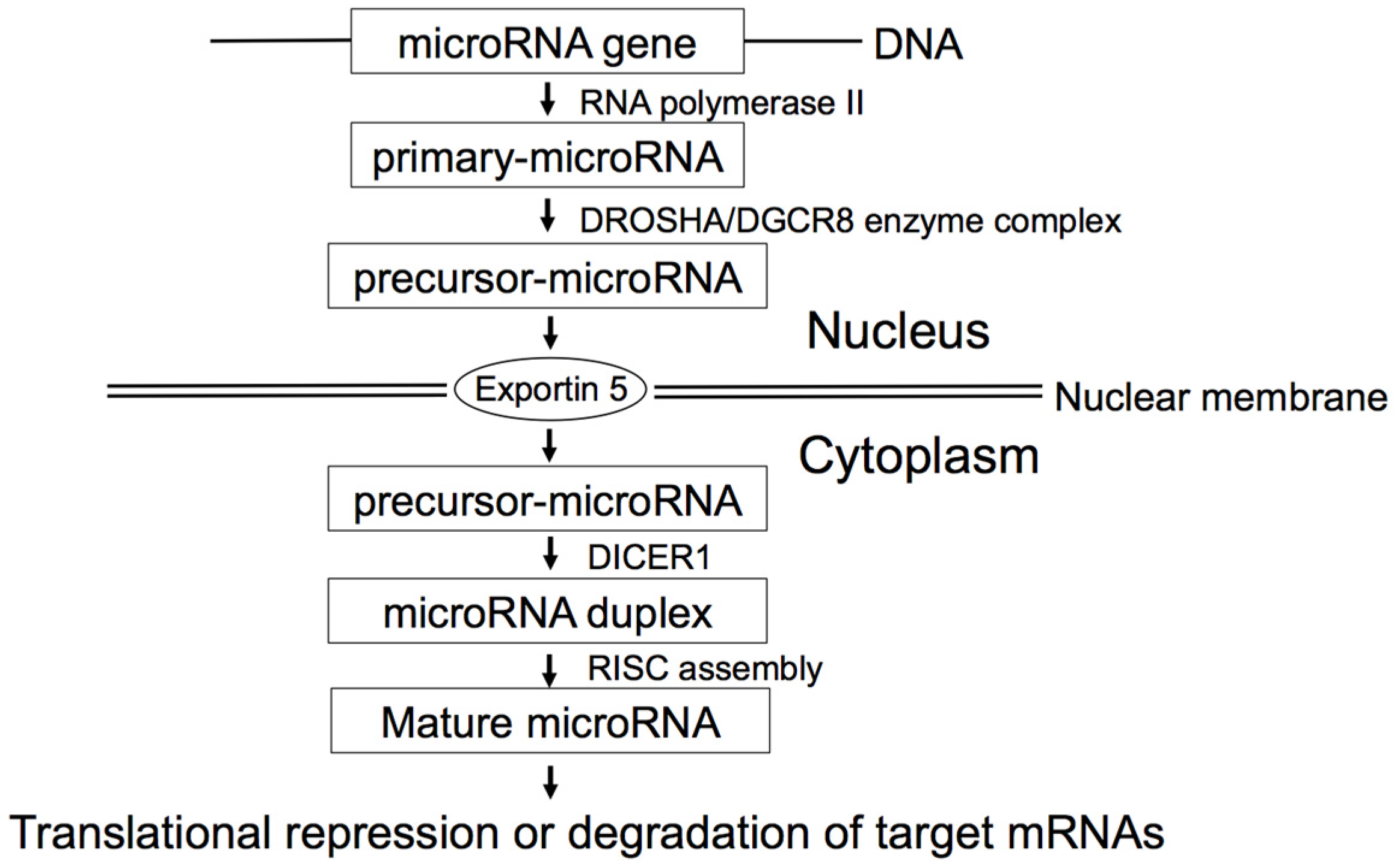
2. MicroRNA Biogenesis

Biogenesis of microRNA (Figure 1) begins with the transcription of primary-microRNA by RNA polymerase II. Then, DROSHA/DGCR8 enzyme complex crops the primary-microRNA into precursor-microRNA, followed by exportin-5-mediated transport from the nucleus to the cytoplasm. Subsequently, DICER1 cleaves the precursor-microRNA to form the microRNA duplex. One strand of the microRNA duplex is selected to function as a mature microRNA and loaded into the RNA-induced silencing complex (RISC), whereas the partner microRNA* strand is preferentially degradated. The mature microRNA leads to translational repression or degradation of target mRNAs.

3. MicroRNAs as Tumor Suppressor Genes and Oncogenes

MicroRNAs play important regulatory roles in carcinogenesis not only as tumor suppressor genes but also as oncogenes. Several microRNAs are dysregulated in lung cancer. Table 1 shows principal microRNAs involved in the development or progression of lung cancer, their gene targets, and associated biological processes.

3.1. Tumor Suppressor microRNAs

3.1.1. Let-7 Family

The let-7 family was the first identified microRNA in humans [17]. In lung cancer, let-7 has been shown to inhibit the expression of oncogenes involved in cellular proliferation, such as RAS, MYC, and HMGA2 [18,19]. Let-7 also inhibits the expression of CDK6, and the reduced expression of let-7, therefore, leads to the promotion of cell cycle progression [19]. Of interest, let-7 directly down-regulates DICER1 expression, suggesting that let-7 may regulate the global production of microRNAs [20].

3.1.2. miR-34 Family

The miR-34 family (miR-34a, miR-34b, and miR-34c) is directly induced by TP53 in response to DNA damage, controlling cell cycle arrest and apoptosis in cancer [21]. The miR-34 family is down-regulated in lung cancer, leading to the up-regulation of miR-34 target genes, such as MET, BCL2, PDGFR-α (PDGFRA), and PDGFR-β (PDGFRB) [22,23,24]. The up-regulation of MET and BCL2 by reduced miR-34 expression leads to cell proliferation. MiR-34 dependent PDGFR-α/β downregulation inhibits tumorigenesis and enhances TRAIL (TNF-related apoptosis-inducing ligand)-induced apoptosis in lung cancer [24].

3.1.3. miR-200 Family

The miR-200 family (miR-200a, miR-200b, miR-200c, and miR-429) plays an important role in the promotion of epithelial-mesenchymal transition (EMT). Through the regulation of ZEB (zinc finger E-box-binding homeobox) transcription factors (ZEB1 and ZEB2), E-cadherin (CDH1), vimentin (VIM), the down-regulated miR-200 family promotes EMT in the progression of lung cancer [25,26].

3.2. Oncogenic microRNAs

3.2.1. miR-21

miR-21 is one of the most representative oncogenic microRNAs, being overexpressed in various types of solid tumors as well as leukemia. miR-21 drives tumorigenesis through inhibition of negative regulators of RAS/MEK/ERK pathway and suppression of apoptosis. Overexpressed miR-21 downregulates the expressions of PTEN [27], PDCD4 [28,29,30], and TPM1 [30], promoting cell proliferation and migration, and inhibiting apoptosis.

3.2.2. miR-17-92 Cluster

The miR-17-92 polycistronic cluster comprises seven different microRNAs (miR-17-3p, miR-17-5p, miR-18a, miR-19a, miR-20a, miR-19b-1, and miR-92a) and resides in intron 3 of the C13orf25 gene at 13q31.3 [31]. The miR-17-92 cluster was reported to be overexpressed in lung cancer, in particular SCLC [31]. Overexpression of miR-17-92 cluster down-regulates E2F1, HIF1A, and PTEN, promoting cell proliferation and cancer progression [32,33].

3.2.3. miR-221/222

miR-221 and miR-222 are involved in the development and progression of lung cancer by targeting PTEN and TIMP3 tumor suppressor genes [34,35]. Overexpressed miR-221/222 inhibits apoptosis and promotes cell migration by down-regulating PTEN and TIMP3.

4. Diagnostic microRNAs

4.1. Diagnostic microRNAs in Tissues

Early detection of lung cancer is prerequisite to reduce lung cancer mortality, because lung cancers are often diagnosed at advanced stages, where clinical treatments are less (or least) effective. MicroRNA expression signatures of lung cancer have been reported by numerous studies, however the reported microRNA profiles were not so consistent. Vosa et al. performed a comprehensive meta-analysis of 20 published microRNA expression studies in lung cancer, including a total of 598 tumor and 528 normal lung tissues [36]. Using a recently published robust rank aggregation method, they identified a statistically significant microRNA meta-signature of seven up-regulated (miR-21, miR-210, miR-182, miR-31, miR-200b, miR-205, and miR-183) and eight down-regulated (miR-126-3p, miR-30a, miR-30d, miR-486-5p, miR-451a, miR-126-5p, miR-143, and miR-145) microRNAs.

4.2. Diagnostic microRNAs in Body Fluids

Importantly, microRNAs are present not only in tissues but also in body fluids, such as blood, plasma, serum, or sputum. By examining body fluids, we may be able to distinguish lung cancer patients from healthy individuals. Jiang’s group conducted several studies to assess the usefulness of microRNAs in body fluids for the lung cancer screening [37,38,39,40]. Using sputum specimens, they examined the expression of miR-21, which is an overexpressed microRNA in lung cancer [37]. The expression of miR-21 in sputum was higher in NSCLC patients, and the detection of miR-21 expression produced 70% sensitivity and 100% specificity in the distinction between 23 NSCLC patients and 17 cancer-free individuals. In another study, using an independent set, they demonstrated that the expression profile of four sputum microRNAs (miR-21, miR-486, miR-375, and miR-200b) demonstrated 81% sensitivity and 92% specificity in the distinction between 64 NSCLC patients and 58 healthy individuals [40]. They also assessed the usefulness for plasma microRNAs as potential biomarkers for NSCLC [39]. They showed that the expression profile of four plasma microRNAs (miR-21, miR-126, miR-210, and miR-486-5p) yielded 86% sensitivity and 97% specificity in distinguishing 58 NSCLC patients from 29 healthy individuals. Furthermore, this panel of microRNAs produced 73% sensitivity and 97% specificity in identifying stage I NSCLC patients, suggesting the usefulness of plasma microRNAs as potential biomarkers to identify even early stage NSCLC patients [39]. In another study, they showed that the expression profile of three plasma microRNAs (miR-21, miR-210, and miR-486-5p) produced 75% sensitivity and 85% specificity in the distinction between 32 patients with malignant solitary pulmonary nodules (SPNs) and 33 individuals with benign SPNs. SPNs have been increasingly diagnosed with the advancement and widespread use of computed tomography (CT) scan. The combination of microRNA testing and CT scan may serve as a minimally invasive method of diagnosing individuals with SPNs. All of these four studies by Jiang’s group [37,38,39,40] included miR-21 as a biomarker to distinguish lung cancer patients from healthy individuals. Because miR-21 is overexpressed in various types of cancer, further studies are warranted to examine miR-21 expression for the distinction between lung cancer patients and patients of the other cancers.

5. MicroRNAs as Biomarkers for Histological Classification

Recent advances in the treatment of NSCLC with new drugs require an appropriate histological subtyping at diagnosis to avoid hazardous side effects. For instance, bevacizumab (brand name: Avastin), a monoclonal antibody which blocks angiogenesis by inhibiting vascular endothelial growth factor A (VEGFA), cannot be used for SqCC patients due to serious hemorrhagic complications. Similarly, pemetrexed (brand name: Alimta), a chemotherapy drug belonging to a class of chemotherapeutic drugs known as folate antimetabolites, cannot be used for SqCC patients due to adverse responses. Several studies have been conducted to distinguish between SqCC and non-SqCC NSCLC, utilizing microRNAs profiles [41,42,43]. Lebanony et al. found that higher expression of miR-205 was specific to SqCC in a test set. In an independent validation set, miR-205 expression in FFPE samples yielded 96% sensitivity and 90% specificity in the distinction between SqCCs and non-SqCC NSCLCs [41]. As a replication study, Bishop et al. showed that the measurement of miR-205 expression in small biopsies/aspirates can distinguish between SqCCs and non-SqCC NSCLCs [42]. Recently, Hamamoto et al. have reported that the expression profile of three microRNAs (miR-205, miR-196b, and miR-375) distinguished between SqCCs and adenocarcinomas with 85% sensitivity and 83% specificity in a validation set [43].
SCLC, one of the neuroendocrine tumors, shows increased expression of ASCL1, which is a master gene of neuroendocrine differentiation. Nishikawa et al. demonstrated that miR-357 expression was induced by ASCL1 in lung neuroendocrine carcinoma [44]. They showed that the increased expression of miR-375 was prerequisite for the neuroendocrine differentiation by ASCL1, and suggested that miR-375 might reduce the YAP1-related proliferative arrest by inhibiting YAP1.

6. Prognostic microRNAs

6.1. Investigation for Prognostic microRNAs

As is the case with mRNA, microRNA profiles have been investigated as potential prognostic biomarkers. In 2004, Takamizawa et al. focused on microRNA let-7 [45], and reported that let-7 expression is lower in lung cancer than in normal lung tissue, and that the lower expression of let-7 in lung cancer was associated with poor prognosis. In addition, overexpression of let-7 in A549 lung adenocarcinoma cell line inhibited cell growth. This study [45] is important with respect to the first report of reduced expression of let-7 and the potential clinical and biological effects of such a microRNA alteration in lung cancer. We examined let-7 expressions in adenocarcinoma in situ (AIS, the new name for BAC (bronchioloalveolar carcinoma) [46]) as well as in invasive adenocarcinoma to investigate the association of let-7 expression with the progression of lung adenocarcinoma [47]. Of note, even in AIS, the expression of let-7 was reduced in comparison to matched normal lung tissue, suggesting that let-7 expression was reduced in the early stage of lung carcinogenesis. AIS is categorized into two subtypes, non-mucinous and mucinous AIS. Interestingly, the expression of let-7 was lower in mucinous AIS than non-mucinous AIS. The differential expression of let-7 between two morphological subtypes of AIS suggests an association between microRNA expression and morphology (even in the same category of AIS). According to this observation, complexity of microRNA expressions depending on morphology as well as cancer progression or driver mutations can be presumed.
In 2006, the first microRNA profiling in lung cancer was reported by Yanaihara et al. [48]. Using 104 NSCLCs (65 adenocarcinomas and 39 SqCCs) and matched normal lung tissues, they conducted profiling analyses of microRNA expression by microarray. They identified the higher expression of miR-155 and the lower expression of let-7a-2 as biomarkers of poor prognosis for NSCLC patients, and confirmed the validity using an independent set by real-time RT-PCR. Subsequently, in 2008, Yu et al. [49] identified a five-microRNA signature (let-7a, miR-221, miR-137, miR-372, and miR-182*) that classified a training set of 56 NSCLC cases into good-prognosis and poor-prognosis group. Then, they confirmed that the five-microRNA signature divided 62 NSCLC testing cases into two groups with good and poor prognosis. Furthermore, they validated the five-microRNAs signature as a prognostic classifier using an independent cohort set of 62 NSCLCs. This five-microRNA signature worked as a prognostic classifier for NSCLCs even with the same stage or SqCCs as well as adenocarcinomas.

6.2. Integrated Prognostic Classifier for Stage I Lung Cancer

MiR-21 is one of the overexpressed microRNAs in various types of cancer, and targets of miR-21 include tumor suppressor genes, such as PTEN [27,50] and PDCD4 [28,29,30]. Harris’ group has identified an integrated prognostic classifier for stage I lung adenocarcinoma based on microRNA, mRNA, and DNA methylation biomarkers, using frozen specimens [51]. This integrated prognostic classifier consisted of miR-21, four protein-coding genes (XPO1, BRCA1, HIF1A, and DLC1), and HOXA9 promoter methylation. miR-21, four protein-coding genes, or HOXA9 promoter methylation could independently classified stage I adenocarcinomas into two groups with a different survival (hazard ratio (HR) = 2.3, p = 0.01; HR = 2.8, p = 0.002; HR = 2.4, p = 0.005, respectively). When combined, the integrated biomarker worked as a much more accurate prognostic classifier for stage I adenocarcinoma (HR = 10.2, p = 3 × 10⁻⁵).
Importantly, abundant undegradaded microRNAs circulate in body fluids, such as blood, plasma, serum, and sputum [8]. There have been several studies to predict survival in NSCLC patients, by using microRNA profiles in body fluids. In 2010, Hu et al. reported that a four-serum-microRNA signature (miR-486, miR-30d, miR-1, and miR-499) was associated with overall survival in NSCLC, utilizing 120 cases for the training set and 123 cases for the testing set [52]. Subsequently, Wang et al. conducted pathway-based serum microRNA profiling and examined the prognostic association in advanced stage NSCLC patients [53]. They focused on microRNAs involved in TGF- β pathway, which plays crucial roles in control of cell proliferation, differentiation, apoptosis, and invasion. They identified 17 microRNAs which were significantly associated with two-year patient survival. Of these 17 microRNAs, miR-16 exhibited the most statistically significant association; high expression of miR-16 was associated with better survival (HR = 0.4, 95% confidence interval (CI), 0.3–0.5). They created 17-microRNA risk score to identify patients at the highest risk of death. NSCLC patients with a high-risk score had a 2.5-fold increased risk of death compared with those with a low-risk score (95% CI, 1.8–3.4; p = 1.1 × 10⁻⁷).

6.3. Let-7, DICER1, and Survival of Lung Cancer

DICER1 has an important role in microRNA biogenesis, converting precursor-microRNAs into mature microRNAs (Figure 1). Inversely, microRNA let-7 directly down-regulates DICER1 expression [20]. According to the study by Karube et al. [54], the reduced expression of DICER1 was associated with poor prognosis in NSCLC, suggesting the involvement of reduced DICER1 expression in the progression of lung cancer.

7. MicroRNA Associated with Driver Mutations and Therapeutic microRNAs

Somatic mutations in tyrosine kinases have recently emerged as driver mutations in carcinogenesis of lung cancer, especially in that of lung adenocarcinoma. Adenocarcinomas with activating mutations of EGFR are responsive to EGFR tyrosine kinase inhibitors (TKIs). However, T790M EGFR mutation and MET amplification lead to the resistance of EGFR TKIs [55]. Other mutually exclusive genetic alterations, which have been reported to work as driver mutations in lung adenocarcinoma, include translocations of ALK, ROS1, RET, NTKR1, NRG2, ERBB4, and BRAF, and mutations of KRAS, BRAF, ERBB2, NRAS, HRAS, MAP2K1, NF1, and RIT1 [56,57,58,59,60,61]. Adenocarcinomas with specific driver mutations sometimes have characteristic clinicopathological features [62,63,64,65,66,67]. For example, ALK-translocation lung adenocarcinomas are characterized by young-onset, never/light smokers, and acinar morphology with mucin/signet-ring cell morphology [64,65,66,67].
There are several studies examining the association between microRNAs expression and driver mutations (especially EGFR and KRAS mutation). miR-21 has been suggested to be an EGFR-regulated anti-apoptotic factor in never-smokers’ lung adenocarcinoma [68]. Recently, Li et al. have reported an association of miR-21 overexpression with acquired resistance of EGFR-TKI in NSCLCs [69]. Dacic et al. [70] examined the microRNA expressions in lung adenocarcinomas with different driver mutations, focusing on EGFR and KRAS mutation, using two EGFR-mutant, two KRAS-mutant, and two EGFR-wild-type/KRAS-wild-type adenocarcinomas and confirmed differentially expressed microRNAs in a validation set of 18 adenocarcinomas. They showed that miR-155, miR-25, and miR-495 were up-regulated only in the EGFR-wild-type/KRAS-wild-type, EGFR-mutant, and KRAS-mutant lung adenocarcinomas, respectively. In 2014, Bjaanaes et al. also examined microRNA profiles according to mutation status of EGFR and KRAS, using 154 lung adenocarcinomas by microarray [71]. The identified profiles were confirmed by real-time RT-PCR, using 103 lung cancer cases. They identified 17 microRNAs that were differentially expressed between EGFR-mutant and EGFR-wild-type adenocarcinomas, and three microRNAs differentially expressed between KRAS-mutant and KRAS-wild-type adenocarcinomas. There was no overlap between Dacic’s and Bjaanaes’ study, possibly due to the small sample size of Dacic’s study, differences in methodologies, or by chance. Further researches with a large sample size are required to elucidate the specific microRNA profiles according to the mutation status of EGFR or KRAS.
Weiss et al. [72] reported that miR-128b could directly regulate EGFR. The loss of heterozygosity (LOH) of miR-128b occurred frequently in NSCLC and correlated significantly with clinical response and survival after EGFR-TKI. Another study showed that microRNA-146a targeted EGFR, suppressed its downstream signaling and regulation of cell growth, and enhanced the cytotoxic effect of EGFR-TKI in five NSCLC cell lines [73].
To understand the role of microRNAs in TKI-resistant NSCLCs, Garofalo et al. examined microRNA changes that were mediated by tyrosine kinase receptors [74]. They demonstrated that this resistance could be overcome by anti-miR-221/222 and anti-miR-30c, which recovered the expression of the pro-apoptotic protein BCL2L11 (BIM) and increased the gefitinib sensitivity of NSCLC both in vitro and in vivo.
Other aspects regarding the role of microRNAs in relation to EGFR-TKI deserve comment. Recently, plasma microRNA profiles (miR-21, miR-27a, and miR-218) have been identified for primary resistance to EGFR-TKIs in advanced NSCLCs with EGFR activating mutation [75]. Pak et al. have found unique microRNAs (miR-34c, miR-183, and miR-210) in lung adenocarcinoma groups according to major TKI sensitive EGFR mutation status [76]. A recent study by Wang et al. suggests that the modulation of specific microRNAs (miR-374a and miR-548b) may provide a therapeutic target to treat or reverse gefitinib resistance in NSCLC with high expression of the Axl kinase [77]. The review by Ricciuti et al. [78] is worth reading because it summarized the existing relationship between microRNAs and resistance to EGFR-TKIs, and also focusing on the possible clinical applications of microRNAs in reverting and overcoming such resistance.
For now, limited evidence can be available for the microRNA profiles of lung cancer with specific driver mutations. Further studies should be conducted to elucidate driver-mutation-specific microRNA profiles. MicroRNA signature may be used as a diagnostic biomarker of lung cancer with a specific driver mutation, and also the combination of TKI and microRNA-based treatment is promising.

8. Prospects from Basic to Clinical Application of microRNAs

Actually, despite enormous efforts, several obstacles remain to be solved for the transition of microRNAs from basic to clinical application as novel biomarkers or potential tools for treatment. These obstacles include the standardization of microRNA detection, improved understanding of how microRNAs interact with other components of the genome, and the development of non-toxic targeted delivery of microRNAs to the lung or metastatic lesions as well as the selection of the proper vehicle for delivery [79]. Therefore, we have a bunch of obstacles to clear away, however we are now trying to solve these issues strenuously [79].

9. Conclusions

In this review, we introduced recent works of microRNAs involved in the development and progression of lung cancer, with a particular interest in microRNAs as novel biomarkers and potential tools for treatment. Of importance, microRNAs are far less degradated in FFPE samples than mRNAs, and therefore, an easy availability of FFPE samples in hospitals enables us to measure accurate microRNA expressions. In addition, microRNAs are also present in body fluids, making microRNAs promising diagnostic biomarkers. Therefore, there exists a great potential in microRNA analyses for cancer research. Further studies are demanded in order to use microRNA profiles as diagnostic markers and conduct microRNA-based therapies in clinical practice.

Acknowledgments

This study was funded, in part, by Grants-in-Aid for scientific Research from the Ministry of Education, Culture, Sports, Science, and Technology, Japan, grants from the Japan Society for the Promotion of Science, the Ministry of Health, Labour, and Welfare, and the Smoking Research Foundation.

Author Contributions

Kentaro Inamura and Yuichi Ishikawa conceived the review, wrote the text, and created the tables and figures.

Conflicts of Interest

The authors declare no conflict of interest.

References

  1. Torre, L.A.; Bray, F.; Siegel, R.L.; Ferlay, J.; Lortet-Tieulent, J.; Jemal, A. Global cancer statistics, 2012. CA Cancer J. Clin. 2015, 65, 87–108. [Google Scholar] [CrossRef] [PubMed]
  2. Bartel, D.P. MicroRNAs: Genomics, biogenesis, mechanism, and function. Cell 2004, 116, 281–297. [Google Scholar] [CrossRef]
  3. Winter, J.; Jung, S.; Keller, S.; Gregory, R.I.; Diederichs, S. Many roads to maturity: MicroRNA biogenesis pathways and their regulation. Nat. Cell Biol. 2009, 11, 228–234. [Google Scholar] [CrossRef] [PubMed]
  4. Joshi, P.; Middleton, J.; Jeon, Y.J.; Garofalo, M. MicroRNAs in lung cancer. World J. Methodol. 2014, 4, 59–72. [Google Scholar] [CrossRef] [PubMed]
  5. Lu, J.; Getz, G.; Miska, E.A.; Alvarez-Saavedra, E.; Lamb, J.; Peck, D.; Sweet-Cordero, A.; Ebert, B.L.; Mak, R.H.; Ferrando, A.A.; et al. MicroRNA expression profiles classify human cancers. Nature 2005, 435, 834–838. [Google Scholar] [CrossRef] [PubMed]
  6. Kishikawa, T.; Otsuka, M.; Ohno, M.; Yoshikawa, T.; Takata, A.; Koike, K. Circulating RNAs as new biomarkers for detecting pancreatic cancer. World J. Gastroenterol. 2015, 21, 8527–8540. [Google Scholar] [CrossRef] [PubMed]
  7. Hollis, M.; Nair, K.; Vyas, A.; Chaturvedi, L.S.; Gambhir, S.; Vyas, D. MicroRNAs potential utility in colon cancer: Early detection, prognosis, and chemosensitivity. World J. Gastroenterol. 2015, 21, 8284–8292. [Google Scholar] [CrossRef] [PubMed]
  8. Gilad, S.; Meiri, E.; Yogev, Y.; Benjamin, S.; Lebanony, D.; Yerushalmi, N.; Benjamin, H.; Kushnir, M.; Cholakh, H.; Melamed, N.; et al. Serum microRNAs are promising novel biomarkers. PLoS ONE 2008, 3, e3148. [Google Scholar] [CrossRef] [PubMed]
  9. Alix-Panabieres, C.; Pantel, K. Challenges in circulating tumour cell research. Nat. Rev. Cancer 2014, 14, 623–631. [Google Scholar] [CrossRef] [PubMed]
  10. Hyun, K.A.; Kim, J.; Gwak, H.; Jung, H.I. Isolation and enrichment of circulating biomarkers for cancer screening, detection, and diagnostics. Analyst 2016, 141, 382–392. [Google Scholar] [CrossRef] [PubMed]
  11. Peterson, M.F.; Otoc, N.; Sethi, J.K.; Gupta, A.; Antes, T.J. Integrated systems for exosome investigation. Methods 2015, 87, 31–45. [Google Scholar] [CrossRef] [PubMed]
  12. Hoshino, A.; Costa-Silva, B.; Shen, T.L.; Rodrigues, G.; Hashimoto, A.; Tesic Mark, M.; Molina, H.; Kohsaka, S.; Di Giannatale, A.; Ceder, S.; et al. Tumour exosome integrins determine organotropic metastasis. Nature 2015, 527, 329–335. [Google Scholar] [CrossRef] [PubMed]
  13. Bettegowda, C.; Sausen, M.; Leary, R.J.; Kinde, I.; Wang, Y.; Agrawal, N.; Bartlett, B.R.; Wang, H.; Luber, B.; Alani, R.M.; et al. Detection of circulating tumor DNA in early- and late-stage human malignancies. Sci. Transl. Med. 2014, 6, 224ra24. [Google Scholar] [CrossRef] [PubMed]
  14. Best, M.G.; Sol, N.; Kooi, I.; Tannous, J.; Westerman, B.A.; Rustenburg, F.; Schellen, P.; Verschueren, H.; Post, E.; Koster, J.; et al. RNA-Seq of Tumor-Educated Platelets Enables Blood-Based Pan-Cancer, Multiclass, and Molecular Pathway Cancer Diagnostics. Cancer Cell 2015, 28, 666–676. [Google Scholar] [CrossRef] [PubMed]
  15. Kosaka, N.; Iguchi, H.; Ochiya, T. Circulating microRNA in body fluid: A new potential biomarker for cancer diagnosis and prognosis. Cancer Sci. 2010, 101, 2087–2092. [Google Scholar] [CrossRef] [PubMed]
  16. Barger, J.F.; Nana-Sinkam, S.P. MicroRNA as tools and therapeutics in lung cancer. Respir. Med. 2015, 109, 803–812. [Google Scholar] [CrossRef] [PubMed]
  17. Pasquinelli, A.E.; Reinhart, B.J.; Slack, F.; Martindale, M.Q.; Kuroda, M.I.; Maller, B.; Hayward, D.C.; Ball, E.E.; Degnan, B.; Muller, P.; et al. Conservation of the sequence and temporal expression of let-7 heterochronic regulatory RNA. Nature 2000, 408, 86–89. [Google Scholar] [PubMed]
  18. Johnson, S.M.; Grosshans, H.; Shingara, J.; Byrom, M.; Jarvis, R.; Cheng, A.; Labourier, E.; Reinert, K.L.; Brown, D.; Slack, F.J. RAS is regulated by the let-7 microRNA family. Cell 2005, 120, 635–647. [Google Scholar] [CrossRef] [PubMed]
  19. Johnson, C.D.; Esquela-Kerscher, A.; Stefani, G.; Byrom, M.; Kelnar, K.; Ovcharenko, D.; Wilson, M.; Wang, X.; Shelton, J.; Shingara, J.; et al. The let-7 microRNA represses cell proliferation pathways in human cells. Cancer Res. 2007, 67, 7713–7722. [Google Scholar] [CrossRef] [PubMed]
  20. Tokumaru, S.; Suzuki, M.; Yamada, H.; Nagino, M.; Takahashi, T. let-7 regulates Dicer expression and constitutes a negative feedback loop. Carcinogenesis 2008, 29, 2073–2077. [Google Scholar] [CrossRef] [PubMed]
  21. He, L.; He, X.; Lim, L.P.; de Stanchina, E.; Xuan, Z.; Liang, Y.; Xue, W.; Zender, L.; Magnus, J.; Ridzon, D.; et al. A microRNA component of the p53 tumour suppressor network. Nature 2007, 447, 1130–1134. [Google Scholar] [CrossRef] [PubMed]
  22. Bommer, G.T.; Gerin, I.; Feng, Y.; Kaczorowski, A.J.; Kuick, R.; Love, R.E.; Zhai, Y.; Giordano, T.J.; Qin, Z.S.; Moore, B.B.; et al. p53-mediated activation of miRNA34 candidate tumor-suppressor genes. Curr. Biol. 2007, 17, 1298–1307. [Google Scholar] [CrossRef] [PubMed]
  23. Kasinski, A.L.; Slack, F.J. MiRNA-34 prevents cancer initiation and progression in a therapeutically resistant K-ras and p53-induced mouse model of lung adenocarcinoma. Cancer Res. 2012, 72, 5576–5587. [Google Scholar] [CrossRef] [PubMed]
  24. Garofalo, M.; Jeon, Y.J.; Nuovo, G.J.; Middleton, J.; Secchiero, P.; Joshi, P.; Alder, H.; Nazaryan, N.; di Leva, G.; Romano, G.; et al. miR-34a/c-Dependent PDGFR-alpha/beta Downregulation Inhibits Tumorigenesis and Enhances TRAIL-Induced Apoptosis in Lung Cancer. PLoS ONE 2013, 8, e67581. [Google Scholar] [CrossRef] [PubMed]
  25. Ceppi, P.; Mudduluru, G.; Kumarswamy, R.; Rapa, I.; Scagliotti, G.V.; Papotti, M.; Allgayer, H. Loss of miR-200c expression induces an aggressive, invasive, and chemoresistant phenotype in non-small cell lung cancer. Mol. Cancer Res. 2010, 8, 1207–1216. [Google Scholar] [CrossRef] [PubMed]
  26. Takeyama, Y.; Sato, M.; Horio, M.; Hase, T.; Yoshida, K.; Yokoyama, T.; Nakashima, H.; Hashimoto, N.; Sekido, Y.; Gazdar, A.F.; et al. Knockdown of ZEB1, a master epithelial-to-mesenchymal transition (EMT) gene, suppresses anchorage-independent cell growth of lung cancer cells. Cancer Lett. 2010, 296, 216–224. [Google Scholar] [CrossRef] [PubMed]
  27. Zhang, J.G.; Wang, J.J.; Zhao, F.; Liu, Q.; Jiang, K.; Yang, G.H. MicroRNA-21 (miR-21) represses tumor suppressor PTEN and promotes growth and invasion in non-small cell lung cancer (NSCLC). Clin. Chim. Acta 2010, 411, 846–852. [Google Scholar] [CrossRef] [PubMed]
  28. Bhatti, I.; Lee, A.; James, V.; Hall, R.I.; Lund, J.N.; Tufarelli, C.; Lobo, D.N.; Larvin, M. Knockdown of microRNA-21 inhibits proliferation and increases cell death by targeting programmed cell death 4 (PDCD4) in pancreatic ductal adenocarcinoma. J. Gastrointest. Surg. 2011, 15, 199–208. [Google Scholar] [CrossRef] [PubMed]
  29. Asangani, I.A.; Rasheed, S.A.; Nikolova, D.A.; Leupold, J.H.; Colburn, N.H.; Post, S.; Allgayer, H. MicroRNA-21 (miR-21) post-transcriptionally downregulates tumor suppressor Pdcd4 and stimulates invasion, intravasation and metastasis in colorectal cancer. Oncogene 2008, 27, 2128–2136. [Google Scholar] [CrossRef] [PubMed]
  30. Zhu, S.; Wu, H.; Wu, F.; Nie, D.; Sheng, S.; Mo, Y.Y. MicroRNA-21 targets tumor suppressor genes in invasion and metastasis. Cell Res. 2008, 18, 350–359. [Google Scholar] [CrossRef] [PubMed]
  31. Hayashita, Y.; Osada, H.; Tatematsu, Y.; Yamada, H.; Yanagisawa, K.; Tomida, S.; Yatabe, Y.; Kawahara, K.; Sekido, Y.; Takahashi, T. A polycistronic microRNA cluster, miR-17-92, is overexpressed in human lung cancers and enhances cell proliferation. Cancer Res. 2005, 65, 9628–9632. [Google Scholar] [CrossRef] [PubMed]
  32. Matsubara, H.; Takeuchi, T.; Nishikawa, E.; Yanagisawa, K.; Hayashita, Y.; Ebi, H.; Yamada, H.; Suzuki, M.; Nagino, M.; Nimura, Y.; et al. Apoptosis induction by antisense oligonucleotides against miR-17–5p and miR-20a in lung cancers overexpressing miR-17-92. Oncogene 2007, 26, 6099–6105. [Google Scholar] [CrossRef] [PubMed]
  33. Osada, H.; Takahashi, T. let-7 and miR-17-92: Small-sized major players in lung cancer development. Cancer Sci. 2011, 102, 9–17. [Google Scholar] [CrossRef] [PubMed]
  34. Garofalo, M.; Quintavalle, C.; di Leva, G.; Zanca, C.; Romano, G.; Taccioli, C.; Liu, C.G.; Croce, C.M.; Condorelli, G. MicroRNA signatures of TRAIL resistance in human non-small cell lung cancer. Oncogene 2008, 27, 3845–3855. [Google Scholar] [CrossRef] [PubMed]
  35. Garofalo, M.; di Leva, G.; Romano, G.; Nuovo, G.; Suh, S.S.; Ngankeu, A.; Taccioli, C.; Pichiorri, F.; Alder, H.; Secchiero, P.; et al. miR-221&222 regulate TRAIL resistance and enhance tumorigenicity through PTEN and TIMP3 downregulation. Cancer Cell 2009, 16, 498–509. [Google Scholar] [PubMed]
  36. Vosa, U.; Vooder, T.; Kolde, R.; Vilo, J.; Metspalu, A.; Annilo, T. Meta-analysis of microRNA expression in lung cancer. Int. J. Cancer 2013, 132, 2884–2893. [Google Scholar] [CrossRef] [PubMed]
  37. Xie, Y.; Todd, N.W.; Liu, Z.; Zhan, M.; Fang, H.; Peng, H.; Alattar, M.; Deepak, J.; Stass, S.A.; Jiang, F. Altered miRNA expression in sputum for diagnosis of non-small cell lung cancer. Lung Cancer 2010, 67, 170–176. [Google Scholar] [CrossRef] [PubMed]
  38. Shen, J.; Todd, N.W.; Zhang, H.; Yu, L.; Lingxiao, X.; Mei, Y.; Guarnera, M.; Liao, J.; Chou, A.; Lu, C.L.; et al. Plasma microRNAs as potential biomarkers for non-small-cell lung cancer. Lab. Investig. 2011, 91, 579–587. [Google Scholar] [CrossRef] [PubMed]
  39. Shen, J.; Liu, Z.; Todd, N.W.; Zhang, H.; Liao, J.; Yu, L.; Guarnera, M.A.; Li, R.; Cai, L.; Zhan, M.; et al. Diagnosis of lung cancer in individuals with solitary pulmonary nodules by plasma microRNA biomarkers. BMC Cancer 2011, 11, 374. [Google Scholar] [CrossRef] [PubMed]
  40. Yu, L.; Todd, N.W.; Xing, L.; Xie, Y.; Zhang, H.; Liu, Z.; Fang, H.; Zhang, J.; Katz, R.L.; Jiang, F. Early detection of lung adenocarcinoma in sputum by a panel of microRNA markers. Int. J. Cancer 2010, 127, 2870–2878. [Google Scholar] [CrossRef] [PubMed]
  41. Lebanony, D.; Benjamin, H.; Gilad, S.; Ezagouri, M.; Dov, A.; Ashkenazi, K.; Gefen, N.; Izraeli, S.; Rechavi, G.; Pass, H.; et al. Diagnostic assay based on hsa-miR-205 expression distinguishes squamous from nonsquamous non-small-cell lung carcinoma. J. Clin. Oncol. 2009, 27, 2030–2037. [Google Scholar] [CrossRef] [PubMed]
  42. Bishop, J.A.; Benjamin, H.; Cholakh, H.; Chajut, A.; Clark, D.P.; Westra, W.H. Accurate classification of non-small cell lung carcinoma using a novel microRNA-based approach. Clin. Cancer Res. 2010, 16, 610–619. [Google Scholar] [CrossRef] [PubMed]
  43. Hamamoto, J.; Soejima, K.; Yoda, S.; Naoki, K.; Nakayama, S.; Satomi, R.; Terai, H.; Ikemura, S.; Sato, T.; Yasuda, H.; et al. Identification of microRNAs differentially expressed between lung squamous cell carcinoma and lung adenocarcinoma. Mol. Med. Rep. 2013, 8, 456–462. [Google Scholar] [PubMed]
  44. Nishikawa, E.; Osada, H.; Okazaki, Y.; Arima, C.; Tomida, S.; Tatematsu, Y.; Taguchi, A.; Shimada, Y.; Yanagisawa, K.; Yatabe, Y.; et al. miR-375 is activated by ASH1 and inhibits YAP1 in a lineage-dependent manner in lung cancer. Cancer Res. 2011, 71, 6165–6173. [Google Scholar] [CrossRef] [PubMed]
  45. Takamizawa, J.; Konishi, H.; Yanagisawa, K.; Tomida, S.; Osada, H.; Endoh, H.; Harano, T.; Yatabe, Y.; Nagino, M.; Nimura, Y.; et al. Reduced expression of the let-7 microRNAs in human lung cancers in association with shortened postoperative survival. Cancer Res. 2004, 64, 3753–3756. [Google Scholar] [CrossRef] [PubMed]
  46. Travis, W.D.; Brambilla, E.; Burke, A.P.; Marx, A.; Nicholson, A.G. WHO Classification of Tumours of the Lung, Pleura, Thymus and Heart, 4th ed.; IARC: Lyon, France, 2015. [Google Scholar]
  47. Inamura, K.; Togashi, Y.; Nomura, K.; Ninomiya, H.; Hiramatsu, M.; Satoh, Y.; Okumura, S.; Nakagawa, K.; Ishikawa, Y. let-7 microRNA expression is reduced in bronchioloalveolar carcinoma, a non-invasive carcinoma, and is not correlated with prognosis. Lung Cancer 2007, 58, 392–396. [Google Scholar] [CrossRef] [PubMed]
  48. Yanaihara, N.; Caplen, N.; Bowman, E.; Seike, M.; Kumamoto, K.; Yi, M.; Stephens, R.M.; Okamoto, A.; Yokota, J.; Tanaka, T.; et al. Unique microRNA molecular profiles in lung cancer diagnosis and prognosis. Cancer Cell 2006, 9, 189–198. [Google Scholar] [CrossRef] [PubMed]
  49. Yu, S.L.; Chen, H.Y.; Chang, G.C.; Chen, C.Y.; Chen, H.W.; Singh, S.; Cheng, C.L.; Yu, C.J.; Lee, Y.C.; Chen, H.S.; et al. MicroRNA signature predicts survival and relapse in lung cancer. Cancer Cell 2008, 13, 48–57. [Google Scholar] [CrossRef] [PubMed]
  50. Inamura, K.; Togashi, Y.; Nomura, K.; Ninomiya, H.; Hiramatsu, M.; Okui, M.; Satoh, Y.; Okumura, S.; Nakagawa, K.; Tsuchiya, E.; et al. Up-regulation of PTEN at the transcriptional level is an adverse prognostic factor in female lung adenocarcinomas. Lung Cancer 2007, 57, 201–206. [Google Scholar] [CrossRef] [PubMed]
  51. Robles, A.I.; Arai, E.; Mathe, E.A.; Okayama, H.; Schetter, A.J.; Brown, D.; Petersen, D.; Bowman, E.D.; Noro, R.; Welsh, J.A.; et al. An Integrated Prognostic Classifier for Stage I Lung Adenocarcinoma Based on mRNA, microRNA, and DNA Methylation Biomarkers. J. Thorac. Oncol. 2015, 10, 1037–1048. [Google Scholar] [CrossRef] [PubMed]
  52. Hu, Z.; Chen, X.; Zhao, Y.; Tian, T.; Jin, G.; Shu, Y.; Chen, Y.; Xu, L.; Zen, K.; Zhang, C.; et al. Serum microRNA signatures identified in a genome-wide serum microRNA expression profiling predict survival of non-small-cell lung cancer. J. Clin. Oncol. 2010, 28, 1721–1726. [Google Scholar] [CrossRef] [PubMed]
  53. Wang, Y.; Gu, J.; Roth, J.A.; Hildebrandt, M.A.; Lippman, S.M.; Ye, Y.; Minna, J.D.; Wu, X. Pathway-based serum microRNA profiling and survival in patients with advanced stage non-small cell lung cancer. Cancer Res. 2013, 73, 4801–4809. [Google Scholar] [CrossRef] [PubMed]
  54. Karube, Y.; Tanaka, H.; Osada, H.; Tomida, S.; Tatematsu, Y.; Yanagisawa, K.; Yatabe, Y.; Takamizawa, J.; Miyoshi, S.; Mitsudomi, T.; et al. Reduced expression of Dicer associated with poor prognosis in lung cancer patients. Cancer Sci. 2005, 96, 111–115. [Google Scholar] [CrossRef] [PubMed]
  55. Bean, J.; Brennan, C.; Shih, J.Y.; Riely, G.; Viale, A.; Wang, L.; Chitale, D.; Motoi, N.; Szoke, J.; Broderick, S.; et al. MET amplification occurs with or without T790M mutations in EGFR mutant lung tumors with acquired resistance to gefitinib or erlotinib. Proc. Natl. Acad. Sci. USA 2007, 104, 20932–20937. [Google Scholar] [CrossRef] [PubMed]
  56. The Cancer Genome Atlas Research Network. Comprehensive molecular profiling of lung adenocarcinoma. Nature 2014, 511, 543–550. [Google Scholar]
  57. Takeuchi, K.; Soda, M.; Togashi, Y.; Suzuki, R.; Sakata, S.; Hatano, S.; Asaka, R.; Hamanaka, W.; Ninomiya, H.; Uehara, H.; et al. RET, ROS1 and ALK fusions in lung cancer. Nat. Med. 2012, 18, 378–381. [Google Scholar] [CrossRef] [PubMed]
  58. Kohno, T.; Ichikawa, H.; Totoki, Y.; Yasuda, K.; Hiramoto, M.; Nammo, T.; Sakamoto, H.; Tsuta, K.; Furuta, K.; Shimada, Y.; et al. KIF5B-RET fusions in lung adenocarcinoma. Nat. Med. 2012, 18, 375–377. [Google Scholar] [CrossRef] [PubMed]
  59. Vaishnavi, A.; Capelletti, M.; Le, A.T.; Kako, S.; Butaney, M.; Ercan, D.; Mahale, S.; Davies, K.D.; Aisner, D.L.; Pilling, A.B.; et al. Oncogenic and drug-sensitive NTRK1 rearrangements in lung cancer. Nat. Med. 2013, 19, 1469–1472. [Google Scholar] [CrossRef] [PubMed]
  60. Fernandez-Cuesta, L.; Plenker, D.; Osada, H.; Sun, R.; Menon, R.; Leenders, F.; Ortiz-Cuaran, S.; Peifer, M.; Bos, M.; Dassler, J.; et al. CD74-NRG1 fusions in lung adenocarcinoma. Cancer Discov. 2014, 4, 415–422. [Google Scholar] [CrossRef] [PubMed]
  61. Nakaoku, T.; Tsuta, K.; Ichikawa, H.; Shiraishi, K.; Sakamoto, H.; Enari, M.; Furuta, K.; Shimada, Y.; Ogiwara, H.; Watanabe, S.; et al. Druggable oncogene fusions in invasive mucinous lung adenocarcinoma. Clin. Cancer Res. 2014, 20, 3087–3093. [Google Scholar] [CrossRef] [PubMed]
  62. Ninomiya, H.; Hiramatsu, M.; Inamura, K.; Nomura, K.; Okui, M.; Miyoshi, T.; Okumura, S.; Satoh, Y.; Nakagawa, K.; Nishio, M.; et al. Correlation between morphology and EGFR mutations in lung adenocarcinomas significance of the micropapillary pattern and the hobnail cell type. Lung Cancer 2009, 63, 235–240. [Google Scholar] [CrossRef] [PubMed]
  63. Inamura, K.; Ninomiya, H.; Ishikawa, Y.; Matsubara, O. Is the epidermal growth factor receptor status in lung cancers reflected in clinicopathologic features? Arch. Pathol. Lab. Med. 2010, 134, 66–72. [Google Scholar] [PubMed]
  64. Inamura, K.; Takeuchi, K.; Togashi, Y.; Nomura, K.; Ninomiya, H.; Okui, M.; Satoh, Y.; Okumura, S.; Nakagawa, K.; Soda, M.; et al. EML4-ALK fusion is linked to histological characteristics in a subset of lung cancers. J. Thorac. Oncol. 2008, 3, 13–17. [Google Scholar] [CrossRef] [PubMed]
  65. Inamura, K.; Takeuchi, K.; Togashi, Y.; Hatano, S.; Ninomiya, H.; Motoi, N.; Mun, M.Y.; Sakao, Y.; Okumura, S.; Nakagawa, K.; et al. EML4-ALK lung cancers are characterized by rare other mutations, a TTF-1 cell lineage, an acinar histology, and young onset. Mod. Pathol. 2009, 22, 508–515. [Google Scholar] [CrossRef] [PubMed]
  66. Shaw, A.T.; Yeap, B.Y.; Mino-Kenudson, M.; Digumarthy, S.R.; Costa, D.B.; Heist, R.S.; Solomon, B.; Stubbs, H.; Admane, S.; McDermott, U.; et al. Clinical features and outcome of patients with non-small-cell lung cancer who harbor EML4-ALK. J. Clin. Oncol. 2009, 27, 4247–4253. [Google Scholar] [CrossRef] [PubMed]
  67. Pan, Y.; Zhang, Y.; Li, Y.; Hu, H.; Wang, L.; Li, H.; Wang, R.; Ye, T.; Luo, X.; Zhang, Y.; et al. ALK, ROS1 and RET fusions in 1139 lung adenocarcinomas: A comprehensive study of common and fusion pattern-specific clinicopathologic, histologic and cytologic features. Lung Cancer 2014, 84, 121–126. [Google Scholar] [CrossRef] [PubMed]
  68. Seike, M.; Goto, A.; Okano, T.; Bowman, E.D.; Schetter, A.J.; Horikawa, I.; Mathe, E.A.; Jen, J.; Yang, P.; Sugimura, H.; et al. miR-21 is an EGFR-regulated anti-apoptotic factor in lung cancer in never-smokers. Proc. Natl. Acad. Sci. USA 2009, 106, 12085–12090. [Google Scholar] [CrossRef] [PubMed]
  69. Li, B.; Ren, S.; Li, X.; Wang, Y.; Garfield, D.; Zhou, S.; Chen, X.; Su, C.; Chen, M.; Kuang, P.; et al. miR-21 overexpression is associated with acquired resistance of EGFR-TKI in non-small cell lung cancer. Lung Cancer 2014, 83, 146–153. [Google Scholar] [CrossRef] [PubMed]
  70. Dacic, S.; Kelly, L.; Shuai, Y.; Nikiforova, M.N. miRNA expression profiling of lung adenocarcinomas: Correlation with mutational status. Mod. Pathol. 2010, 23, 1577–1582. [Google Scholar] [CrossRef] [PubMed]
  71. Bjaanaes, M.M.; Halvorsen, A.R.; Solberg, S.; Jorgensen, L.; Dragani, T.A.; Galvan, A.; Colombo, F.; Anderlini, M.; Pastorino, U.; Kure, E.; et al. Unique microRNA-profiles in EGFR-mutated lung adenocarcinomas. Int. J. Cancer 2014, 135, 1812–1821. [Google Scholar] [CrossRef] [PubMed]
  72. Weiss, G.J.; Bemis, L.T.; Nakajima, E.; Sugita, M.; Birks, D.K.; Robinson, W.A.; Varella-Garcia, M.; Bunn, P.A., Jr.; Haney, J.; Helfrich, B.A.; et al. EGFR regulation by microRNA in lung cancer: Correlation with clinical response and survival to gefitinib and EGFR expression in cell lines. Ann. Oncol. 2008, 19, 1053–1059. [Google Scholar] [CrossRef] [PubMed]
  73. Chen, G.; Umelo, I.A.; Lv, S.; Teugels, E.; Fostier, K.; Kronenberger, P.; Dewaele, A.; Sadones, J.; Geers, C.; de Greve, J. miR-146a inhibits cell growth, cell migration and induces apoptosis in non-small cell lung cancer cells. PLoS ONE 2013, 8, e60317. [Google Scholar] [CrossRef] [PubMed]
  74. Garofalo, M.; Romano, G.; Di Leva, G.; Nuovo, G.; Jeon, Y.J.; Ngankeu, A.; Sun, J.; Lovat, F.; Alder, H.; Condorelli, G.; et al. EGFR and MET receptor tyrosine kinase-altered microRNA expression induces tumorigenesis and gefitinib resistance in lung cancers. Nat. Med. 2011, 18, 74–82. [Google Scholar] [CrossRef] [PubMed]
  75. Wang, S.; Su, X.; Bai, H.; Zhao, J.; Duan, J.; An, T.; Zhuo, M.; Wang, Z.; Wu, M.; Li, Z.; et al. Identification of plasma microRNA profiles for primary resistance to EGFR-TKIs in advanced non-small cell lung cancer (NSCLC) patients with EGFR activating mutation. J. Hematol. Oncol. 2015, 8, 127. [Google Scholar] [CrossRef] [PubMed]
  76. Pak, M.G.; Lee, C.H.; Lee, W.J.; Shin, D.H.; Roh, M.S. Unique microRNAs in lung adenocarcinoma groups according to major TKI sensitive EGFR mutation status. Diagn. Pathol. 2015, 10, 99. [Google Scholar] [CrossRef] [PubMed]
  77. Wang, Y.; Xia, H.; Zhuang, Z.; Miao, L.; Chen, X.; Cai, H. Axl-altered microRNAs regulate tumorigenicity and gefitinib resistance in lung cancer. Cell Death Dis. 2014, 5, e1227. [Google Scholar] [CrossRef] [PubMed]
  78. Ricciuti, B.; Mecca, C.; Cenci, M.; Leonardi, G.C.; Perrone, L.; Mencaroni, C.; Crino, L.; Grignani, F.; Baglivo, S.; Chiari, R.; et al. miRNAs and resistance to EGFR-TKIs in EGFR-mutant non-small cell lung cancer: Beyond “traditional mechanisms” of resistance. Ecancermedicalscience 2015, 9, 569. [Google Scholar] [CrossRef] [PubMed]
  79. Brown, D.; Rahman, M.; Nana-Sinkam, S.P. MicroRNAs in respiratory disease. A clinician’s overview. Ann. Am. Thorac. Soc. 2014, 11, 1277–1285. [Google Scholar] [CrossRef] [PubMed]
Figure 1. MicroRNA biogenesis. MicroRNAs are initially transcribed by RNA polymerase II as primary-microRNAs with hairpin structures. DROSHA/DGCR8 enzyme complex then cleaves primary-microRNAs into precursor-microRNAs, which are transported to cytoplasm by Exportin 5, and cleaved by DICER to form microRNA duplexes. One strand is selected to function as a mature microRNA and loaded into the RNA-induced silencing complex (RISC), whereas the partner microRNA* is preferentially degraded. The mature microRNA with RISC binds to 3′UTR of target mRNA resulting in translational repression or degradation.
Jcm 05 00036 g001
Table 1. Principal microRNAs involved in the development or progression of lung cancer.
microRNAsGene TargetsBiological Processes
Tumor suppressor microRNAs with down-regulation in lung cancer
let-7 familyRAS, HMGA2, CDK6, MYC, DICER1(i) Cell proliferation (RAS, MYC, HMGA2)
(ii) Cell cycle regulation (CDK6)
(iii) microRNA maturation (DICER1)
miR-34 familyMET, BCL2, PDGFRA, PDGFRBTRAIL-induced cell death and cell proliferation
miR-200 familyZEB1, ZEB2, E-cadherin (CDH1), vimentin (VIM)Promotion of EMT and metastasis
Oncogenic microRNAs with up-regulation in lung cancer
miR-21PTEN, PDCD4, TPM1Apoptosis, cell proliferation, and migration
miR-17-92 clusterE2F1, PTEN, HIF1ACell proliferation and carcinogenesis
miR-221/222PTEN, TIMP3Apoptosis and cell migration
TRAIL: TNF-related apoptosis-inducing ligand; EMT: epithelial-mesenchymal transition.

