Salacinol and Related Analogs: New Leads for Type 2 Diabetes Therapeutic Candidates from the Thai Traditional Natural Medicine Salacia chinensis

 , ,
, ,
Abstract
:1. Introduction

2. Experimental Section
2.1. Materials
2.1.1. Plant Material
2.1.2. Animals
2.2. Methods
2.2.1. Effects of SCE and Sulfonium Constituents (1, 3, and 4) on Blood Glucose Levels in Starch-Loaded Rats
2.2.2. Effects on Blood Glucose Levels in SCE-Pretreated Starch-Loaded Rats
2.2.3. Effects of Blood Glucose and HbA1c Levels after Three Weeks Administration of SCE in CE-2 Diet-fed KK-Ay Mice
2.2.4. Effects of HbA1c Levels after Chronic Administration of SCE in AIN93M Purified and AIN93M/Glc (All Digestible Glucides Replaced with Glucose) Diet-fed KK-Ay Mice

| AIN93M Purified | AIN93M/Glc | |
|---|---|---|
| d-Glucose | – | 72.0692% |
| Corn starch | 46.5692% | – |
| Dextrin | 15.50% | – |
| Sucrose | 10.00% | – |
| Casein | 14.00% | 14.00% |
| Powdered cellulose | 5.00% | 5.00% |
| Soybean oil | 4.00% | 4.00% |
| AIN-93 mineral mixture | 3.50% | 3.50% |
| AIN-93 vitamin mixture | 1.00% | 1.00% |
| l-Cystine | 0.18% | 0.18% |
| Choline bitartrate | 0.25% | 0.25% |
| tert-Butylhydroquinone | 0.0008% | 0.0008% |
| Total | 100% | 100% |
2.2.5. Effects on Human Intestinal α-Glucosidase
2.2.6. Stability of Sulfonium Constituents (1–4) in Artificial Gastric Juice
2.2.7. In Situ Intestinal Absorption Study of Sulfonium Constituents (1–4) Using Rat Ligated Intestinal Loop Model
2.3. Statistics
3. Results and Discussion
| Group | Dose | Blood Glucose (mg/dL) | iAUC0-2 h | ED50 | ||||
|---|---|---|---|---|---|---|---|---|
| (mg/kg) | 0 h | 0.5 h | 1.0 h | 2.0 h | 3.0 h | (mg·h/dL) | (mg/kg) | |
| Normal | — | 64.6 ± 2.1 | 69.9 ± 2.0 b | 68.5 ± 3.6 b | 61.5 ± 5.1 | 62.0 ± 4.2 | 0.0 ± 6.0 b | |
| Control | — | 63.9 ± 3.8 | 126.1 ± 6.3 | 107.6 ± 11.4 | 65.9 ± 7.9 | 54.3 ± 5.1 | 59.5 ± 14.5 | |
| SCE | 10 | 67.8 ± 3.8 | 124.1 ± 6.3 | 106.0 ± 6.3 | 69.8 ± 4.9 | 65.6 ± 3.2 | 60.2 ± 9.0 | 94.0 |
| 30 | 72.9 ± 2.8 | 101.9 ± 3.9 a | 96.8 ± 4.7 | 66.8 ± 2.6 | 61.3 ± 4.1 | 41.9 ± 5.7 | ||
| 100 | 68.0 ± 2.5 | 86.5 ± 7.5 b | 90.5 ± 4.7 | 68.8 ± 7.1 | 66.9 ± 6.0 | 29.3 ± 9.8 | ||
| 300 | 67.1 ± 2.1 | 81.1 ± 2.1 b | 79.6 ± 3.3 b | 63.1 ± 3.5 | 56.5 ± 2.9 | 15.4 ± 4.7 b | ||
| 1 | 0.21 | 65.9 ± 3.7 | 107.3 ± 8.1 | 99.6 ± 6.3 | 69.4 ± 3.4 | 59.6 ± 3.4 | 46.3 ± 7.8 | >2.06 |
| 0.69 | 68.3 ± 2.1 | 101.5 ± 5.1 a | 107.4 ± 3.3 | 73.3 ± 4.9 | 56.0 ± 3.5 | 51.8 ± 4.3 | ||
| 2.06 | 69.6 ± 3.9 | 88.3 ± 4.7 b | 93.1 ± 3.0 | 71.4 ± 3.0 | 54.5 ± 4.2 | 33.8 ± 4.4 | ||
| 3 | 0.15 | 67.3 ± 2.5 | 110.1 ± 6.1 | 100.0 ± 6.8 | 68.0 ± 4.4 | 61.6 ± 2.5 | 47.7 ± 10.2 | 0.62 |
| 0.49 | 69.9 ± 1.9 | 94.9 ± 3.1 b | 86.5 ± 4.1 | 65.1 ± 4.6 | 62.1 ± 3.2 | 29.1 ± 6.1 | ||
| 1.48 | 69.9 ± 2.3 | 84.1 ± 3.6 b | 81.0 ± 3.0 b | 68.8 ± 3.9 | 66.3 ± 1.8 | 21.4 ± 5.7 b | ||
| 4 | 0.07 | 66.8 ± 2.9 | 104.5 ± 6.1 | 108.4 ± 4.7 | 67.0 ± 4.9 | 56.0 ± 5.4 | 50.5 ± 7.9 | 0.54 |
| 0.20 | 64.1 ± 2.7 | 99.6 ± 6.0 b | 102.3 ± 3.5 | 65.0 ± 2.6 | 54.6 ± 4.4 | 41.8 ± 4.2 | ||
| 0.68 | 64.8 ± 3.9 | 89.9 ± 5.1 b | 88.9 ± 6.0 | 63.4 ± 4.8 | 55.6 ± 2.4 | 26.3 ± 8.6 a | ||
| Group | Time of Administration | Blood Glucose (mg/dL) | |||||
|---|---|---|---|---|---|---|---|
| Before Starch Loading | Before SCE Loading | After Starch Loading | |||||
| 0 h | 0.5 h | 1.0 h | 2.0 h | 3.0 h | |||
| Normal | ― | ― | 64.4 ± 2.4 | 62.4 ± 3.9 a | 60.5 ± 3.8 a | 56.5 ± 2.9 | 57.3 ± 3.7 |
| Control | ― | ― | 63.8 ± 2.0 | 126.8 ± 4.5 | 93.1 ± 2.2 | 63.9 ± 2.8 | 64.0 ± 2.6 |
| SCE (75 mg/kg) | 0 h | ― | 63.8 ± 1.4 | 88.5 ± 2.4 a | 87.0 ± 4.6 | 62.6 ± 4.7 | 62.0 ± 3.1 |
| 0.5 h | 65.4 ± 3.7 | 71.1 ± 3.2 | 109.4 ± 8.0 | 92.5 ± 3.9 | 63.5 ± 1.9 | 58.3 ± 3.2 | |
| 1.0 h | 65.8 ± 3.0 | 68.3 ± 2.1 | 124.3 ± 6.8 | 94.9 ± 6.0 | 65.9 ± 2.3 | 55.3 ± 3.5 | |
| 2.0 h | 63.3 ± 2.0 | 63.4 ± 3.1 | 124.1 ± 10.6 | 100.3 ± 6.6 | 64.5 ± 3.3 | 63.6 ± 5.3 | |
| Group | Dose (%) | Average Food Intake (g/day) | Body Weight (g) | ||||
|---|---|---|---|---|---|---|---|
| 0–21 Days | 0 Day | 15 Days | 21 Days | ||||
| Control | ― | 7.5 ± 0.2 | 29.9 ± 0.6 | 36.6 ± 0.9 | 39.7 ± 1.1 | ||
| SCE | 0.10 | 6.9 ± 0.4 | 29.8 ± 0.5 | 35.9 ± 1.3 | 38.4 ± 1.5 | ||
| 0.25 | 6.7 ± 0.2 | 30.0 ± 0.6 | 37.4 ± 1.0 | 40.5 ± 1.3 | |||
| 0.50 | 6.8 ± 0.1 | 30.1 ± 0.5 | 35.5 ± 0.6 | 39.2 ± 0.8 | |||
| Group | Dose (%) | Blood Glucose (mg/dL) | HbA1c (%) | ||||
| 0 Day | 15 Days | 21 Days | 0 Day | 15 Days | 21 Days | ||
| Control | ― | 206.2 ± 15.7 | 502.0 ± 33.7 | 576.3 ± 15.1 | 3.2 ± 0.1 | 5.9 ± 0.2 | 6.9 ± 0.2 |
| SCE | 0.10 | 197.8 ± 17.9 | 402.3 ± 66.4 | 436.7 ± 67.6 | 3.2 ± 0.1 | 5.0 ± 0.5 | 5.5 ± 0.6 |
| 0.25 | 217.7 ± 38.3 | 335.7 ± 59.5 | 382.5 ± 64.2 | 3.2 ± 0.1 | 4.5 ± 0.2 a | 4.8 ± 0.4 a | |
| 0.50 | 209.3 ± 22.8 | 217.8 ± 41.4 a | 281.7 ± 54.2 a | 3.2 ± 0.1 | 4.1 ± 0.1 a | 4.3 ± 0.3 a | |
| Group | (%) | Body Weight (g) | Blood Glucose (mg/dL) | HbA1c (%) | ||||||
|---|---|---|---|---|---|---|---|---|---|---|
| (A) | ||||||||||
| 0 Day | 11 Days | 18 Days | 0 Day | 11 Days | 18 Days | 0 Day | 11 Days | 18 Days | ||
| Control | ― | 27.5 ± 0.3 | 33.2 ± 0.7 | 35.4 ± 1.0 | 243.8 ± 26.8 | 447.0 ± 44.2 | 389.8 ± 47.5 | 3.8 ± 0.1 | 6.1 ± 0.4 | 7.2 ± 0.5 |
| SCE | 0.03 | 26.8 ± 0.5 | 33.2 ± 0.6 | 35.8 ± 0.9 | 279.0 ± 46.8 | 459.0 ± 59.1 | 415.7 ± 49.7 | 3.9 ± 0.1 | 5.6 ± 0.4 | 6.8 ± 0.5 |
| (B) | ||||||||||
| 0 Day | 11 Days | 18 Days | 0 Day | 11 Days | 18 Days | 0 Day | 11 Days | 18 Days | ||
| Control | ― | 27.3 ± 0.4 | 32.6 ± 0.5 | 34.1 ± 0.5 | 231.7 ± 50.3 | 451.7 ± 33.3 | 432.5 ± 40.2 | 3.9 ± 0.1 | 5.7 ± 0.3 | 6.7 ± 0.4 |
| SCE | 0.06 | 27.1 ± 0.3 | 32.3 ± 0.7 | 34.6 ± 0.6 | 210.7 ± 29.4 | 220.5 ± 32.7 b | 171.7 ± 6.6 b | 4.0 ± 0.1 | 4.7 ± 0.1 a | 5.1 ± 0.2 b |
| (C) | ||||||||||
| 0 Day | 13 Days | 27 Days | 0 Day | 13 Days | 27 Days | 0 Day | 13 Days | 27 Days | ||
| Control | ― | 26.2 ± 0.4 | 33.1 ± 0.4 | 38.0 ± 0.5 | 247.3 ± 22.6 | 300.1 ± 38.6 | 305.9 ± 39.8 | 4.0 ± 0.0 | 5.1 ± 0.1 | 5.9 ± 0.2 |
| SCE | 0.12 | 26.9 ± 0.3 | 32.3 ± 0.3 | 34.7 ± 0.6 b | 299.4 ± 51.3 | 156.6 ± 9.4 b | 184.4 ± 9.5 a | 4.0 ± 0.0 | 4.6 ± 0.1 b | 4.7 ± 0.2 b |


| IC50 (μM) | |
|---|---|
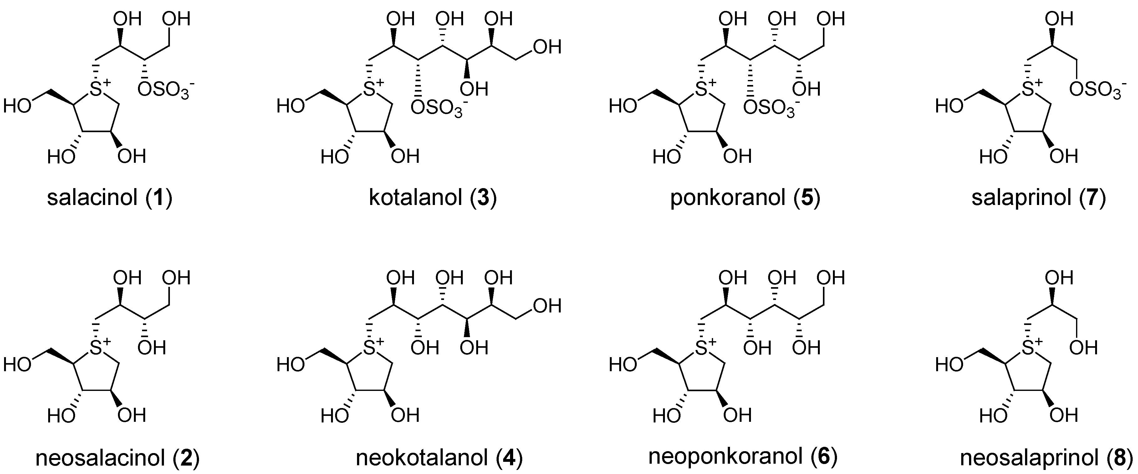
| Salacinol (1) | 4.9 |
| Neosalacinol (2) | 9.0 |
| Kotalanol (3) | 3.9 |
| Neokotalanol (4) | 3.9 |
| Ponkoranol (5) | 5.0 |
| Neoponkoranol (6) | 4.0 |
| Voglibose | 1.3 |
| Acarbose | 15.2 |
| Miglitol | 3.7 |
| Relative Content (% of 0 h) | |||
|---|---|---|---|
| 0 h | 1.0 h | 3.0 h | |
| Salacinol (1) | 100.0 ± 4.6 | 100.0 ± 6.9 | 92.5 ± 6.1 |
| Neosalacinol (2) | 100.0 ± 6.0 | 96.5 ± 5.1 | 93.2 ± 6.2 |
| Kotalanol (3) | 100.0 ± 4.1 | 97.3 ± 6.6 | 91.4 ± 4.6 |
| Neokotalanol (4) | 100.0 ± 3.3 | 97.4 ± 3.0 | 96.5 ± 4.7 |
| Relative Content (% of 0 h) | |||
|---|---|---|---|
| 0 h | 0.5 h | 2.0 h | |
| Salacinol (1) | 100.0 ± 2.4 | 98.7 ± 2.5 | 97.6 ± 1.8 |
| Neosalacinol (2) | 100.0 ± 3.9 | 101.3 ± 3.0 | 94.5 ± 1.8 |
| Kotalanol (3) | 100.0 ± 3.0 | 98.1 ± 2.6 | 99.7 ± 2.7 |
| Neokotalanol (4) | 100.0 ± 2.1 | 100.0 ± 2.6 | 96.6 ± 1.7 |
| Miglitol | 100.0 ± 1.4 | 87.6 ± 1.4 | 52.2 ± 4.7 |
| Glucose | 100.0 ± 3.1 | 32.5 ± 0.5 | 9.1 ± 0.4 |
4. Conclusions
Acknowledgments
Author Contributions
Conflicts of Interest
References
- Evert, A.B.; Boucher, J.L.; Cypress, M.; Dunbar, S.A.; Franz, M.J.; Mayer-Davis, E.J.; Neumiller, J.J.; Nwankwo, R.; Verdi, C.L.; Urbanski, P.; et al. Nutrition therapy recommendations for the management of adults with diabetes. Diabetes Care 2013, 36, 3821–3842. [Google Scholar] [CrossRef] [PubMed]
- Perera, P.K.; Li, Y. Functional herbal food ingredients used in type 2 diabetes mellitus. Pharmacogn. Rev. 2012, 6, 37–45. [Google Scholar] [CrossRef] [PubMed]
- Jayaweera, D.M.A. Medicinal Plants in Ceylon Part. 1; National Science Council of Sri Lanka: Colombo, Sri Lanka, 1981; p. 77. [Google Scholar]
- Vaidyartanam, P.S. In Indian Medicinal Plants: A Compendium of 500 Species; Warrier, P.K., Nambiar, V.P.K., Ramankutty, C., Eds.; Orient Longman: Madras, India, 1993; pp. 47–48. [Google Scholar]
- Chuakul, W.; Saralamp, P.; Paonil, W.; Temsiririkkul, R.; Clayton, T. Medicinal Plants in Thailand (Volume II); Department of Pharmaceutical Botany Faculty of Pharmacy, Mahidol University: Bangkok, Thailand, 1997; pp. 192–193. [Google Scholar]
- Matsuda, H.; Yoshikawa, M.; Morikawa, T.; Tanabe, G.; Muraoka, O. Antidiabetogenic constituents from Salacia species. J. Trad. Med. 2005, 22, 145–153. [Google Scholar]
- Yoshikawa, M.; Murakami, T.; Shimada, H.; Matsuda, H.; Yamahara, J.; Tanabe, G.; Muraoka, O. Salacinol, potent antidiabetic principle with unique thiosugar sulfonium sulfate structure from the Ayurvedic traditional medicine Salacia reticulata in Sri Lanka and India. Tetrahedron Lett. 1997, 38, 8367–8370. [Google Scholar] [CrossRef]
- Yoshikawa, M.; Morikawa, T.; Matsuda, H.; Tanabe, G.; Muraoka, O. Absolute stereostructure of potent α-glucosidase inhibitor, salacinol, with unique thiosugar sulfonium sulfate inner salt structure from Salacia reticulata. Bioorg. Med. Chem. 2002, 10, 1547–1554. [Google Scholar] [CrossRef] [PubMed]
- Tanabe, G.; Yoshikai, K.; Hatanaka, T.; Yamamoto, M.; Shao, Y.; Minematsu, T.; Muraoka, O.; Wang, T.; Matsuda, H.; Yoshikawa, M. Biological evaluation of de-O-sulfonated analogs of salacinol, the role of sulfate anion in the side chain on the α-glucosidase inhibitory activity. Bioorg. Med. Chem. 2007, 15, 3926–3937. [Google Scholar] [CrossRef] [PubMed]
- Tanabe, G.; Xie, W.; Ogawa, A.; Cao, C.; Minematsu, T.; Yoshikawa, M.; Muraoka, O. Facile synthesis of de-O-sulfated salacinols: Revision of the structure of neosalacinol, a potent α-glucosidase inhibitor. Bioorg. Med. Chem. Lett. 2009, 19, 2195–2198. [Google Scholar] [CrossRef] [PubMed]
- Yoshikawa, M.; Murakami, T.; Yashiro, K.; Matsuda, H. Kotalanol, a potent α-glucosidase inhibitor with thiosugar sulfonium sulfate structure, from antidiabetic Ayurvedic medicine Salacia reticulata. Chem. Pharm. Bull. 1998, 46, 1339–1340. [Google Scholar] [CrossRef] [PubMed]
- Muraoka, O.; Xie, W.; Osaki, S.; Kagawa, A.; Tanabe, G.; Amer, M.F.A.; Minematsu, T.; Morikawa, T.; Yoshikawa, M. Characteristic alkaline catalyzed degradation of kotalanol, a potent α-glucosidase inhibitor isolated from Ayurvedic traditional medicine Salacia reticulata, leading to anhydroheptitols: Another structural proof. Tetrahedron 2010, 66, 3717–3722. [Google Scholar] [CrossRef]
- Muraoka, O.; Xie, W.; Tanabe, G.; Amer, M.F.A.; Minematsu, T.; Yoshikawa, M. On the structure of the bioactive constituent from Ayurvedic medicine Salacia reticulata: Revision of the literature. Tetrahedron Lett. 2008, 49, 7315–7317. [Google Scholar] [CrossRef]
- Yoshikawa, M.; Xu, F.; Nakamura, S.; Wang, T.; Matsuda, H.; Tanabe, G.; Muraoka, O. Salaprinol and ponkoranol with thiosugar sulfonium sulfate structure from Salacia prinoides and α-glucosidase inhibitory activity of ponkoranol and kotalanol desulfate. Heterocycles 2008, 75, 1397–1405. [Google Scholar] [CrossRef]
- Xie, W.; Tanabe, G.; Akaki, J.; Morikawa, T.; Ninomiya, K.; Minematsu, T.; Yoshikawa, M.; Wu, X.; Muraoka, M. Isolation, structure identification and SAR studies on thiosugar sulfonium salts, neosalaprinol and neoponkoranol, as potent α-glucosidase inhibitors. Bioorg. Med. Chem. 2011, 19, 2015–2022. [Google Scholar] [CrossRef] [PubMed]
- Tanabe, G.; Sakano, M.; Minematsu, T.; Matsuda, H.; Yoshikawa, M.; Muraoka, O. Synthesis and elucidation of absolute stereochemistry of salaprinol, another thiosugar sulfonium sulfate from the Ayurvedic traditional medicine Salacia prinoides. Tetrahedron 2008, 64, 10080–10086. [Google Scholar] [CrossRef]
- Shimoda, H.; Kawamori, S.; Kawahara, Y. Effects of an aqueous extract of Salacia reticulata, a useful plant in Sri Lanka, on postprandial hyperglycemia in rats and humans. Nippon Eiyo Shokuryo Gakkaishi 1998, 151, 279–287. [Google Scholar] [CrossRef]
- Yoshikawa, M.; Pongpiriyadacha, Y.; Kishi, A.; Kageura, T.; Wang, T.; Morikawa, T.; Matsuda, H. Biological activities of Salacia chinensis originating in Thailand: The quality evaluation guided by α-glucosidase inhibitory activity. Yakugaku Zasshi 2003, 123, 871–880. [Google Scholar] [CrossRef] [PubMed]
- Beppu, H.; Kiuchi, Y.; Kishino, E.; Ito, T.; Fujita, K.; Shimpo, K.; Ozaki, S.; Chihara, T.; Itani, Y.; Sonoda, S.; et al. Effects of a hydrothermal extract of Salacia reticulata on oral saccharide tolerance tests in mice, rats and humans—Evaluations of the effects of a mixture with cyclodextrin on the hypoglycemic effect and variations in the blood glucose level in combination use of oral antidiabetic agents. J. JSMUFF 2005, 3, 25–30. [Google Scholar]
- Oe, H.; Ozaki, S. Hypoglycemic effect of 13-membered ring thiocyclitol, a novel α-glucosidase inhibitor from Kothala-himbutu (Salacia reticulata). Biosci. Biotechnol. Biochem. 2008, 72, 1962–1964. [Google Scholar] [CrossRef] [PubMed]
- Kajimoto, O.; Kawamori, S.; Shimoda, H.; Kawahara, Y.; Hirata, H.; Takahashi, T. Effects of a diet containing Salacia reticulata on mild type 2 diabetes in humans: A placebo-controlled, cross-over trial. Nippon Eiyo Shokuryo Gakkaishi 2000, 53, 199–205. [Google Scholar] [CrossRef]
- Shimoda, H.; Fujimura, T.; Makino, K.; Yoshijima, K.; Naito, K.; Ihota, H.; Miwa, Y. Safety profile of extractive from trunk of Salacia reticulata (Celastraceae). J. Food Hyg. Soc. Jpn. 1999, 40, 198–205. [Google Scholar] [CrossRef]
- Williams, J.A.; Choe, Y.S.; Noss, M.J.; Baumgartner, C.J.; Mustad, V.A. Extract of Salacia oblonga lowers acute glycemia in patients with type 2 diabetes. Am. J. Clin. Nutr. 2007, 86, 124–130. [Google Scholar] [PubMed]
- Kobayashi, M.; Akaki, J.; Yamashita, K.; Morikawa, T.; Ninomiya, K.; Yoshikawa, M.; Muraoka, O. Suppressive effect of the tablet containing Salacia chinensis extract on postprandial blood glucose. Jpn. Pharmacol. Ther. 2010, 38, 545–550. [Google Scholar]
- Tanimura, C.; Terada, I.; Hiramatu, K.; Ikeda, T.; Kasagi, T.; Kishino, E.; Ito, T.; Fujita, K. Effect of a mixture of aqueous extract from Salacia reticulata (Kotala himbutu) and cyclodextrin on the serum glucose and the insulin levels in sucrose tolerance test and on serum glucose level changes and gastro-intestinal disorder by massive ingestion. Yonago Igaku Zasshi 2005, 56, 85–93. [Google Scholar]
- Jihong, Y.; Shaozhong, L.; Jingfeng, S.; Kobayashi, M.; Akaki, J.; Yamashita, K.; Tamesada, M.; Umemura, T. Effects of Salacia chinensis extract on reproductive outcome in rats. Food Chem. Toxicol. 2011, 49, 57–60. [Google Scholar] [CrossRef] [PubMed]
- Muraoka, O.; Morikawa, T.; Miyake, S.; Akaki, J.; Ninomiya, K.; Yoshikawa, M. Quantitative determination of potent α-glucosidase inhibitors, salacinol and kotalanol, in Salacia species using liquid chromatography-mass spectrometry. J. Pharm. Biomed. Anal. 2010, 52, 770–773. [Google Scholar] [CrossRef] [PubMed]
- Akaki, J.; Morikawa, T.; Miyake, S.; Ninomiya, K.; Okada, M.; Tanabe, G.; Pongpiriyadacha, Y.; Yoshikawa, O.; Muraoka, O. Evaluation of Salacia species as anti-diabetic natural resources based on quantitative analysis of eight sulphonium constituents: A new class of α-glucosidase inhibitors. Phytochem. Anal. 2014, 25, 544–550. [Google Scholar] [CrossRef] [PubMed]
- Muraoka, O.; Morikawa, T.; Miyake, S.; Akaki, J.; Ninomiya, K.; Pongpiriyadacha, Y.; Yoshikawa, M. Quantitative analysis of neosalacinol and neokotalanol, another two potent α-glucosidase inhibitors from Salacia species, by LC-MS with ion pair chromatography. J. Nat. Med. 2011, 65, 142–148. [Google Scholar] [CrossRef] [PubMed]
- Nakamura, K.; Akaki, J.; Ishibushi, F.; Tani, K.; Morikawa, T.; Pongpiriyadacha, Y.; Muraoka, O.; Hayakawa, T.; Kakutani, K. Discrimination of genus Salacia plants based on the DNA sequence of the internal transcribed spacer region. Shoyakugaku Zasshi 2015. submitted. [Google Scholar]
- Kashiwagi, A.; Kasuga, M.; Araki, E.; Oka, Y.; Hanafusa, T.; Hiroshi, I.; Tominaga, M.; Oikawa, S.; Noda, M.; Kawamura, T.; et al. International clinical harmonization of glycated hemoglobin in Japan: From Japan diabetes society to national glycohemoglobin standardization program Valu. J. Diabetes Investig. 2012, 3, 39–40. [Google Scholar] [CrossRef] [PubMed]
- Morikawa, T.; Ninomiya, K.; Imamura, M.; Akaki, J.; Fujikura, S.; Pan, Y.; Yuan, D.; Yoshikawa, M.; Jia, X.; Li, Z.; et al. Acylated phenylethanoid glycosides, echinacoside and acteoside from Cistanche. tubulosa, improve glucose tolerance in mice. J. Nat. Med. 2014, 68, 561–566. [Google Scholar] [CrossRef] [PubMed]
- Wang, Y.; Sun, G.; Sun, J.; Liu, S.; Wang, J.; Xu, X.; Miao, L. Spontaneous type 2 diabetic rodent models. J. Diabetes Res. 2013, 2013, 401723. [Google Scholar] [CrossRef] [PubMed]
- Nasi, R.; Patrick, B.O.; Sim, L.; Rose, D.R.; Pinto, B.M. Studies directed toward the stereochemical structure determination of the naturally occurring glucosidase inhibitor, kotalanol: Synthesis and inhibitory activities against human maltase glucoamylase of seven-carbon, chain-extended homologues of salacinol. J. Org. Chem. 2008, 73, 6172–6181. [Google Scholar] [CrossRef] [PubMed]
- Mohan, S.; Pinto, B.M. Towards the elusive structure of kotalanol, a naturally occurring glucosidase inhibitor. Nat. Prod. Rep. 2010, 27, 481–488. [Google Scholar] [CrossRef] [PubMed]
- Sim, L.; Jayakanthan, K.; Mohan, S.; Nasi, R.; Johnston, B.D.; Pinto, B.M.; Rose, D.R. New glucosidase inhibitors from an Ayurvedic herbal treatment for type 2 diabetes: structures and inhibition of human intestinal maltase-glucoamylase with compounds from Salacia reticulata. Biochemistry 2010, 49, 443–451. [Google Scholar] [CrossRef] [PubMed]
- Mohan, S.; Eskandari, R.; Pinto, B.M. Naturally occurring sulfonium-ion glucosidase inhibitors and their derivatives: A promising class of potential antidiabetic agents. Acc. Chem. Res. 2014, 47, 211–225. [Google Scholar] [CrossRef] [PubMed]
© 2015 by the authors; licensee MDPI, Basel, Switzerland. This article is an open access article distributed under the terms and conditions of the Creative Commons Attribution license (http://creativecommons.org/licenses/by/4.0/).
Share and Cite
Morikawa, T.; Akaki, J.; Ninomiya, K.; Kinouchi, E.; Tanabe, G.; Pongpiriyadacha, Y.; Yoshikawa, M.; Muraoka, O. Salacinol and Related Analogs: New Leads for Type 2 Diabetes Therapeutic Candidates from the Thai Traditional Natural Medicine Salacia chinensis. Nutrients 2015, 7, 1480-1493. https://doi.org/10.3390/nu7031480
Morikawa T, Akaki J, Ninomiya K, Kinouchi E, Tanabe G, Pongpiriyadacha Y, Yoshikawa M, Muraoka O. Salacinol and Related Analogs: New Leads for Type 2 Diabetes Therapeutic Candidates from the Thai Traditional Natural Medicine Salacia chinensis. Nutrients. 2015; 7(3):1480-1493. https://doi.org/10.3390/nu7031480
Chicago/Turabian StyleMorikawa, Toshio, Junji Akaki, Kiyofumi Ninomiya, Eri Kinouchi, Genzoh Tanabe, Yutana Pongpiriyadacha, Masayuki Yoshikawa, and Osamu Muraoka. 2015. "Salacinol and Related Analogs: New Leads for Type 2 Diabetes Therapeutic Candidates from the Thai Traditional Natural Medicine Salacia chinensis" Nutrients 7, no. 3: 1480-1493. https://doi.org/10.3390/nu7031480