In a bifurcation intervention, coronary wire re-crossing through an appropriate cell of the jailing strut is important before kissing balloon post-dilation (KBPD). We assessed the re-crossing position of the wire employing 3-dimensional optical coherence tomography (3D-OCT) image reconstruction.
A 69-year-old male who had presented with angina pectoris underwent percutaneous coronary intervention (PCI). Baseline coronary angiography revealed a napkin-ring narrowing at the ostium of the left circumflex coronary artery (LCx, Figure 1A). After deployment of a 3.0/15 mm zotarolimus-eluting Endeavor® stent (Medronic Inc., Santa Rosa, CA, USA) across the left anterior descending coronary artery (LAD), two wires were re-crossed into the LAD and the high lateral (HL) branch (Figure 1B).
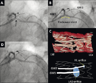
Figure 1. Coronary angiography and on-site 3D-OCT reconstruction. A) Baseline angiography shows a napkin-ring stenosis at the ostium of the circumflex coronary artery (LCx) (Arrow). B) After stent deployment, two wires were passed into the left anterior descending coronary artery (LAD) and the high lateral (HL) branch. C) On-site 3D-OCT image and schematic illustration. 3D-OCT shows that the wire was passed into the LAD, apparently through the appropriate cell. D) Final angiogram.
Fourier-domain OCT images (ILUMIEN; St Jude Medical Inc., St. Paul, MN, USA) were obtained at pullback speeds of 10 mm/s. Automatic strut detection (in-house software from Yamaguchi University, Ube, Japan) and 3D-OCT image reconstruction (INTAGE realia; Cybernet, Tokyo, Japan) for the bifurcation segment were achieved using the dedicated off-line workstation. The 3D-OCT clearly demonstrated that the coronary wire had been successfully passed into the LAD through the distal cell of the jailing strut (Figures 1C and 2A, Moving image 1). After KBPD, OCT images were obtained at the pullback speed of 10 mm/s. 3D-OCT demonstrated that the jailing strut was adequately opened and no protruding struts were seen in front of the LAD ostium (Figure 2B, Moving image 2). The final angiogram confirmed a good result (Figure 1D).
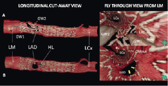
Figure 2. 3D-OCT reconstruction after procedure. A) After guidewire (GW) re-cross. The LAD ostium is jailed by the strut. Moving image 1. B) After kissing balloon post-dilation. The jailing struts are adequately opened with scaffolding to the vessel wall against the carina (yellow arrow). Moving image 2.
In order to visualise the stent clearly in 3D-OCT, an enhancement of the stent strut is necessary. However, manual strut detection is time-consuming, usually requiring one to two hours per stent. The novel automatic strut detecting program allows rapid 3D stent image reconstructions (about 10 minutes) during procedure. 3D-OCT guidance of the wire re-crossing the jailed side branch before KBPD is feasible in the interventional suite, and may improve bifurcation stenting.
Conflict of interest statement
The authors have no conflict of interest to declare.
Online data supplement
Moving image 1. 3D-OCT after guidewire re-cross.
Moving image 2. 3D-OCT after kissing balloon post-dilation.