Spontaneous Rupture of Pseudopancreatic Cyst Causing Massive Pleural Effusion

Sanjay Marwah, Kapil Dev Shivaran and Jaiprakash Gurawalia

Department of Surgery, Post graduate Institute of Medical Sciences, Rohtak, Haryana, India

Academic Editor: Kit Fai Lee

Cite this Article as:

Sanjay Marwah, Kapil Dev Shivaran and Jaiprakash Gurawalia (2013), "Spontaneous Rupture of Pseudopancreatic Cyst Causing Massive Pleural Effusion," International Journal of Case Reports in Medicine, Vol. 2013 (2013), Article ID 832910, DOI: 10.5171/2013.832910

Copyright © 2013 Sanjay Marwah, Kapil Dev Shivaran and Jaiprakash Gurawalia. Distributed under Creative Commons CC-BY 3.0

Abstract

Background: Pancreatic pseudocyst is a well known complication that develops 3-4 weeks after onset of attack of acute pancreatitis. Sudden onset massive pleural effusion represents an unusual pulmonary complication of pseudopancreatic cyst. Method: We are reporting a case of 48 years old male who presented in emergency with pseudopancratic cyst and developed acute respiratory distress during his stay. The timely diagnosis of ruptured pseudocyst into the pleural cavity and prompt intervention with intercostal tube drainage saved the patient’s life. Conclusion: Spontaneous rupture of pseudopancreatic cyst into the pleural cavity is a rare complication that can be potentially fatal if not managed in time.

Keywords: Pseudocyst pancreas; pleural effusion; pancreatico-pleural fistula.

Introduction

In acute pancreatitis, a self limiting reactionary left sided pleural effusion commonly occurs in about 20% of the patients, many of them also develop pseudocyst (Maringhini et al, 1996). Massive pleural effusion associated with pancreaticopleural fistula is a rare complication and it should be suspected on the basis of clinical picture and analysis of pleural fluid following thoracocentesis which reveals an extremely elevated pleural fluid amylase level (King et al, 2010). We are reporting one such case that presented with acute onset dyspnoea.

Case Report

A 48-year-old man presented in emergency with complaint of severe epigastric pain radiating to back, fever and vomiting for the last four days. He was chronic smoker as well as alcoholic and was admitted in emergency multiple times during the last six months with similar complains and was discharged after conservative treatment. On examination, there was a mildly tender, tense cystic, fixed lump in the epigastrium measuring 15 x 10 cms having transmitted pulsations. His total leucocyte count was 17,000 cells/cm3.Ultrasound of the abdomen revealed fluid collection in the lesser sac with normal gallbladder and pancreas could not be visualized. He was treated conservatively and started improving. On third day of admission, he suddenly developed persistent dry cough and became dyspnoic. On examination, his trachea was shifted to right side; respiratory rate was 32 per minute, chest examination revealed bilateral crepitations and rhonchi with diminished air entry on left side. On abdominal examination, there was tenderness in the epigastrium, but epigastric lump had almost disappeared. There was severe hypoxia and metabolic acidosis on blood gas analysis. His chest X-ray revealed massive left hydropneumothorax (Fig 1) and on aspiration, free flow of haemorrhagic fluid, characteristic of pancreatitis, was aspirated from the thoracic cavity.

832910-fig-1

Fig 1: Chest X- Ray Showing Massive Left Hydropneumothorax with Mediastinal Shift to the Right Side (White Arrow). There are Multiple Air Fluid Levels in the Left Hemithorax and Left Dome of Diaphragm is Not Outlined. The Right Lung Shows Hilar Prominence with Few Bullae (Red Arrow)


The fluid amylase level was 2142 IU/ml and it was sterile on culture. These findings made the diagnosis of ruptured pseudopancreatic cyst. The patient was treated with left intercostal intubation and hemorrhagic fluid was drained in the underwater seal drain (Fig 2A). His respiration improved and repeat chest X-ray showed partially expanded left lung and decreased hemopneumothorax (Fig 2B).


832910-fig-2

Fig 2A: Underwater Seal Draining Hemorrhagic Fluid

Fig 2B: Repeat X-Ray Chest Showing Expanded Left Lung (Arrow) with Chest Tube in Situ

 
CT chest and abdomen confirmed the diagnosis of intra-thoracic rupture of abdominal pseudopancreatic cyst (Fig 3). The cyst in the abdomen resolved after the rupture. The patient was treated with antibiotics, chest physiotherapy and octreotide. His left lung expanded and volume of drainage fluid gradually decreased. His intercostal tube was removed after four weeks and he was discharged. At three months follow up, he was normal without any cyst recurrence.

832910-fig-3

Fig 3: CECT Chest and Upper Abdomen Showing Collapse of Left Lung with Areas of Consolidation and Left Hydropneumothorax. A Peripherally Enhanced Sub-Diaphragmatic Collection is Seen in Postero-Superior Aspect of the Left Kidney (Red Arrow)

 
Discussion

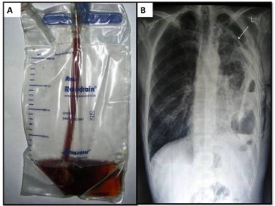
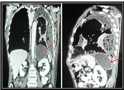
Spontaneous rupture of abdominal pseudopancreatic cyst into pleural cavity is extremely unusual complication of pancreatitis. The ruptured cyst passes either through the aortic or oesophageal hiatus or directly through diaphragm into pleural cavity leading to pancreaticopleural fistula (Tombroff et al, 1973). The enzyme-rich pleural fluid is due to disruption of the dorsal pancreatic ductal system (Traverso & Kozarek, 1999).Typically, the patients are middle-aged males(40—50 years) with chronic pancreatitis related mainly to alcohol abuse.  As reported by Ali et al (2009) and King et al (2010), due to predominant pulmonary symptoms in form of cough and dyspnoea, the diagnosis is often delayed and the patients are usually worked up extensively for pulmonary pathology.Chest X-ray is the first line of investigation but it only gives information of pleural effusion. However majority of the cases give past history of recurrent attacks of pancreatitis and pleural fluid aspirate is characteristically haemorrhagic in appearance with very high amylase level.A CT scan of the chest and abdomen confirms the diagnosis.Ali et al (2009) and Megibow  et al (2001) observed that endoscopic retrograde cholangiopancreatography (ERCP) and magnetic resonance cholangiopancreatography (MRCP) are useful diagnostic modalities in delineating pancreatic ductal anatomy and in localizing the site of leak before operative intervention.

Regarding treatment of such cases, Ali et al (2009) and King et al (2010) suggested that in acute cases presenting with respiratory distress due to massive pleural effusion, urgent thoracocentasis/tube thoracostomy should be done. It helps in relief of symptoms and encourages apposition of serosal surfaces. Medical management with octreotide significantly reduces fistula output and decreases the time to fistula closure. If fistula persists despite conservative treatment, ERCP, sphincterotomy and stent placement will decompress the duct and helps in fistula closure. In resistant cases, surgical intervention in form of distal pancreatectomy or Roux-en-Y pancreaticojejunostomy at the site of leak may be required.

References

Ali, T., Srinivasan, N., Le, V., Chimpiri, A. R. & Tierney, W. M. (2009). “Pancreaticopleural Fistula,” Pancreas, 38:e26—31.
PublisherGoogle Scholar

King, J. C., Reber, H. A., Shiraga, S. & Hines, O. J. (2010). “Pancreatic-Pleural Fistula is Best Managed by Early Operative Intervention,” Surgery, 147:154—59.
PublisherGoogle Scholar

Maringhini, A., Ciambra, M., Patti, R., Randazzo, M. A., Dardanoni, G., Mancuso, L., Termini, A. & Pagliaro, L. (1996). “Ascites, Pleural, and Pericardial Effusions in Acute Pancreatitis. A Prospective Study of Incidence, Natural History, and Prognostic Role,” Dig Dis Sci, 41(5): 848-52.
PublisherGoogle Scholar – British Library Direct

Megibow, A. J., Lavelle, M. T. & Rofsky, N. M. (2001). “MR Imaging of the Pancreas,” Surg Clin North Am, 81: 307—20.
PublisherGoogle Scholar – British Library Direct

Tombroff, M., Loicq, A., de Koster, J.- P., Engleholm, L. & Govaerts, J.- P. (1973). “Pleural Effusion with Pancreatico-Pleural Fistula,” Br Med J, i: 330—1.
PublisherGoogle Scholar

Traverso, L. W. & Kozarek, R. A. (1999). “Interventional Management of Pancreatic Fluid Collections,” Surg Clin North Am, 79: 745—57.
PublisherGoogle Scholar – British Library Direct

Shares