Erschienen in: Thrombosis Journal 1/2022

Open Access 01.12.2022 | COVID-19 | Correspondence

NETosis and SARS-COV-2 infection related thrombosis: a narrative review

verfasst von: Mahin Behzadifard, Masoud Soleimani

Erschienen in: Thrombosis Journal | Ausgabe 1/2022

Abstract

Background

Coronavirus disease 2019 (COVID-19) infection is related to immune hyperactivity, the release of inflammatory cytokines, and immunothrombosis. Among the underlying mechanisms in COVID-19 thrombosis, neutrophil extracellular traps (NETs) formation, NETosis, may have a significant role. COVID-19 thrombi obtained from extracorporeal membrane oxygenation contained an accumulation of neutrophils and in a higher amount of NETs when compared with non-COVID-19 thrombi specimens.

Main body

During sepsis and inflammatory status, NETs released from neutrophils and histones and nucleosomes extruded into the extracellular space and take part in the host innate immunity defense, inflammation, and thrombosis. Excessive NETosis is related to clinical progression and respiratory failure in infections and sepsis. NETosis act as a scaffold for thrombus formation, and new associative data support the relation between deregulated immune responses with thrombus formation and organ failure. NETosis is reported in COVID-19 patients. In COVID-19 infection, overproduction of tissue factor (TF) by neutrophils has a role in immunothrombosis. Additionally, NETs can trap TF pathway inhibitor (TFPI) as the only endogenous protein that effectively inhibits the activity of the significant proteases– complexes, TF–FVIIa and prothrombinase.

Conclusion

Because of NETosis can induce intrinsic and extrinsic coagulation cascade activation through the production of TF, activation of FXII, and inhibition of TFPI and fibrinolysis and induce immunothrombosis, targeting NETosis may diminish thrombus formation related to NETs in COVID-19 patients.
Hinweise

Publisher’s Note

Springer Nature remains neutral with regard to jurisdictional claims in published maps and institutional affiliations.
Abkürzungen
SARS-CoV-2
Severe acute respiratory syndrome coronavirus 2
COVID19
Coronavirus disease
TF
Tissue factor
NETs
Neutrophil extracellular traps
FSAP
Factor seven activating protease
TFPI
Tissue factor pathway inhibitor
ARDS
Acute respiratory distress syndrome
ACE2
Angiotensin converting enzyme

Introduction

The pathophysiology of COVID-19 infection-related thrombosis is still poorly understood. Endothelial damage and subsequently activation of coagulation cascade can lead to widespread microvascular thrombi formation in the lungs and other organs. The lungs injury increases alveolar and capillary permeability that then will be shown as ground-glass appearance in the chest X-ray, and clearly ARDS [1]. Elevated evidence has been shown an increased risk of thrombosis in severe COVID-19 patients and suggested microvascular thrombosis as a main pathophysiologic parameter in COVID-19 infection [2, 3]. The complement system, inflammatory cytokines, and neutrophil extracellular traps (NETs) formation, NETosis, can contribute to coagulation activation [4, 5]. Various pathogens such as bacteria, fungi, protozoa, and viruses can activate NETosis. Cytokines and chemokines like IL-8 and TNF, antibodies and immune complexes, and microcrystals can induce NETs formation [68].
NETs are actively released from neutrophils into the extracellular space [9]. NETs formation is a part of innate immunity and is described as responsible for trapping the pathogens, killing the microbes, and inducing inflammation [10]. The NETosis contributes to sepsis and acute respiratory distress syndrome(ARDS) pathogenesis and causes vascular tissue damage, thrombosis, multiorgan failure, and death [11, 12]. Increased NETs formation correlates with COVID-19 related ARDS and is a potential biomarker for the disease severity [13, 14]. SARS-COV-2 may directly infect monocytes/ macrophages and induce tissue factor (TF) expression/release from these cells that may play a critical role in developing COVID-19 coagulopathy [15]. NETs used several pathways that support fibrin formation and enhance platelets activation and thrombosis. NETs can stimulate thrombosis in a platelet-dependent manner by adhesion and activation of platelets and binding the cells to von Willebrand factor (VWF) and fibrinogen or directly by coagulation cascade activation [16]. Neutrophils and platelets release microparticles that contain TF that NETs can trap. TF has been detected in NETs inside venous thrombi in vivo as a factor of induce thrombosis [17, 18]. NETs are involved in fibrinolysis inhibition by tissue plasminogen activator (tPA) inhibition [19]. Additionally NETs promote intrinsic coagulation cascade by activation of FXII by nucleic acids and phosphates and increase fibrin formation. NETs induce tissue factor pathway inhibitor (TFPI) degradation as the main source of extrinsic pathway inhibitor and increase the chance of blood coagulation [20].

NETosis

NETosis/ETosis described as a form of necrosis that is associated with neutrophils (NETosis) and other granulocytes or macrophages (ETosis). In sepsis, after neutrophil stimulation with IL-8, lipopolysaccharide (LPS), TNF-α, and complement the decondensed chromatin networks or NETs are released by neutrophils [68]. Additionally, TLR2/4 and complement are responsible for initiating NETs formation and associated with dysregulated innate immune response and subsequent tissue injury and organ dysfunction [21].
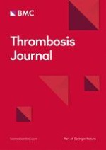
In the NETosis process, neutrophil granules contents translocate to the nucleus, decondense the chromatin, and induce a neutrophil extracellular trap (NETs). In NETosis, the internal, granular, and nuclear membranes are broken down and the cell content extruded to the extracellular space, but cytoplasmic membrane integrity is maintained [22]. NETs contain decondensed chromatin, histones, and proteolytic peptides such as myeloperoxidase (MPO), neutrophil elastase (NE), high mobility group protein B1 (HMGB1), matrix metalloproteinases (MMPs), proteinase 3 (PR3), and cathepsins that trap, immobilize, and kill microorganisms and activate other immune cells. Additionally, Peptidyl arginine deaminase 4 (PAD4) is transferred from the cytoplasm into the nucleus to catalyze citrullination of histones, leading to decondensation of chromatin. Viral infections stimulate NETs formation (Fig. 1). Virus-induced NETs can lead to hyperactivation of immune system response and produce cytokines, chemokines, immune complexes, and inflammation [2225].
In vivo, plasma DNases degrade NETs and are subsequently cleared by macrophages [26, 27]. The NETs clearance importance is demonstrated in deficient DNase 1 and DNase 3 mice models. After a few days of inducing neutrophil activation, vessel occlusion occurred due to a large number of NETs, leading to death in the models.

NETosis and intrinsic and extrinsic coagulation cascade

The coagulation cascade can be activated via extrinsic or intrinsic pathways. The extrinsic pathway is started by tissue factor (TF), which is normally expressed as a transmembrane protein in cells that are not directly in contact with the blood flow. When a blood vessel is damaged, TF is exposed to the blood and attaches to factor VII that is presented in blood and starts a common coagulation pathway. Activation of factor FXII initiates the intrinsic pathway upon binding to negatively charged surfaces, such as collagen and phospholipid. Subsequently, cleavage of FXI and FIX can use active FX and lead to fibrin formation [28].
Plasmin degraded fibrin is the important regulator preventing clot formation and thrombosis. Fibrin clots like NETs can capture bacteria and prevent their invasion. In contrast to fibrin clots, NETs contain antimicrobial proteins that enable them to prevent the pathogens from spreading and kill them [9, 29]. In an experiment in which whole blood was contained NETs, platelets recruited to NETs [10]. The platelets recruitment may be due to binding to C3b deposits on NETs because platelets express complement receptor 1(CR1) on their membrane and or mediated by histones [10, 30, 31]. NETs might lead to platelet aggregation as an essential step in clots formation. NETs contain histones, especially the type of H4 can activate platelets. Subsequently, the activated platelets releaseHMGB1that stimulate NETosis in a positive feedback loop. Additionally, histones stimulate platelets to secrete polyP from α-granules that activate FXII and blood coagulation's intrinsic pathway [32, 33]. Decorated NETs with platelets may be suitable scaffolds for thrombus formation. In this regard, experiments on NETs in mice treated with DNase showed the formation of smaller thrombi [34, 35]. The exact mechanism of coagulation activation by NETs remains unknown, and the separate components of NETs, including DNA and histones, have been demonstrated to induce thrombin formation [32]. Experiments demonstrated that neutrophils treated with cytokines can upregulate TF mRNA and release TF on their NETs, resulting in thrombin formation [36]. However, TF exposure on NETs apparently depends on the type of stimulation used to induce NETosis. Consequently, not all experiments have been shown NETs could produce TF and initiate coagulation [9]. NETs can directly activate intrinsic coagulation cascade because NETs have negative charges and could bind and activate FXII and induce thrombin generation [37]. In this regard, an in vitro study showed that inhibition of FXII or FXI in NETs reduced thrombin formation [38]. So that NETs can start the initiation of coagulation via either intrinsic or extrinsic pathways[9]. NE and, to a minor extent, cathepsin G as the NET-associated protein may contribute to fibrin formation on NETs because they could degrade TFPI. TFPI is the major extrinsic coagulation pathway inhibitor that recruited through nucleosome into the site of injury. [39] The fibrin clots mixed with NETs have more resistant to fibrinolysis by plasmin that might be a critical point in NETs related thrombosis. [40].

NETosis and immune complexes

A major responsible factor to immunothrombosis development, a feature observed in COVID-19 infection that induces ARDS and disease severity, is NETs formation. A well-known mechanism for NETs formation is stimulation of the FcγRIIA and downstream SYK signaling pathway [41]. Immune complexes are the primary mediators of FcγRIIA signaling and NETosis. COVID-19 patients with robust antibody responses have been associated with poor clinical outcomes that raise the possibility of the contribution of immune complexes in NETs formation [42, 43]. Additionally, endogenous stimuli such as inflammatory cytokines, damage-associated molecular patterns (DAMPs), and pathogen-associated molecular patterns (PAMPs) could stimulate the release of NETs and the contribution of their role in COVID-19 infection remains to be clarified [44].

NETosis and Factor Seven Activating Protease (FSAP) activation

Hyaluronan-binding protein 2 or factor VII activating protease is a serine protease produced by the liver, kidney, and pancreas. It is present in the circulation in the form of zymogen (pro-FSAP). It is known to activate coagulation factor-VII independent tissue factor and urokinase single-chain plasminogen activator[45, 46]. Several in vitro and patient-based studies have been shown a link between the levels of FSAP, inflammation, and disease. FSAP level is elevated in lung endothelial cells of acute lung injury induced by lipopolysaccharide and in the lungs of patients with acute respiratory distress syndrome [4749]. In pathologic situations like acute coronary disease, ischemic stroke, and symptomatic carotid stenosis, FSAP levels increased. In addition, FSAP can activate inflammation pathways in non-immune cells like smooth muscle and endothelial cells and myeloid cells through NF-kB mediated proinflammatory cytokine production [5052]. Elevated FSAP levels may indicate systemic inflammation that increases the risk of thrombosis in the COVID-19 patients.

NETs and antiphospholipid antibodies

NETs and their associated components are known to stimulate thrombosis because intravascular NETosis can initiate thrombotic events in arteries, veins, and especially microvasculature and has a critical role in thrombosis formation in COVID-19 [53]. Antiphospholipid antibodies (aPLs) are known as one of the mechanisms of thrombosis through NETs formation. APLs promote NETs release in a manner dependent on Reactive oxygen species (ROS) and TLR4 [54]. NETs are essential in antiphospholipid syndrome (APS) because in APS, neutrophils are prone to spontaneous release of NETs. NETs are an essential activator of the coagulation cascade in APS [55]. NETs in APS are the main source of tissue factors (TFs), platelet activation, and aggregation and play a vital role in forming atherosclerosis and arterial thrombosis. In APS, neutrophils seem to have a greater adhesion potential, enhancing neutrophil-endothelium interaction and NETs diffusion [56]. Thus, neutrophils and NETosis are directly or indirectly involved in APS pathogenesis [57]. Many studies have reported low/moderate aPL titers in COVID-19 patients. These antibodies mainly target β2GP1 but indicate epitope characteristics different from APS antibodies [58, 59]. There is currently limited information on the role and importance of aPLs in COVID-19 pathogenesis, and evidence suggests that aPLs may have little clinical association with prolonged activated partial-thromboplastin time (aPTT) and thrombosis in COVID-19 patients [60, 61].

NETosis inhibition

NETosis stimulates coagulation activation and fibrinolysis inhibition in various pathways. Targeting NETs formation might be a feasible and valuable therapeutic strategy to prevent thrombus formation and improve clinical outcomes of COVID-19 infection. Anti-cytokine therapy against IL-1β is widely used in various inflammatory and autoimmune diseases, preventing activation and accumulation of neutrophils and subsequently NETosis. The recombinant anakinra protein as IL-1β receptor antagonist may be a potential target to COVID-19 treatment and is currently undergoing clinical trials (https://​clinicaltrials.​gov: NCT04324021, NCT04330638, NCT02735707). Additionally, using inhibitors that target involved components of NETs formation process like NE, PAD4, and gasdermin D protein (GSDMD) during inflammation can be another approach. The NETosis prevention by nocodazole as a microtubule inhibitor is reported in in vitro experiments [62]. In this regard, clinical trials (https://​clinicaltrials.​gov: NCT04326790, NCT04328480, NCT04322565, NCT04322682) that test the efficacy of colchicine against COVID-19 are currently underway [63]. Drugs inhibitors of the NETosis axis such as glucocorticoids can block neutrophils' function and prevent NETs formation. Additionally, using exogenous DNase treatment improves NETs clearance, and recombinant human DNase I is currently under investigation for safety and efficacy in clinical trials in COVID-19.

Conclusion

NETs formation in COVID-19 infection that is related to dysregulated immune response, releasing of inflammatory cytokines, and development of pathogenic microvascular thrombi. COVID-19 patients seem especially prone to excessive NETs formation and disease severity parallel increasing markers of NETosis [13, 64]. In this regard NETs, inhibitors may dampen the severity of SARS-COV-2 infection.

Acknowledgements

Not applicable

Declarations

The authors have consent for publication

Competing interests

The authors declare that they have no competing interests
Open AccessThis article is licensed under a Creative Commons Attribution 4.0 International License, which permits use, sharing, adaptation, distribution and reproduction in any medium or format, as long as you give appropriate credit to the original author(s) and the source, provide a link to the Creative Commons licence, and indicate if changes were made. The images or other third party material in this article are included in the article's Creative Commons licence, unless indicated otherwise in a credit line to the material. If material is not included in the article's Creative Commons licence and your intended use is not permitted by statutory regulation or exceeds the permitted use, you will need to obtain permission directly from the copyright holder. To view a copy of this licence, visit http://​creativecommons.​org/​licenses/​by/​4.​0/​. The Creative Commons Public Domain Dedication waiver (http://​creativecommons.​org/​publicdomain/​zero/​1.​0/​) applies to the data made available in this article, unless otherwise stated in a credit line to the data.

Publisher’s Note

Springer Nature remains neutral with regard to jurisdictional claims in published maps and institutional affiliations.
Literatur
1.
Zurück zum Zitat Leisman DE, Deutschman CS, Legrand M. Facing COVID-19 in the ICU: vascular dysfunction, thrombosis, and dysregulated inflammation. Intensive Care Medicine. 2020;46(6):1105–8.PubMedPubMedCentralCrossRef Leisman DE, Deutschman CS, Legrand M. Facing COVID-19 in the ICU: vascular dysfunction, thrombosis, and dysregulated inflammation. Intensive Care Medicine. 2020;46(6):1105–8.PubMedPubMedCentralCrossRef
2.
Zurück zum Zitat Pons S, Fodil S, Azoulay E, Zafrani L. The vascular endothelium: the cornerstone of organ dysfunction in severe SARS-CoV-2 infection. Critical care. 2020;24(1):1–8.CrossRef Pons S, Fodil S, Azoulay E, Zafrani L. The vascular endothelium: the cornerstone of organ dysfunction in severe SARS-CoV-2 infection. Critical care. 2020;24(1):1–8.CrossRef
3.
Zurück zum Zitat Rovas A, Osiaevi I, Buscher K, Sackarnd J, Tepasse P-R, Fobker M, et al. Microvascular dysfunction in COVID-19: the MYSTIC study. Angiogenesis. 2021;24(1):145–57.PubMedCrossRef Rovas A, Osiaevi I, Buscher K, Sackarnd J, Tepasse P-R, Fobker M, et al. Microvascular dysfunction in COVID-19: the MYSTIC study. Angiogenesis. 2021;24(1):145–57.PubMedCrossRef
4.
Zurück zum Zitat Geisbert TW, Young HA, Jahrling PB, Davis KJ, Kagan E, Hensley LE. Mechanisms underlying coagulation abnormalities in ebola hemorrhagic fever: overexpression of tissue factor in primate monocytes/macrophages is a key event. The Journal of infectious diseases. 2003;188(11):1618–29.PubMedCrossRef Geisbert TW, Young HA, Jahrling PB, Davis KJ, Kagan E, Hensley LE. Mechanisms underlying coagulation abnormalities in ebola hemorrhagic fever: overexpression of tissue factor in primate monocytes/macrophages is a key event. The Journal of infectious diseases. 2003;188(11):1618–29.PubMedCrossRef
5.
Zurück zum Zitat Chan LL, Nicholls JM, Peiris JM, Lau YL, Chan MC, Chan RW. Host DNA released by NETosis in neutrophils exposed to seasonal H1N1 and highly pathogenic H5N1 influenza viruses. Respiratory research. 2020;21(1):1–10.CrossRef Chan LL, Nicholls JM, Peiris JM, Lau YL, Chan MC, Chan RW. Host DNA released by NETosis in neutrophils exposed to seasonal H1N1 and highly pathogenic H5N1 influenza viruses. Respiratory research. 2020;21(1):1–10.CrossRef
6.
Zurück zum Zitat Vorobjeva N, Pinegin B. Neutrophil extracellular traps: mechanisms of formation and role in health and disease. Biochemistry (Moscow). 2014;79(12):1286–96.CrossRef Vorobjeva N, Pinegin B. Neutrophil extracellular traps: mechanisms of formation and role in health and disease. Biochemistry (Moscow). 2014;79(12):1286–96.CrossRef
7.
Zurück zum Zitat Ravindran M, Khan MA, Palaniyar N. Neutrophil extracellular trap formation: physiology, pathology, and pharmacology. Biomolecules. 2019;9(8):365.PubMedCentralCrossRef Ravindran M, Khan MA, Palaniyar N. Neutrophil extracellular trap formation: physiology, pathology, and pharmacology. Biomolecules. 2019;9(8):365.PubMedCentralCrossRef
8.
Zurück zum Zitat Yousefi S, Simon D, Stojkov D, Karsonova A, Karaulov A, Simon H-U. In vivo evidence for extracellular DNA trap formation. Cell death & disease. 2020;11(4):1–15.CrossRef Yousefi S, Simon D, Stojkov D, Karsonova A, Karaulov A, Simon H-U. In vivo evidence for extracellular DNA trap formation. Cell death & disease. 2020;11(4):1–15.CrossRef
9.
Zurück zum Zitat de Bont CM, Boelens WC, Pruijn GJ. NETosis, complement, and coagulation: a triangular relationship. Cellular & molecular immunology. 2019;16(1):19–27.CrossRef de Bont CM, Boelens WC, Pruijn GJ. NETosis, complement, and coagulation: a triangular relationship. Cellular & molecular immunology. 2019;16(1):19–27.CrossRef
10.
Zurück zum Zitat Fuchs TA, Brill A, Duerschmied D, Schatzberg D, Monestier M, Myers DD, et al. Extracellular DNA traps promote thrombosis. Proceed Natl Acad Sci. 2010;107(36):15880–5.CrossRef Fuchs TA, Brill A, Duerschmied D, Schatzberg D, Monestier M, Myers DD, et al. Extracellular DNA traps promote thrombosis. Proceed Natl Acad Sci. 2010;107(36):15880–5.CrossRef
11.
Zurück zum Zitat Czaikoski PG, Mota JMSC, Nascimento DC, Sônego F, Castanheira FVeS, Melo PH, et al. Neutrophil extracellular traps induce organ damage during experimental and clinical sepsis. PloS one. 2016;11(2):e0148142.PubMedPubMedCentralCrossRef Czaikoski PG, Mota JMSC, Nascimento DC, Sônego F, Castanheira FVeS, Melo PH, et al. Neutrophil extracellular traps induce organ damage during experimental and clinical sepsis. PloS one. 2016;11(2):e0148142.PubMedPubMedCentralCrossRef
12.
Zurück zum Zitat Lefrançais E, Mallavia B, Zhuo H, Calfee CS, Looney MR. Maladaptive role of neutrophil extracellular traps in pathogen-induced lung injury. JCI insight. 2018;3(3). Lefrançais E, Mallavia B, Zhuo H, Calfee CS, Looney MR. Maladaptive role of neutrophil extracellular traps in pathogen-induced lung injury. JCI insight. 2018;3(3).
13.
Zurück zum Zitat Zuo Y, Yalavarthi S, Shi H, Gockman K, Zuo M, Madison JA, et al. Neutrophil extracellular traps in COVID-19. JCI insight. 2020;5(11). Zuo Y, Yalavarthi S, Shi H, Gockman K, Zuo M, Madison JA, et al. Neutrophil extracellular traps in COVID-19. JCI insight. 2020;5(11).
14.
Zurück zum Zitat Barnes BJ, Adrover JM, Baxter-Stoltzfus A, Borczuk A, Cools-Lartigue J, Crawford JM, et al. Targeting potential drivers of COVID-19: Neutrophil extracellular traps. Journal of Experimental Medicine. 2020;217(6). Barnes BJ, Adrover JM, Baxter-Stoltzfus A, Borczuk A, Cools-Lartigue J, Crawford JM, et al. Targeting potential drivers of COVID-19: Neutrophil extracellular traps. Journal of Experimental Medicine. 2020;217(6).
15.
Zurück zum Zitat Eslamifar Z, Behzadifard M, Soleimani M, Behzadifard S. Coagulation abnormalities in SARS-CoV-2 infection: overexpression tissue factor. Thrombosis journal. 2020;18(1):1–4.CrossRef Eslamifar Z, Behzadifard M, Soleimani M, Behzadifard S. Coagulation abnormalities in SARS-CoV-2 infection: overexpression tissue factor. Thrombosis journal. 2020;18(1):1–4.CrossRef
16.
Zurück zum Zitat Martinod K, Wagner DD. Thrombosis: tangled up in NETs. Blood, The Journal of the American Society of Hematology. 2014;123(18):2768–76. Martinod K, Wagner DD. Thrombosis: tangled up in NETs. Blood, The Journal of the American Society of Hematology. 2014;123(18):2768–76.
17.
Zurück zum Zitat Maroney SA, Mast AE. Expression of tissue factor pathway inhibitor by endothelial cells and platelets. Transfusion and Apheresis Science. 2008;38(1):9–14.PubMedPubMedCentralCrossRef Maroney SA, Mast AE. Expression of tissue factor pathway inhibitor by endothelial cells and platelets. Transfusion and Apheresis Science. 2008;38(1):9–14.PubMedPubMedCentralCrossRef
18.
Zurück zum Zitat Novotny WF, Girard TJ, Miletich JP, Broze Jr GJ. Platelets secrete a coagulation inhibitor functionally and antigenically similar to the lipoprotein associated coagulation inhibitor. Blood. 1988;72(6):2020–5. Novotny WF, Girard TJ, Miletich JP, Broze Jr GJ. Platelets secrete a coagulation inhibitor functionally and antigenically similar to the lipoprotein associated coagulation inhibitor. Blood. 1988;72(6):2020–5.
19.
Zurück zum Zitat Miesbach W. Pathological role of angiotensin II in severe COVID-19. TH open. 2020;4(02):e138-e44. Miesbach W. Pathological role of angiotensin II in severe COVID-19. TH open. 2020;4(02):e138-e44.
20.
Zurück zum Zitat Broze GJ Jr, Miletich JP. Characterization of the inhibition of tissue factor in serum. Blood. 1987;69(1)150–5. Broze GJ Jr, Miletich JP. Characterization of the inhibition of tissue factor in serum. Blood. 1987;69(1)150–5.
21.
Zurück zum Zitat Clark SR, Ma AC, Tavener SA, McDonald B, Goodarzi Z, Kelly MM, et al. Platelet TLR4 activates neutrophil extracellular traps to ensnare bacteria in septic blood. Nature medicine. 2007;13(4):463–9.PubMedCrossRef Clark SR, Ma AC, Tavener SA, McDonald B, Goodarzi Z, Kelly MM, et al. Platelet TLR4 activates neutrophil extracellular traps to ensnare bacteria in septic blood. Nature medicine. 2007;13(4):463–9.PubMedCrossRef
22.
Zurück zum Zitat Mozzini C, Girelli D. The role of Neutrophil Extracellular Traps in Covid-19: Only an hypothesis or a potential new field of research? Thrombosis research. 2020;191:26–7.PubMedPubMedCentralCrossRef Mozzini C, Girelli D. The role of Neutrophil Extracellular Traps in Covid-19: Only an hypothesis or a potential new field of research? Thrombosis research. 2020;191:26–7.PubMedPubMedCentralCrossRef
23.
Zurück zum Zitat Bonaventura A, Liberale L, Carbone F, Vecchié A, Diaz-Cañestro C, Camici GG, et al. The pathophysiological role of neutrophil extracellular traps in inflammatory diseases. Thrombosis and haemostasis. 2018;118(01):006–27.CrossRef Bonaventura A, Liberale L, Carbone F, Vecchié A, Diaz-Cañestro C, Camici GG, et al. The pathophysiological role of neutrophil extracellular traps in inflammatory diseases. Thrombosis and haemostasis. 2018;118(01):006–27.CrossRef
24.
Zurück zum Zitat Bardoel BW, Kenny EF, Sollberger G, Zychlinsky A. The balancing act of neutrophils. Cell host & microbe. 2014;15(5):526–36.CrossRef Bardoel BW, Kenny EF, Sollberger G, Zychlinsky A. The balancing act of neutrophils. Cell host & microbe. 2014;15(5):526–36.CrossRef
25.
Zurück zum Zitat Brinkmann V, Reichard U, Goosmann C, Fauler B, Uhlemann Y, Weiss DS, et al. Neutrophil extracellular traps kill bacteria. science. 2004;303(5663):1532–5.PubMedCrossRef Brinkmann V, Reichard U, Goosmann C, Fauler B, Uhlemann Y, Weiss DS, et al. Neutrophil extracellular traps kill bacteria. science. 2004;303(5663):1532–5.PubMedCrossRef
26.
Zurück zum Zitat Jiménez-Alcázar M, Rangaswamy C, Panda R, Bitterling J, Simsek YJ, Long AT, et al. Host DNases prevent vascular occlusion by neutrophil extracellular traps. Science. 2017;358(6367):1202–6.PubMedCrossRef Jiménez-Alcázar M, Rangaswamy C, Panda R, Bitterling J, Simsek YJ, Long AT, et al. Host DNases prevent vascular occlusion by neutrophil extracellular traps. Science. 2017;358(6367):1202–6.PubMedCrossRef
27.
Zurück zum Zitat Farrera C, Fadeel B. Macrophage clearance of neutrophil extracellular traps is a silent process. The Journal of Immunology. 2013;191(5):2647–56.PubMedCrossRef Farrera C, Fadeel B. Macrophage clearance of neutrophil extracellular traps is a silent process. The Journal of Immunology. 2013;191(5):2647–56.PubMedCrossRef
28.
Zurück zum Zitat Esmon CT. The regulation of natural anticoagulant pathways. Science. 1987;235(4794):1348–52.PubMedCrossRef Esmon CT. The regulation of natural anticoagulant pathways. Science. 1987;235(4794):1348–52.PubMedCrossRef
29.
Zurück zum Zitat Schulz C, Engelmann B, Massberg S. Crossroads of coagulation and innate immunity: the case of deep vein thrombosis. Journal of thrombosis and haemostasis. 2013;11:233–41.PubMedCrossRef Schulz C, Engelmann B, Massberg S. Crossroads of coagulation and innate immunity: the case of deep vein thrombosis. Journal of thrombosis and haemostasis. 2013;11:233–41.PubMedCrossRef
30.
Zurück zum Zitat Hamzeh-Cognasse H, Damien P, Chabert A, Pozzetto B, Cognasse F, Garraud O. Platelets and infections–complex interactions with bacteria. Frontiers in immunology. 2015;6:82.PubMedPubMedCentralCrossRef Hamzeh-Cognasse H, Damien P, Chabert A, Pozzetto B, Cognasse F, Garraud O. Platelets and infections–complex interactions with bacteria. Frontiers in immunology. 2015;6:82.PubMedPubMedCentralCrossRef
31.
Zurück zum Zitat van der Maten E, de Bont CM, de Groot R, de Jonge MI, Langereis JD, van der Flier M. Alternative pathway regulation by factor H modulates Streptococcus pneumoniae induced proinflammatory cytokine responses by decreasing C5a receptor crosstalk. Cytokine. 2016;88:281–6.PubMedCrossRef van der Maten E, de Bont CM, de Groot R, de Jonge MI, Langereis JD, van der Flier M. Alternative pathway regulation by factor H modulates Streptococcus pneumoniae induced proinflammatory cytokine responses by decreasing C5a receptor crosstalk. Cytokine. 2016;88:281–6.PubMedCrossRef
32.
Zurück zum Zitat Noubouossie DF, Whelihan MF, Yu Y-B, Sparkenbaugh E, Pawlinski R, Monroe DM, et al. In vitro activation of coagulation by human neutrophil DNA and histone proteins but not neutrophil extracellular traps. Blood, The Journal of the American Society of Hematology. 2017;129(8):1021–9. Noubouossie DF, Whelihan MF, Yu Y-B, Sparkenbaugh E, Pawlinski R, Monroe DM, et al. In vitro activation of coagulation by human neutrophil DNA and histone proteins but not neutrophil extracellular traps. Blood, The Journal of the American Society of Hematology. 2017;129(8):1021–9.
33.
Zurück zum Zitat Semeraro F, Ammollo CT, Morrissey JH, Dale GL, Friese P, Esmon NL, et al. Extracellular histones promote thrombin generation through platelet-dependent mechanisms: involvement of platelet TLR2 and TLR4. Blood, The Journal of the American Society of Hematology. 2011;118(7):1952–61. Semeraro F, Ammollo CT, Morrissey JH, Dale GL, Friese P, Esmon NL, et al. Extracellular histones promote thrombin generation through platelet-dependent mechanisms: involvement of platelet TLR2 and TLR4. Blood, The Journal of the American Society of Hematology. 2011;118(7):1952–61.
34.
Zurück zum Zitat Brill A, Fuchs T, Savchenko A, Thomas G, Martinod K, De Meyer S, et al. Neutrophil extracellular traps promote deep vein thrombosis in mice. Journal of Thrombosis and Haemostasis. 2012;10(1):136–44.PubMedPubMedCentralCrossRef Brill A, Fuchs T, Savchenko A, Thomas G, Martinod K, De Meyer S, et al. Neutrophil extracellular traps promote deep vein thrombosis in mice. Journal of Thrombosis and Haemostasis. 2012;10(1):136–44.PubMedPubMedCentralCrossRef
35.
Zurück zum Zitat McDonald B, Davis RP, Kim S-J, Tse M, Esmon CT, Kolaczkowska E, et al. Platelets and neutrophil extracellular traps collaborate to promote intravascular coagulation during sepsis in mice. Blood, The Journal of the American Society of Hematology. 2017;129(10):1357–67. McDonald B, Davis RP, Kim S-J, Tse M, Esmon CT, Kolaczkowska E, et al. Platelets and neutrophil extracellular traps collaborate to promote intravascular coagulation during sepsis in mice. Blood, The Journal of the American Society of Hematology. 2017;129(10):1357–67.
36.
Zurück zum Zitat Kambas K, Mitroulis I, Apostolidou E, Girod A, Chrysanthopoulou A, Pneumatikos I, et al. Autophagy mediates the delivery of thrombogenic tissue factor to neutrophil extracellular traps in human sepsis. 2012;e45427. Kambas K, Mitroulis I, Apostolidou E, Girod A, Chrysanthopoulou A, Pneumatikos I, et al. Autophagy mediates the delivery of thrombogenic tissue factor to neutrophil extracellular traps in human sepsis. 2012;e45427.
37.
Zurück zum Zitat von Brühl M-L, Stark K, Steinhart A, Chandraratne S, Konrad I, Lorenz M, et al. Monocytes, neutrophils, and platelets cooperate to initiate and propagate venous thrombosis in mice in vivo. Journal of Experimental Medicine. 2012;209(4):819–35.CrossRef von Brühl M-L, Stark K, Steinhart A, Chandraratne S, Konrad I, Lorenz M, et al. Monocytes, neutrophils, and platelets cooperate to initiate and propagate venous thrombosis in mice in vivo. Journal of Experimental Medicine. 2012;209(4):819–35.CrossRef
38.
Zurück zum Zitat Gould TJ, Vu TT, Swystun LL, Dwivedi DJ, Mai SH, Weitz JI, et al. Neutrophil extracellular traps promote thrombin generation through platelet-dependent and platelet-independent mechanisms. Arteriosclerosis, thrombosis, and vascular biology. 2014;34(9):1977–84.PubMedCrossRef Gould TJ, Vu TT, Swystun LL, Dwivedi DJ, Mai SH, Weitz JI, et al. Neutrophil extracellular traps promote thrombin generation through platelet-dependent and platelet-independent mechanisms. Arteriosclerosis, thrombosis, and vascular biology. 2014;34(9):1977–84.PubMedCrossRef
39.
Zurück zum Zitat Massberg S, Grahl L, von Bruehl M-L, Manukyan D, Pfeiler S, Goosmann C, et al. Reciprocal coupling of coagulation and innate immunity via neutrophil serine proteases. Nature medicine. 2010;16(8):887–96.PubMedCrossRef Massberg S, Grahl L, von Bruehl M-L, Manukyan D, Pfeiler S, Goosmann C, et al. Reciprocal coupling of coagulation and innate immunity via neutrophil serine proteases. Nature medicine. 2010;16(8):887–96.PubMedCrossRef
40.
Zurück zum Zitat Thammavongsa V, Kim HK, Missiakas D, Schneewind O. Staphylococcal manipulation of host immune responses. Nature Reviews Microbiology. 2015;13(9):529–43.PubMedPubMedCentralCrossRef Thammavongsa V, Kim HK, Missiakas D, Schneewind O. Staphylococcal manipulation of host immune responses. Nature Reviews Microbiology. 2015;13(9):529–43.PubMedPubMedCentralCrossRef
41.
Zurück zum Zitat Perdomo J, Leung HH, Ahmadi Z, Yan F, Chong JJ, Passam FH, et al. Neutrophil activation and NETosis are the major drivers of thrombosis in heparin-induced thrombocytopenia. Nature communications. 2019;10(1):1–14.CrossRef Perdomo J, Leung HH, Ahmadi Z, Yan F, Chong JJ, Passam FH, et al. Neutrophil activation and NETosis are the major drivers of thrombosis in heparin-induced thrombocytopenia. Nature communications. 2019;10(1):1–14.CrossRef
42.
Zurück zum Zitat Wang Y, Zhang L, Sang L, Ye F, Ruan S, Zhong B, et al. Kinetics of viral load and antibody response in relation to COVID-19 severity. The Journal of clinical investigation. 2020;130(10):5235–44.PubMedPubMedCentralCrossRef Wang Y, Zhang L, Sang L, Ye F, Ruan S, Zhong B, et al. Kinetics of viral load and antibody response in relation to COVID-19 severity. The Journal of clinical investigation. 2020;130(10):5235–44.PubMedPubMedCentralCrossRef
43.
Zurück zum Zitat Liu L, Wei Q, Lin Q, Fang J, Wang H, Kwok H, et al. Anti–spike IgG causes severe acute lung injury by skewing macrophage responses during acute SARS-CoV infection. JCI insight. 2019;4(4). Liu L, Wei Q, Lin Q, Fang J, Wang H, Kwok H, et al. Anti–spike IgG causes severe acute lung injury by skewing macrophage responses during acute SARS-CoV infection. JCI insight. 2019;4(4).
44.
Zurück zum Zitat Papayannopoulos V. Neutrophil extracellular traps in immunity and disease. Nature Reviews Immunology. 2018;18(2):134–47.PubMedCrossRef Papayannopoulos V. Neutrophil extracellular traps in immunity and disease. Nature Reviews Immunology. 2018;18(2):134–47.PubMedCrossRef
45.
Zurück zum Zitat Choi-Miura N-H, Tobe T, Sumiya J-i, Nakano Y, Sano Y, Mazda T, et al. Purification and characterization of a novel hyaluronan-binding protein (PHBP) from human plasma: it has three EGF, a kringle and a serine protease domain, similar to hepatocyte growth factor activator. The journal of biochemistry. 1996;119(6):1157–65.PubMedCrossRef Choi-Miura N-H, Tobe T, Sumiya J-i, Nakano Y, Sano Y, Mazda T, et al. Purification and characterization of a novel hyaluronan-binding protein (PHBP) from human plasma: it has three EGF, a kringle and a serine protease domain, similar to hepatocyte growth factor activator. The journal of biochemistry. 1996;119(6):1157–65.PubMedCrossRef
46.
Zurück zum Zitat Römisch J, Feussner A, Vermöhlen S, Stöhr H. A protease isolated from human plasma activating factor VII independent of tissue factor. Blood coagulation & fibrinolysis: an international journal in haemostasis and thrombosis. 1999;10(8):471–9.CrossRef Römisch J, Feussner A, Vermöhlen S, Stöhr H. A protease isolated from human plasma activating factor VII independent of tissue factor. Blood coagulation & fibrinolysis: an international journal in haemostasis and thrombosis. 1999;10(8):471–9.CrossRef
47.
Zurück zum Zitat Mambetsariev N, Mirzapoiazova T, Mambetsariev B, Sammani S, Lennon FE, Garcia JG, et al. Hyaluronic acid binding protein 2 is a novel regulator of vascular integrity. Arteriosclerosis Thrombosis Vasc Biol. 2010;30(3):483–90.CrossRef Mambetsariev N, Mirzapoiazova T, Mambetsariev B, Sammani S, Lennon FE, Garcia JG, et al. Hyaluronic acid binding protein 2 is a novel regulator of vascular integrity. Arteriosclerosis Thrombosis Vasc Biol. 2010;30(3):483–90.CrossRef
48.
Zurück zum Zitat Ware LB, Matthay MA. The acute respiratory distress syndrome. New England Journal of Medicine. 2000;342(18):1334–49.CrossRef Ware LB, Matthay MA. The acute respiratory distress syndrome. New England Journal of Medicine. 2000;342(18):1334–49.CrossRef
49.
Zurück zum Zitat Wygrecka M, Markart P, Fink L, Guenther A, Preissner KT. Raised protein levels and altered cellular expression of factor VII activating protease (FSAP) in the lungs of patients with acute respiratory distress syndrome (ARDS). Thorax. 2007;62(10):880–8.PubMedPubMedCentralCrossRef Wygrecka M, Markart P, Fink L, Guenther A, Preissner KT. Raised protein levels and altered cellular expression of factor VII activating protease (FSAP) in the lungs of patients with acute respiratory distress syndrome (ARDS). Thorax. 2007;62(10):880–8.PubMedPubMedCentralCrossRef
50.
Zurück zum Zitat Parahuleva MS, Hölschermann H, Zandt D, Pons-Kühnemann J, Parviz B, Weiskirchen R, et al. Circulating factor VII activating protease (FSAP) is associated with clinical outcome in acute coronary syndrome. Circulation Journal. 2012:CJ-11-1502. Parahuleva MS, Hölschermann H, Zandt D, Pons-Kühnemann J, Parviz B, Weiskirchen R, et al. Circulating factor VII activating protease (FSAP) is associated with clinical outcome in acute coronary syndrome. Circulation Journal. 2012:CJ-11-1502.
51.
Zurück zum Zitat Hanson E, Kanse S, Joshi A, Jood K, Nilsson S, Blomstrand C, et al. Plasma factor VII-activating protease antigen levels and activity are increased in ischemic stroke. Journal of thrombosis and haemostasis. 2012;10(5):848–56.PubMedCrossRef Hanson E, Kanse S, Joshi A, Jood K, Nilsson S, Blomstrand C, et al. Plasma factor VII-activating protease antigen levels and activity are increased in ischemic stroke. Journal of thrombosis and haemostasis. 2012;10(5):848–56.PubMedCrossRef
52.
Zurück zum Zitat Byskov K, Boettger T, Ruehle PF, Nielsen NV, Etscheid M, Kanse SM. Factor VII activating protease (FSAP) regulates the expression of inflammatory genes in vascular smooth muscle and endothelial cells. Atherosclerosis. 2017;265:133–9.PubMedCrossRef Byskov K, Boettger T, Ruehle PF, Nielsen NV, Etscheid M, Kanse SM. Factor VII activating protease (FSAP) regulates the expression of inflammatory genes in vascular smooth muscle and endothelial cells. Atherosclerosis. 2017;265:133–9.PubMedCrossRef
53.
Zurück zum Zitat Wang Y, Luo L, Braun OÖ, Westman J, Madhi R, Herwald H, et al. Neutrophil extracellular trap-microparticle complexes enhance thrombin generation via the intrinsic pathway of coagulation in mice. Scientific reports. 2018;8(1):1–14. Wang Y, Luo L, Braun OÖ, Westman J, Madhi R, Herwald H, et al. Neutrophil extracellular trap-microparticle complexes enhance thrombin generation via the intrinsic pathway of coagulation in mice. Scientific reports. 2018;8(1):1–14.
54.
Zurück zum Zitat Jariwala MP, Laxer RM. NETosis in rheumatic diseases. Current rheumatology reports. 2021;23(2):1–12.CrossRef Jariwala MP, Laxer RM. NETosis in rheumatic diseases. Current rheumatology reports. 2021;23(2):1–12.CrossRef
55.
Zurück zum Zitat Tambralli A, Gockman K, Knight JS. NETs in APS: current knowledge and future perspectives. Current Rheumatology Reports. 2020;22(10):1–12.CrossRef Tambralli A, Gockman K, Knight JS. NETs in APS: current knowledge and future perspectives. Current Rheumatology Reports. 2020;22(10):1–12.CrossRef
57.
Zurück zum Zitat Bruschi M, Petretto A, Bertelli R, Galetti M, Bonanni A, Pratesi F, et al. Post-translational modified proteins are biomarkers of autoimmune-processes: NETosis and the inflammatory–autoimmunity connection. Clinica chimica acta. 2017;464:12–6.CrossRef Bruschi M, Petretto A, Bertelli R, Galetti M, Bonanni A, Pratesi F, et al. Post-translational modified proteins are biomarkers of autoimmune-processes: NETosis and the inflammatory–autoimmunity connection. Clinica chimica acta. 2017;464:12–6.CrossRef
58.
Zurück zum Zitat Borghi MO, Beltagy A, Garrafa E, Curreli D, Cecchini G, Bodio C, et al. Anti-phospholipid antibodies in COVID-19 are different from those detectable in the anti-phospholipid syndrome. Frontiers in immunology. 2020:2692. Borghi MO, Beltagy A, Garrafa E, Curreli D, Cecchini G, Bodio C, et al. Anti-phospholipid antibodies in COVID-19 are different from those detectable in the anti-phospholipid syndrome. Frontiers in immunology. 2020:2692.
59.
Zurück zum Zitat Xiao M, Zhang Y, Zhang S, Qin X, Xia P, Cao W, et al. Antiphospholipid antibodies in critically Ill patients with COVID-19. Arthritis & Rheumatology. 2020;72(12):1998–2004.CrossRef Xiao M, Zhang Y, Zhang S, Qin X, Xia P, Cao W, et al. Antiphospholipid antibodies in critically Ill patients with COVID-19. Arthritis & Rheumatology. 2020;72(12):1998–2004.CrossRef
60.
Zurück zum Zitat Mehta S, Bhandari S, Mehta S. Cautious interpretation of antiphospholipid antibodies in COVID-19. Clinica Chimica Acta; International Journal of Clinical Chemistry. 2020;509:166.PubMed Mehta S, Bhandari S, Mehta S. Cautious interpretation of antiphospholipid antibodies in COVID-19. Clinica Chimica Acta; International Journal of Clinical Chemistry. 2020;509:166.PubMed
61.
Zurück zum Zitat Galeano-Valle F, Oblitas C, Ferreiro-Mazón M, Alonso-Muñoz J, Del Toro-Cervera J, Di Natale M, et al. Antiphospholipid antibodies are not elevated in patients with severe COVID-19 pneumonia and venous thromboembolism. Thrombosis research. 2020;192:113–5.PubMedPubMedCentralCrossRef Galeano-Valle F, Oblitas C, Ferreiro-Mazón M, Alonso-Muñoz J, Del Toro-Cervera J, Di Natale M, et al. Antiphospholipid antibodies are not elevated in patients with severe COVID-19 pneumonia and venous thromboembolism. Thrombosis research. 2020;192:113–5.PubMedPubMedCentralCrossRef
62.
Zurück zum Zitat Neeli I, Dwivedi N, Khan S, Radic M. Regulation of extracellular chromatin release from neutrophils. Journal of innate immunity. 2009;1(3):194–201.PubMedPubMedCentralCrossRef Neeli I, Dwivedi N, Khan S, Radic M. Regulation of extracellular chromatin release from neutrophils. Journal of innate immunity. 2009;1(3):194–201.PubMedPubMedCentralCrossRef
63.
Zurück zum Zitat Vorobjeva N, Chernyak B. NETosis: molecular mechanisms, role in physiology and pathology. Biochemistry (Moscow). 2020;85(10):1178–90.CrossRef Vorobjeva N, Chernyak B. NETosis: molecular mechanisms, role in physiology and pathology. Biochemistry (Moscow). 2020;85(10):1178–90.CrossRef
64.
Zurück zum Zitat Middleton EA, He X-Y, Denorme F, Campbell RA, Ng D, Salvatore SP, et al. Neutrophil extracellular traps contribute to immunothrombosis in COVID-19 acute respiratory distress syndrome. Blood. 2020;136(10):1169–79.PubMedCrossRef Middleton EA, He X-Y, Denorme F, Campbell RA, Ng D, Salvatore SP, et al. Neutrophil extracellular traps contribute to immunothrombosis in COVID-19 acute respiratory distress syndrome. Blood. 2020;136(10):1169–79.PubMedCrossRef
Metadaten
Titel
NETosis and SARS-COV-2 infection related thrombosis: a narrative review
verfasst von
Mahin Behzadifard
Masoud Soleimani
Publikationsdatum
01.12.2022
Verlag
BioMed Central
Schlagwort
COVID-19
Erschienen in
Thrombosis Journal / Ausgabe 1/2022
Elektronische ISSN: 1477-9560
DOI
https://doi.org/10.1186/s12959-022-00375-1

Weitere Artikel der Ausgabe 1/2022

Thrombosis Journal 1/2022 Zur Ausgabe

Leitlinien kompakt für die Innere Medizin

Mit medbee Pocketcards sicher entscheiden.

Seit 2022 gehört die medbee GmbH zum Springer Medizin Verlag

Auf Antibiotika verzichten? Was bei unkomplizierter Zystitis hilft

15.05.2024 Harnwegsinfektionen Nachrichten

Welche Antibiotika darf man bei unkomplizierter Zystitis verwenden und wovon sollte man die Finger lassen? Welche pflanzlichen Präparate können helfen? Was taugt der zugelassene Impfstoff? Antworten vom Koordinator der frisch überarbeiteten S3-Leitlinie, Prof. Florian Wagenlehner.

Schadet Ärger den Gefäßen?

14.05.2024 Arteriosklerose Nachrichten

In einer Studie aus New York wirkte sich Ärger kurzfristig deutlich negativ auf die Endothelfunktion gesunder Probanden aus. Möglicherweise hat dies Einfluss auf die kardiovaskuläre Gesundheit.

Intervallfasten zur Regeneration des Herzmuskels?

14.05.2024 Herzinfarkt Nachrichten

Die Nahrungsaufnahme auf wenige Stunden am Tag zu beschränken, hat möglicherweise einen günstigen Einfluss auf die Prognose nach akutem ST-Hebungsinfarkt. Darauf deutet eine Studie an der Uniklinik in Halle an der Saale hin.

Klimaschutz beginnt bei der Wahl des Inhalators

14.05.2024 Klimawandel Podcast

Auch kleine Entscheidungen im Alltag einer Praxis können einen großen Beitrag zum Klimaschutz leisten. Die neue Leitlinie zur "klimabewussten Verordnung von Inhalativa" geht mit gutem Beispiel voran, denn der Wechsel vom klimaschädlichen Dosieraerosol zum Pulverinhalator spart viele Tonnen CO2. Leitlinienautor PD Dr. Guido Schmiemann erklärt, warum nicht nur die Umwelt, sondern auch Patientinnen und Patienten davon profitieren.

Zeitschrift für Allgemeinmedizin, DEGAM

Update Innere Medizin

Bestellen Sie unseren Fach-Newsletter und bleiben Sie gut informiert.