Ataxia-telangiectasia is an autosomal-recessive disease that affects neuro-immunological functions, associated with increased susceptibility to malignancy, chromosomal instability and hypersensitivity to ionizing radiation. Although ataxia-telangiectasia mutated (ATM) heterozygous deficiency has been proposed to increase susceptibility to breast cancer, some studies have not found excess risk. In experimental animals, increased susceptibility to breast cancer is not observed in the Atm heterozygous deficient mice (Atm+/-) carrying a knockout null allele. In order to determine the effect of Atm heterozygous deficiency on mammary tumourigenesis, we generated a series of Atm+/- mice on the p53+/- background with a certain predisposition to spontaneous development of mammary carcinomas, and we examined the development of the tumours after X-irradiation.
Methods
BALB/cHeA-p53+/- mice were crossed with MSM/Ms-Atm+/- mice, and females of the F1 progeny ([BALB/cHeA × MSM/Ms]F1) with four genotypes were used in the experiments. The mice were exposed to X-rays (5 Gy; 0.5 Gy/min) at age 5 weeks.
Results
We tested the effect of haploinsufficiency of the Atm gene on mammary tumourigenesis after X-irradiation in the p53+/- mice of the BALB/cHeA × MSM/Ms background. The singly heterozygous p53+/- mice subjected to X-irradiation developed mammary carcinomas at around 25 weeks of age, and the final incidence of mammary carcinomas at 39 weeks was 31% (19 out of 61). The introduction of the heterozygous Atm knockout alleles into the background of the p53+/- genotype significantly increased the incidence of mammary carcinoma to 58% (32 out of 55) and increased the average number of mammary carcinomas per mouse. However, introduction of Atm alleles did not change the latency of development of mammary carcinoma.
Conclusion
Our results indicate a strong enhancement in mammary carcinogenesis by Atm heterozygous deficiency in p53+/- mice. Thus, doubly heterozygous mice represent a useful model system with which to analyze the interaction of heterozygous genotypes for p53, Atm and other genes, and their effects on mammary carcinogenesis.
The online version of this article (doi:10.1186/bcr968) contains supplementary material, which is available to authorized users.
Competing interests
The author(s) declare that they have no competing interests.
Authors' contributions
SU carried out design of the study, observed mammary carcinoma development and other diseases, and drafted a manuscript. KF prepared tissue specimens and conducted histopathological examinations (veterinarian). SI carried out DNA isolation and genotyping of mice. NM carried out X-irradiation of mice and statistical analysis. MT performed DNA isolation and genotyping of mice. DH carried out production of heterozygous deficient mice. CS participated in designing the study and discussion of data on mammary carcinoma development. SH contributed to histopathological examination. SI conducted histopathological examinations (medical doctor). ON participated in discussion of data and contributed to preparation of the final manuscript. MO carried out design of the study and X-irradiation, and wrote the final manuscript. All authors read and approved the final manuscript.
Abkürzungen
PCR
polymerase chain reaction.
Introduction
Ataxia-telangiectasia is an autosomal-recessive disease that affects neuro-immunologic functions, and is associated with increased susceptibility to malignancy, chromosomal instability and hypersensitivity to ionizing radiation [1, 2]. ATM (ataxia-telangiectasia mutated) heterozygous deficiency has been proposed to increase susceptibility to breast cancer [3‐7]. However, those early studies were limited by the lack of reliable assays with which to identify carriers [8]. In fact, in a later study a lack of association of heterozygous ATM mutations with early onset of breast cancer was found [9]. More recent epidemiological studies suggested that missense mutation in the ATM gene, rather than a protein-truncating mutation, which accounts for the majority of mutations in patients with ataxia-telangiectasia, confers increased risk for breast cancer [10]. Thus, cancer risk in ATM heterozygotes varies depending on the mutation type (i.e. some missense-type mutations are associated with early onset of breast carcinoma whereas truncation-type mutations are not) [11, 12]. Recently, epidemiological studies on excess risk for breast cancer in ATM heterozygosity were reported [13].
In experimental animals, no tumours were observed in Atm heterozygous mice carrying a knockout null allele of Atm [14]. In contrast, Atm knock-in heterozygous mice harbouring an in-frame deletion corresponding to the human mutation exhibit increased susceptibility to a wide variety of tumours [14]. Thus, data in humans and mice suggest that the type of Atm mutation determines susceptibility to cancer in heterozygous individuals. Heterozygosity for a null knockout allele of Atm in mice and protein-truncating alleles of ATM in humans was thought not to increase susceptibility to mammary cancer. On the other hand, haploinsufficiency at the Atm gene has a phenotype of increased sensitivity to ionizing radiation in mice [15].
Anzeige
In contrast to the Atm gene, the p53 null allele exhibits haploinsufficiency for the development of tumours in mice, mainly lympho-haematopoietic malignancies [16, 17]. The p53 heterozygotes of BALB/c genetic background develop mammary tumours [18‐20]. Mice doubly null for the p53 and Atm genes were reported to exhibit a dramatic acceleration in tumour formation relative to singly null mice, indicating that the genes cooperate in a significant manner to prevent tumourigenesis [21]. However, the authors noted no mammary carcinoma in any of the four genotypes studied (p53+/+Atm-/-, p53+/-Atm-/-, p53-/-Atm+/-, and p53-/-Atm-/-). Thus, the significance of haploinsufficiency of the Atm null allele in mammary carcinogenesis is obscure at present.
In order to determine the effect of Atm heterozygous deficiency on mammary tumourigenesis, we generated a series of Atm+/- mice on the background of p53+/- mice with a certain predisposition to spontaneous development of mammary carcinomas, and we examined the development of tumours after X-irradiation. Our results indicate a strong enhancement of mammary carcinogenesis in the Atm heterozygous deficient mice under the p53 heterozygous deficiency.
Materials and Methods
Mice
The p53 targeted allele generated by Donehower and coworkers [22] was introduced into the BALB/cHeA mouse at The Netherlands Cancer Institute (Amsterdam). The p53 heterozygous deficient mice (p53+/-) were repeatedly backcrossed to BALB/cHeA mice more than 30 times, and maintained at the animal facility of Osaka Prefecture University. The Atm targeted mouse (129/SvEv-Atmtm1Awb/+ mouse) was originally generated in the Jackson Laboratory [23]. The Atm heterozygous deficient mice (Atm+/-) were repeatedly backcrossed more than 10 times to MSM/Ms mice. The BALB/cHeA-p53+/- mice were crossed with MSM/Ms-Atm+/- mice, and females of the F1 progeny ([BALB/cHeA × MSM/Ms]F1) with four genotypes (i.e. p53+/-Atm+/-, p53+/-Atm+/+, p53+/+Atm+/- and p53+/+Atm+/+) were used in the experiments. The conditions for breeding were described previously [24].
X-irradiation
Mice were exposed at 5 weeks of age to X-rays (5 Gy; 260 kV, 12.0 mA, 0.3 mm Cu + 0.5 mm Al filter; 0.5 Gy/min) from an X-ray generator (Radioflex 350; Rigaku Industrial Co., Takatsuki, Japan). All animal experiments were carried out in accordance with the standards relating to the care and management of experimental animals (Japan) and Osaka Prefecture University's guidelines for animal care and use.
Anzeige
Histopathological examination
Moribund mice were killed by cervical dislocation for autopsy. In cases of thymic lymphoma, the enlarged thymuses were examined as previously described [25]. In tumour-bearing mice the tumours were fixed in 10% buffered formalin, processed histologically, and stained with haematoxylin and eosin. The processed tumour specimens were evaluated by medical and veterinary pathologists using the Annapolis guidelines established by Cardiff and coworkers [26].
DNA isolation and genotyping
Normal and mammary carcinoma tissues were removed. Isolation of DNA, PCR amplification, electrophoresis of PCR products and assessment of allelic losses were performed according to a procedure described previously [24]. Genotypes for the wild-type and targeted alleles of p53 and Atm genes were determined by analyzing the PCR products for these alleles. Amplification for the p53 alleles was done as described elsewhere [27]. The wild-type and the targeted alleles of the p53 gene were amplified by PCR using primers p53-4F (5'-CGACCTCCGTTCTCTCTCCTCTCTT-3') and p53-6R (5'-AGACGCACAAACCAAAACAAAATTACA-3'), and primers p53-NF (5'-GCCTTCTATCGCCTTCTTGACGAGT-3') and p53-6R, respectively. Similarly, amplification of the wild-type and the targeted allele of the Atm gene were performed by using primers IMR0640F (5'-GCTGCCATACTTGATCCATG-3') and IMR0641R (5'-TCCGAATTTGCAGGAGTTG-3'), and primers IMR0640F and AtmNeo410R (5'-CGGTGGATGTGGAATGTGTG-3'), respectively.
Statistical analysis
Statistical significance was evaluated for the incidence of mammary carcinoma and number of carcinomas per mouse by χ2 analysis and Mann–Whitney U-test, respectively. Comparison of latency in mammary carcinoma development was examined by unpaired Student's t-test.
Results and discussion
Histopathological features of tumours developed in F1 mice doubly heterozygous for p53 and Atmnull alleles
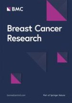
Mammary carcinomas occurred in BALB/c mice of the p53+/- genotype, and tumours of similar macroscopic morphologies were also observed in the (BALB/c × MSM/Ms)F1–p53+/- mice. The histological features of these tumours in the mammary glands are shown in Fig. 1a,1b,1c,1d,1e,1f, which indicates that they are adenocarcinomas. The histopathology of the tumours in nonirradiated mice (Fig. 1a,1b) exhibited more hyperplastic lesions than did those in irradiated mice (Fig. 1c,1d,1e,1f), but basically there were no marked differences between the two groups. There was also no remarkable difference in histopathological features between Atm+/- (Fig. 1c,1d) and Atm+/+ mice (Fig. 1e,1f) in the irradiated groups. These tumours formed glands lined by highly pleomorphic cells exhibiting frequent mitosis, and were classified as glandular adenocarcinomas, high grade according to the Annapolis Pathology Classification [26].
Figure 1
Histopathology of tumours in (BALB/cHeA × MSM/Ms)F1 mice with genotypes p53+/-Atm+/-, p53+/-Atm+/+ or p53+/+Atm+/-. (a, b) Spontaneously developing mammary adenocarcinoma from p53+/-Atm+/- mouse. (c, d) Mammary adenocarcinoma from X-irradiated p53+/-Atm+/- mouse. (e, f) Mammary adenocarcinoma from X-irradiated p53+/-Atm+/+ mouse. (g) Osteosarcoma observed in nonirradiated p53+/-Atm+/- mouse. (h) Thymic lymphoma from p53+/+Atm+/- mouse exposed to X-rays. Panels a, c and e: 40×. Panels b, d, f, g and h: 100×.
×
Lymphomas (Fig. 1g), mainly thymic lymphomas, were efficiently induced by exposure to X-rays, regardless of p53 and Atm genotype, although only a few lymphomas were observed in nonirradiated mice. Ovarian carcinomas, osteosarcomas (Fig. 1h) and hepatomas developed spontaneously. Squamous cell carcinomas, basal cell carcinomas, histiocytic sarcomas and granulocytic leukaemias were also observed in irradiated mice.
Spontaneous tumour development in mice with four genotypes for p53 and Atm
Twenty-eight p53+/-Atm+/-, 22 p53+/-Atm+/+, 11 p53+/+Atm+/- and 25 p53+/+Atm+/+ mice were examined for spontaneous development of tumours until age 26 months (113 weeks). Fourteen out of 28 p53+/-Atm+/- mice (50%) and seven out of 22 p53+/-Atm+/+ mice (32%) developed mammary carcinomas during the period of observation (Table 1, Fig. 2). The incidence of mammary carcinomas in p53+/-Atm+/- mice appeared to be higher than that in p53+/-Atm+/+ mice, but the incidences did not differ significantly between Atm+/- and Atm+/+ genotypes (P = 0.32, by Fisher's exact probability test). Among the tumours that developed in the p53+/- mice, mammary carcinoma were the most common. These mammary carcinomas were mainly observed at 41–77 weeks after birth (Fig. 2). No significant difference in latency in the nonirradiated groups was observed between doubly heterozygous mice and p53 singly heterozygous mice (P = 0.47, by unpaired Student's t-test). None of 11 p53+/+Atm+/- mice and 25 p53+/+Atm+/+ mice developed mammary carcinomas. Several lymphomas and a few other tumours developed in the four genotypes (Table 1). Thus, mammary carcinoma development depended strongly on p53 heterozygous deficiency in (BALB/c × MSM/Ms)F1 mice, and p53+/- mice of both Atm+/+ and Atm+/- genotypes developed mammary carcinoma.
Table 1
Incidence of tumours developing spontaneously in (BALB/cHeA × MSM/Ms)F1 female mice that were heterozygously deficient for p53 and/or Atm genes
Genotypes
p53+/-
p53+/+
Atm+/- (n = 28)
Atm+/+ (n = 22)
Atm+/- (n = 11)
Atm+/+ (n = 25)
Mammary carcinoma
14 (50%)a
7 (32%)
0 (0%)
0 (0%)
Lymphoma
3 (11%)
3 (14%)
2 (18%)
4 (16%)
Ovarian carcinoma
1 (4%)
1 (5%)
0 (0%)
1 (4%)
Osteosarcoma
1 (4%)
0(0%)
0 (0%)
0 (0%)
Hepatoma
1 (4%)
0(0%)
0 (0%)
0 (0%)
Number of mammary carcinomas/mouseb
14/28 (0.50)
7/22 (0.32)
0/11 (0.00)
0/25 (0.00)
aNumber of mice with tumours. bTotal number of mammary carconomas developing in mice with given genotypes/number of mice examined; numbers in parentheses are average numbers of tumours per mouse.
Figure 2
Cumulative incidence of mammary carcinomas in irradiated and nonirradiated (BALB/cHeA × MSM/Ms)F1 mice with p53+/-Atm+/- or p53+/-Atm+/+ genotype.
×
Enhancement of mammary carcinogenesis in Atmheterozygous deficient mice by X-irradiation
To test the effect of haploinsufficiency of the Atm gene on mammary carcinogenesis after X-irradiation in p53+/- mice, 55 p53+/-Atm+/-, 61 p53+/-Atm+/+, 47 p53+/+Atm+/- and 53 p53+/+Atm+/+ mice (for a total of 216 mice) were exposed to X-rays (5 Gy) at age 5 weeks. Only one out of 53 p53+/+Atm+/+ mice and none of 47 p53+/+Atm+/- mice developed mammary carcinoma, indicating that almost all p53+/+ mice fail to develop mammary carcinomas, despite X-irradiation and irrespective of Atm gene status. In contrast, 32 out of 55 p53+/-Atm+/- mice (58%) and 19 out of 61 p53+/-Atm+/+ mice (31%) developed mammary carcinomas (Table 2, Fig. 2). The proportion of mice developing mammary carcinomas in the p53+/-Atm+/- group was significantly greater than that in the p53+/-Atm+/+ group (P = 0.0034, by χ2 test). A total of 52 mammary carcinomas developed in 55 p53+/-Atm+/- mice (average number of mammary carcinomas/mouse = 0.95), whereas 28 mammary carcinoma developed in 61 p53+/-Atm+/+ mice (average number of mammary carcinomas/mouse = 0.46; Table 2). The average number of mammary carcinomas per mouse in the p53+/-Atm+/- group was significantly greater than that in p53+/-Atm+/+ mice (P = 0.0052, by Mann–Whitney U-test). Thus, Atm heterozygous deficiency enhanced development of mammary carcinoma in irradiated p53 heterozygous knockout mice. Spring and coworkers [14] observed no tumours in Atm knockout (Atm+/-) heterozygous mice. Mice bearing a knockout allele of Atm and humans carrying a mutant allele of truncated type in the ATM gene have been shown not to have obviously elevated susceptibility to mammary carcinogenesis. Our findings show that heterozygosity for a null knockout allele of Atm enhances mammary carcinogenesis under p53+/- status, although the Atm mutation is not a dominant-negative type. Heterozygous deficiency of p53 might make clear the effect on mammary carcinogenesis of haploinsufficiency in the Atm gene.
Table 2
Incidence of tumours in irradiated (BALB/cHeA × MSM/Ms) F1 female mice that were heterozygously deficient for p53 and/or Atm genes
Genotypes
p53+/-
p53+/+
Atm+/- (n = 55)
Atm+/+ (n = 61)
Atm+/- (n = 47)
Atm+/+ (n = 53)
Mammary carcinoma
32 (58%)a*
19 (31%)
0 (0%)
1 (2%)
Lymphoma
24 (44%)b
29 (48%)
26 (55%)
22 (42%)
Squamous cell carcinoma
1 (2%)
1 (2%)
0 (0%)
0 (0%)
Histiocytic sarcoma
1 (2%)c
0 (0%)
0 (0%)
0 (0%)
Basal cell carcinoma
0 (0%)
1 (2%)
0 (0%)
0 (0%)
Granulocytic leukaemia
0 (0%)
1 (2%)
0 (0%)
0 (0%)
Ovarian carcinoma
0 (0%)
1 (2%)
0 (0%)
0 (0%)
Nonthymic lymphoma (NOS)
0 (0%)
1 (2%)
3 (6%)
0 (0%)
Solid tumour (NOS)
1 (2%)d
1 (2%)e
1 (2%)
1 (2%)
Number of mammary carcinomas/mousee
52/55 (0.95)**
28/61 (0.46)
0/47 (0.00)
1/53 (0.02)
aNumber of mice with tumours; percentage in parenthesis is the proportion of mice developing mammary carcinoma. bThree animals developed both lymphomas and mammary carcinoma. cOne animal developed both histiocytic sarcoma and mammary carcinoma. dTumour in abdomen. eTotal number of mammary carcinomas developing in mice with given genotypes/number of mice examined; numbers in parentheses are average numbers of tumours per mouse. *The proportion of mice developing tumours is significantly greater than that in p53+/-Atm+/+ mice (P = 0.0034 by c2 test). **The average number of tumours/mouse is significantly greater than that in p53+/-Atm+/+ mice (P = 0.0052 by Mann–Whitney U-test). NOS, not otherwise specified.
These mammary carcinomas were observed significantly earlier (at 18–38 weeks after irradiation; i.e. 23–43 weeks of age) than in the nonirradiated group (age 41–75 weeks; Fig. 2). In particular, mammary carcinomas frequently developed 23–28 weeks after irradiation. The mean (± standard deviation) latency periods were 32.6 ± 4.8 and 29.8 ± 3.6 weeks in p53+/-Atm+/- and p53+/-Atm+/+ mice, respectively. Thus, X-irradiation at 5 Gy at age 5 weeks considerably shortened the latency period of mammary carcinoma development in these two groups with different genotypes. As shown in Tables 1 and 2, the incidences of mammary carcinoma in p53+/-Atm+/- mice were 58% (32 out of 55) and 50% (14 out of 28) in irradiated and nonirradiated groups, respectively; in p53+/-Atm+/+ mice the incidence in the irradiated group was 31% (19 out of 61) and that in the nonirradiated group was 32% (7 out of 22). The incidence of mammary carcinoma for each genotype was similar between irradiated and non-irradiated groups. Thus, irradiation may not elevate the incidence of the tumours.
Anzeige
Altogether, irradiation markedly hastened mammary carcinoma development in the p53+/- mice, in which mammary carcinomas developed spontaneously. Furthermore, irradiation also induced lymphomas, mainly thymic lymphomas, in all four genotypes of mice. The incidence of the lymphomas did not differ significantly among the four groups, with different genotypes for p53 and Atm genes. A high incidence of thymic lymphoma was observed in previous studies performed using p53 heterozygous deficient F1 mice [17, 28]. In the present study an extremely high incidence of tumours, most of which were mammary carcinomas and thymic lymphomas, was observed in irradiated p53+/-Atm+/- mice.
Status of wild-type alleles of p53 and Atmin mammary carcinoma
The wild-type alleles of the p53 and Atm genes were examined in mammary carcinoma tissue from heterozygous mice. The wild-type p53 allele was lost in 25 (96%) out of 26 mammary carcinomas from irradiated p53+/- mice and in all of 15 mammary carcinomas from nonirradiated p53+/- mice, regardless of Atm gene status. Only one mammary carcinoma in irradiated p53+/+Atm+/+ mice (Table 2) was found to retain the p53 wild-type allele. On the other hand, wild-type Atm allele was preserved in all of 17 mammary carcinomas from irradiated Atm+/- mice and in all of 10 mammary carcinomas from irradiated Atm+/+ mice, regardless of p53 gene status. Wild-type Atm allele was also retained in nine out of 10 mammary carcinomas from nonirradiated Atm+/- mice and in all of five mammary carcinomas from nonirradiated Atm+/+ mice. These results suggest that the homozygous loss of the p53 allele was a necessary condition for the development of mammary carcinomas, whereas the Atm null allele exhibited haploinsufficiency.
Haploinsufficiency for tumour development was reported for the Nbn knockout mice, the mouse homologue of NBS1. Heterozygosity for the Nbn knockout allele rendered the mice susceptible to tumour development, yet the Nbn wild-type allele was fully retained in all 12 tumours examined [29]. p27 heterozygotes are also predisposed to tumours in multiple tissues when challenged with irradiation or a chemical carcinogen, and in the developed tumours the remaining wild-type allele is neither mutated nor silenced, indicating that p27 is haploinsufficient for tumour suppression [30]. In that study, heterozygous Atm knockout enhanced mammary carcinoma development in p53-heterozygous deficient mice, but the effect of Atm deficiency was not as profound. A previous study showed that heterozygosity for the Atm knockout allele did not enhance tumour development but that dominant-negative type missense mutations in the Atm gene did [14]. In humans, heterozygosity of the truncation-type mutations, which represent the majority of Atm mutations that occur in humans, had no effect on carcinogenesis, suggesting that enhancement in tumourigenesis depends strongly on mutation type. However, in the present study we demonstrated that haploinsufficiency does occur for the Atm null allele in combination with heterozygous deficiency in the p53 gene. The p53 heterozygous deficient BALB/c mice, which developed mammary carcinomas early and efficiently, may represent a useful model for the study of effects of genes other than Atm on mammary carcinogenesis. F1 mice between different subspecies may also provide an experimental system for precise genome-wide allelotype analysis of genes that cooperate with p53.
Conclusion
Tumourigenesis is strongly enhanced in mice with homogeneous deficiency in the p53 or Atm gene. In the present study we tested the effect of haploinsufficiency of the Atm gene on mammary tumourigenesis after X-irradiation in p53+/- mice of the BALB/cHeA × MSM/Ms background. Singly heterozygous p53+/- mice X-irradiated (5 Gy) at age 5 weeks developed mammary carcinomas at around 25 weeks of age, and the final incidence of mammary carcinoma at 39 weeks was 31% (19 out of 61). Introduction of the heterozygous Atm alleles into the background of the p53+/- genotype significantly increased the incidence of mammary carcinomas to 58% (32 out of 55) and increased the average number of mammary carcinomas per mouse. However, it apparently did not change the latency of mammary carcinoma development. In nonirradiated mice, introduction of the Atm+/- allele into p53+/- mice also tended to increase spontaneous incidence of mammary carcinoma. In contrast, almost none of the p53+/+ mice developed mammary carcinoma, regardless of the Atm gene status and whether mice were subjected to irradiation. In almost all of the spontaneous and radiation-induced mammary carcinomas, the wild-type p53 allele was found to be lost whereas the wild-type Atm allele was retained, suggesting haploinsufficiency of the latter gene in mammary carcinoma development. Thus, doubly heterozygous mice represent a useful model system with which to analyze the interaction of heterozygous genotypes for p53, Atm and other genes and their effect on mammary carcinogenesis.
Anzeige
Acknowledgements
We are grateful to Mr M Ikeda and Mr U Ujihara for their skilful animal care. This study was performed in part by Grants-in-Aid for scientific research No. 13480172 (to MO) and No. 14580571 (to NM) from the Ministry of Education, Science, Sports and Culture of Japan, and a Grant-in-Aid from the Japan Atomic Energy Research Institute under contract to the Nuclear Safety Research Association.
Competing interests
The author(s) declare that they have no competing interests.
Authors' contributions
SU carried out design of the study, observed mammary carcinoma development and other diseases, and drafted a manuscript. KF prepared tissue specimens and conducted histopathological examinations (veterinarian). SI carried out DNA isolation and genotyping of mice. NM carried out X-irradiation of mice and statistical analysis. MT performed DNA isolation and genotyping of mice. DH carried out production of heterozygous deficient mice. CS participated in designing the study and discussion of data on mammary carcinoma development. SH contributed to histopathological examination. SI conducted histopathological examinations (medical doctor). ON participated in discussion of data and contributed to preparation of the final manuscript. MO carried out design of the study and X-irradiation, and wrote the final manuscript. All authors read and approved the final manuscript.
Nun gibt es auch Resultate zum Gesamtüberleben: Eine adjuvante Pembrolizumab-Therapie konnte in einer Phase-3-Studie das Leben von Menschen mit Nierenzellkarzinom deutlich verlängern. Die Sterberate war im Vergleich zu Placebo um 38% geringer.
Das Risiko für Rezidiv oder Tod von Patienten und Patientinnen mit reseziertem ALK-positivem NSCLC ist unter einer adjuvanten Therapie mit dem Tyrosinkinase-Inhibitor Alectinib signifikant geringer als unter platinbasierter Chemotherapie.
Patienten, die zur Behandlung ihres Prostatakarzinoms eine Androgendeprivationstherapie erhalten, entwickeln nicht selten eine Anämie. Wer ältere Patienten internistisch mitbetreut, sollte auf diese Nebenwirkung achten.
Müssen sich Schwangere einer Krebstherapie unterziehen, rufen Immuncheckpointinhibitoren offenbar nicht mehr unerwünschte Wirkungen hervor als andere Mittel gegen Krebs.
Update Onkologie
Bestellen Sie unseren Fach-Newsletterund bleiben Sie gut informiert.