During my study of constipation, I encountered patients who had achalasia of the esophagus (AE) as well. The possibility of an existing relationship between the 2 conditions was studied.
Method
Investigations to study the anorectal motility in 9 AE patients included: the intestinal transit time, anorectal manometry, rectoanal inhibitory reflex, defecography and electromyography (EMG) of external anal sphincter and levator ani muscle. Anorectal biopsy was done. The study comprised 8 healthy volunteers as controls.
Results
6/9 AE patients had constipation presenting as strainodynia (excessive prolonged straining at stool). Rectocele was present in 4 of them. The 6 constipated patients showed significantly high rectal neck pressure (p < 0.05), absent rectoanal inhibitory reflex and aganglionosis in the anorectal biopsy. The EMG revealed diminished activity in 4 of the 6 constipated patients. The remaining 3 patients with AE had normal anorectal function. Heller's myotomy with Nissen's fundoplication improved the dysphagia, but not the constipation which was, however, relieved after performance of anorectal myectomy.
Conclusion
The high incidence of constipation with AE postulates a relationship between the 2 conditions. Both have the same pathologic lesion which is aganglionosis. This study is preliminary and requires further studies on a larger number of patients.
The online version of this article (doi:10.1186/1471-230X-3-28) contains supplementary material, which is available to authorized users.
Competing interests
None declared.
Abkürzungen
(AE)
achalasia of the esophagus
(EMG)
electromyogram
(DRE)
Digital rectal examination
Background
Achalasia of the esophagus (AE) is a motility disorder of controversial etiology [1‐4]. Myenteric plexus degeneration impairing the lower esophageal sphincter innervation is put forward as a factor in achalasia genesis. Viral damage of the plexus is suggested as another factor [5].
During examination of 194 subjects with constipation, I came across three patients who had in addition achalasia of the esophagus. As the two conditions, AE and constipation, involve the gut, and as both are representative of impeded passage of its contents, we postulated a relationship between them. The current study aimed at exploring this hypothesis so as to add to the understanding of the etiology of both conditions and assist in the planning for a sound treatment.
Anzeige
Methods
The study comprised 9 patients (8 women, 1 man) with achalasia of the esophagus. Their clinical and investigative data are shown in table 1. The patients gave an informed consent prior to the tests.
Table 1
Clinical and investigative data of the 9 patients with achalasia of the esophagus.
Patients
Age
Sex
Duration of symptoms (months)
Radiologic findings
Constipation
Dysphagia
Rectocele
1
34
F
44
38
-
2
36
F
-
40
-
3
41
F
66
55
+
4
40
F
40
36
-
5
35
F
52
46
+
6
38
F
60
45
+
7
36
F
-
52
-
8
42
F
54
44
+
9
42
M
-
38
-
Barium swallow studies had provided the radiographic evidence of AE which included absent primary peristalsis, dilated body of the esophagus and a conically narrow cardioesophageal junction. Manometric studies [6] showed high pressure within, and lack of relaxation of, the lower esophageal sphincter. Esophagoscopic biopsy revealed the absence or disintegration of the ganglion cells of the Auerbach's plexus.
The anorectal motility was evaluated by means of the standardized questionnaire for anorectal symptoms, anorectal examination, intestinal transit time [7], anorectal manometry [8] rectoanal inhibitory reflex [9], defecography [10] and EMG activity of the external anal sphincter and levator ani muscle [11]. Suction rectal biopsies (Model SBT-100, Medical Measurements, Inc., Hackensack, NJ) were examined for the presence of ganglia in the myenteric plexus of nerves. The biopsies were stained with hematoxylin and eosin, and with silver impregnation.
Eight healthy volunteers from our laboratory, matching the test subjects in gender and age, were included in the study as controls after having given an informed consent. They had had no swallowing or anorectal complaints in the past or at the time of presentation. The clinical and investigative data are shown in table 2.
Table 2
Clinical data and pressure measurements of lower esophagus, rectum and rectal neck as well as rectoanal inhibitory reflex of the 8 control healthy volunteers.
Patients
Age
Sex
Stool frequency (per week)
Pressure (cm H2O) LES Rectal neck
Rectoanal inhibitory reflex
LES
Rectal neck
Rectal
1
32
F
8
22
5
73
+
2
38
F
9
20
7
72
+
3
37
F
10
20
6
67
+
4
44
F
11
26
6
66
+
5
40
F
10
24
8
76
+
6
35
F
8
28
9
70
+
7
42
F
12
27
7
68
+
8
39
M
7
26
6
72
+
Anzeige
The results were analyzed statistically using the Student's t test. Differences assumed significance at p < 0.05 and values were given as mean ± standard deviation. The study complies with the Helsinki Declaration and was approved by our Faculty Review Board and Ethics Committee.
Results
No complications were encountered during or after the performance of the tests and all of the 9 patients were evaluated.
Six of the 9 patients with AE complained of constipation necessitating excessive straining over a long period to evacuate the bowel. Yet, the stools were soft and had a mean frequency of 8.7 ± 1.8 per week (range 7–12), which did not differ significantly from the frequency in the control subjects (p > 0.05; table 2). The remaining 3 patients defecated normally and had a mean stool frequency of 9.2 week (range 8–13). Digital rectal examination (DRE) and anoscopy were negative in the 9 patients; no hemorrhoids, fissures or fistulas were detected. In 4 of the 6 constipated patients DRE and defecography revealed a rectocele which differed in size from one patient to the other. The intestinal transit test showed accumulation of the pellets at the anorectal junction.
Manometric measurements showed a significantly high pressure in the lower esophageal sphincter of the 9 AE patients compared to the normal controls (p < 0.05: tables 2,3). The mean rectal neck pressure in the 6 constipated patients was significantly higher than in the control subjects (p < 0.05, tables 2,3), whereas the rectal pressure showed no significant difference (p > 0.05). The rectal and rectal neck pressures in the remaining 3 patients were normal (p > 0.05). The rectoanal inhibitory reflex was absent in the 6 constipated patients whose anorectal biopsy examination showed aganglionosis (fig. 1).
Table 3
Lower esophageal, rectal and rectal neck pressures as well as rectoanal inhibitory reflex of the 9 patients with achalasia of the esophagus+.
Patients
Pressure (cm H2O)
Rectoanal inhibitory reflex
LES
Rectal
Rectal neck
1
48
6
126
-
2
62
7
72
+
3
58
5
112
-
4
46
7
98
-
5
50
8
115
-
6
63
9
108
-
7
45
7
68
+
8
53
6
102
-
9
58
6
70
+
+ The numerical arrangement of the patients is the same as in table 1.
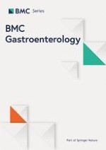
Figure 1
Microphotograph showing aganglionosis of an anorectal biopsy from a patient with achalasia of the esophagus. Silver stain × 200. Arrows point to the muscle fibers; no ganglia.
×
The EMG of the external anal sphincter was normal in all the 9 patients (fig. 2) while the levator ani showed diminished activity in 4 of the 6 constipated patients (fig. 3).
Figure 2
EMG of the external anal sphincter showing normal activity a- at rest. b- upon contraction.
Figure 3
EMG of the levator ani muscle showing diminished activity. a- at rest. b- upon contraction.
×
×
Treatment
Heller's myotomy [12] with Nissen's fundoplication were performed for the 9 patients with AE. The dysphagia improved in all the subjects. However, the constipation in the 6 patients persisted after the operation.
Considering the absence of the rectoanal inhibitory reflex and the presence of aganglionosis and high rectal neck pressure, we decided to perform anorectal myectomy for the 6 constipated patients, who gave an informed consent. Anorectal myectomy like the one done for the short segment Hirschsprung's disease [13] was carried out. The excised muscle strip showed aganglionosis. Excessive straining at stool disappeared. The rectal neck pressure decreased to a mean of 74.6 ± 7.3 cm H2O (range 66–86) which matched with the normal pressures in our laboratory.
Discussion
Of the 9 patients with AE, 6 had constipation (66.6%). This high incidence of constipation in AE patients postulates a relationship between the 2 conditions. Both AE and constipation had started later in life but constipation preceded dysphagia by a few months. This seems to rule out the assumption that constipation could result from dysphagia and diminished food intake which occur in AE.
Anzeige
We suggest that constipation in these patients is the result of anorectal aganglionosis. This is evidenced by the absent ganglia in both the anorectal biopsy and the strip excised at myectomy operation. Furthermore, absent rectoanal inhibitory reflex and high rectal neck pressure are encountered in anorectal aganglionosis [14]. Also, the improvement achieved by myectomy is an evidence that the constipation might be due to the anorectal aganglionosis. The normality of the EMG activity of the external anal sphincter excludes the possibility of anismus as a cause of the constipation. The accumulation of the pellets at the anorectal junction negates that the rest of the gut could be the cause. The diminished EMG activity of the levator ani muscle seems to be the result of excessive straining at stool and not a primary cause of constipation.
Operative correction of AE did not improve the anorectal manifestations. This might be viewed as indicating that there is no direct relation between the esophagus and the anorectal lesions. However, the presence of aganglionosis in both AE and constipated patients warrants consideration.
Although the high incidence of constipation in AE as well as the presence of aganglionosis in both conditions suggest a relationship between the esophageal and anorectal lesions, the current study could not elucidate its nature. The questions requiring an answer include: 1. Why does aganglionosis occur in the two opposite and outermost segments of the gut? 2. Why did aganglionic manifestations develop later in life, thereby negating a possible congenital origin as occurs in Hirschsprung's disease? However, an acquired aganglionosis caused by cytomegalovirus or ischemia may be considered. 3. Does aganglionosis involve other parts of the gut although the intestinal transit time was normal in the rest of the gut?
It is known that the myenteric plexus of nerves extends along the whole gut [15]. Impulses can extend along this plexus from one segment of the gut to the other. Furthermore, it was found that distension of the lower esophageal sphincter induces rectal contractions mediated through an esophago-rectal reflex [16]; it was put forward that impulses extend from the esophagus to the rectum along the myenteric nerve plexus.
Anzeige
In conclusion, the high incidence of constipation in AE in the current study would draw the attention to the possible existence of a relationship between the 2 conditions which we call "esophago-rectal syndrome". This relationship needs further investigations to answer the above mentioned questions. We suggest that achalasia patients be asked about the history of constipation and vice versa constipated patients for disorders in swallowing. In the meantime, the findings of the current study warrant publication although the number of cases is small.
Acknowledgment
- Professor Abdulla Khalil, Professor of Pathology at the Faculty of Medicine, Cairo University, examined the esophageal and anorectal biopsies.
- Margot Yehia and Waltraut Reichelt assisted in preparing the manuscript.
Die Therapie von Echinokokkosen sollte immer in spezialisierten Zentren erfolgen. Eine symptomlose Echinokokkose kann – egal ob von Hunde- oder Fuchsbandwurm ausgelöst – konservativ erfolgen. Wenn eine Op. nötig ist, kann es sinnvoll sein, vorher Zysten zu leeren und zu desinfizieren.
Seit November 2023 gibt es evidenzbasierte Empfehlungen zum perioperativen Management bei gastrointestinalen Tumoren (POMGAT) auf S3-Niveau. Vieles wird schon entsprechend der Empfehlungen durchgeführt. Wo es im Alltag noch hapert, zeigt eine Umfrage in einem Klinikverbund.
Mit dem demographischen Wandel versorgt auch die Chirurgie immer mehr betagte Menschen. Von Entwicklungen wie Fast-Track können auch ältere Menschen profitieren und bei proximaler Humerusfraktur können selbst manche 100-Jährige noch sicher operiert werden.
Worauf kommt es beim Management von Personen mit infektiöser Endokarditis an? Eine Kardiologin und ein Kardiologe fassen die zehn wichtigsten Punkte der neuen ESC-Leitlinie zusammen.
Update Innere Medizin
Bestellen Sie unseren Fach-Newsletter und bleiben Sie gut informiert.


