Dental radiographic findings in 18 individuals with SATB2-associated syndrome
verfasst von:
John Scott, Chad Adams, Kirt Simmons, Andrea Feather, John Jones, Larry Hartzell, Lucia Wesley, Adam Johnson, Jennifer Fish, Katherine Bosanko, Stephen Beetstra, Yuri A. Zarate
To characterize the radiographic dental phenotype of individuals with SATB2-associated syndrome (SAS).
Materials and methods
Participants were evaluated by a multidisciplinary team during a concurrent clinic conducted during the 1st international SAS family meeting held in 2017 at a single institution. Whenever possible, panoramic and/or periapical radiographs were obtained in clinic or previously obtained and provided by the caregiver.
Results
Of the 37 individuals evaluated, 18 (12 males, median age 8.5 years) underwent radiographic examination. Dental radiographs revealed anomalies in all individuals starting at 2 years of age. The most consistent finding was delayed development of the mandibular second bicuspids (83%) with other common radiographic findings including delayed development of the roots of the permanent teeth (78%), severely rotated (56%) or malformed teeth (44%), and taurodontism (44%).
Conclusions
Dental anomalies are fully penetrant and can be documented radiographically in all individuals with SAS.
Clinical relevance
Dental radiographic findings of delayed second premolar development and delayed development of permanent root formation, especially concurrent with findings of taurodontism and malformed teeth, support a clinical suspicion for SAS and should help differentiate SAS from other neurodevelopmental syndromes.
Introduction
The precise mechanism of tooth development is a complex embryologic process that involves well-regulated interactions between the dental epithelium and the neural crest-derived mesenchyme [1]. Alterations in genes important in these epithelial–mesenchymal interactions during different stages of odontogenesis can result in a variety of morphologic, numeric, or physiologic dental anomalies alone or in combination with other systemic features [2‐5].
SATB2 is a transcription factor and high level regulator of several gene regulatory networks with critical roles in multiple developmental processes including those of the jaw, brain, and skeleton [6, 7]. The critical role of SATB2 in odontogenesis has been documented in the past [8]. Studies looking at a variety of murine expression patterns at different stages of embryologic and post-natal tooth development as well as immunohistochemical patterns in human healthy teeth have shown that SATB2 has a role in the differentiation of ameloblasts and odontoblasts, dentin formation, and the regulation of dental pulp physiological functions [8]. In mice, full functional loss of Satb2 and Satb2 heterozygotes results in several craniofacial anomalies as well as potential variable incisor hypodontia and/or anodontia and a reduction in dentary length [9, 10].
Anzeige
SATB2-associated syndrome (SAS; Glass syndrome, OMIM 612313) is a rare disorder caused by alterations in the special AT-rich sequence-binding protein 2 (SATB2; MIM 608148) [11]. Predominantly described as a neurodevelopmental disorder, craniofacial anomalies, including palatal and dental abnormalities, are commonly seen [12‐14]. Although cleft palate and micrognathia have variable expression, we recently reported dental anomalies to be universally present after the first year of life in a large cohort of individuals with SAS [15]. In that report, intraoral dental problems included crowding (96%), large upper incisors (87%), aberrant dental shape (87%), hypodontia (44%), delayed tooth eruption (14%), fused teeth (9%), and supernumerary teeth (6%). In this report, we present the first description of dental radiographic findings in a cohort of individuals with SAS evaluated at a single institution.
Methods
Patients
Participants were first recruited into the SAS clinical registry through a referral by a treating clinician, a facilitated inquiry by the testing laboratory, direct contact by a caregiver, or via the SAS support group as previously described [15]. As patients are enrolled into the clinical SAS registry, a unique ID number is assigned sequentially and henceforth used in this manuscript. As part of the 1st international SAS family meeting held in 2017 at Arkansas Children’s Hospital, a concurrent multidisciplinary clinic took place. As part of the clinical assessment, participants were evaluated by providers from the following specialties: Otolaryngology, Dentistry, Orthodontics, Maxillofacial surgery, Genetics, Audiology, Speech, and Neuropsychology. All individuals included in this study had a molecularly confirmed diagnosis of SAS. All families reported herein agreed to share clinical information and were enrolled under a research clinical registry protocol approved by the Institutional Review Board of the University of Arkansas for Medical Sciences. Detailed clinical and molecular description of all individuals is available in previous reports [13, 15].
Evaluations
Whenever possible, panoramic and/or periapical radiographs were obtained in clinic and reviewed by the same dental team members (J.S., C.A., K.S., A.F., J.J., S.B.). For one individual, previously obtained images were provided by the caregiver. The following dental problems were routinely evaluated: delayed development, malformations of the crown and/or roots, missing teeth as well as taurodontism, tooth size and shape mismatches, malpositions, pulp stones, and dental fusion. Delayed development of tooth structure (crown/root) or eruption was determined according to the individual’s age at assessment when compared to published norms [16]. Delayed root development was identified where portions of the dental unit were missing or lack development. First, molars with less than a 1:1 crown to root ratio by the age of 9–10 were identified as having delayed root development.
Results
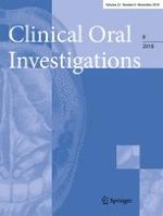
Of the 102 enrolled into the SAS clinical registry with complete medical records available at the moment of writing, 37 were clinically evaluated during the 2017 family meeting. Of these, 18 (12 males) were cooperative enough to undergo radiographic characterization at our institution (17) or elsewhere (1) with panoramic and/or periapical radiographs. The median age was 8.5 years (range 2.6–20.5 years). Panoramic images were obtained and generated using Planmeca Romexis® software at 64 to 66 Kilovolts (kV), 6 to 8 milliamperes (mA), and 22.1 s of exposure time (Fig. 1). The dental radiographs revealed anomalies in all individuals as summarized in Table 1. The most consistent finding, delayed formation of the second mandibular bicuspids, was identified from ages 2 to as late as age 20. Other common radiographic findings included delayed formation of the roots of permanent teeth, malformed teeth, and taurodontism.
Fig. 1
Dental radiographic anomalies in individuals with SAS. a SATB2-38. b SATB2-39. c SATB-04. d SATB2-53. e SATB2-21. f SATB2-17. Common findings include delayed/missing maxillary 2nd bicuspids (triangle), delayed/missing mandibular 2nd bicuspids (arrow), large teeth (star), delayed root formation (diamond), or taurodontism (chevron). Pulp stones are circled
Table 1
Dental radiographic description of 18 individuals with SATB2-associated syndrome
SATB2 has been suggested to have a role in several odontogenic processes, including epithelial–mesenchymal interactions during early odontogenesis, odontoblast and ameloblast differentiation, and dentin matrix mineralization [8, 17]. In mice, homozygous mutants display abnormalities of the anterior part of the mandible and incisors correlating with the SATB2 expression pattern. The molars, on the other hand, do not express SATB2 and are unaffected in mutant embryos [7]. In this study, we documented abnormalities in dental position, number, size, and shape in individuals with SATB2 alterations.
Thus far, the exact mechanism of how SATB2 regulates tooth formation is unclear. Other molecular pathways related to SATB2 have been implicated, including RUNX2 and sonic hedgehog (Shh) [6, 8, 18]. Cleidocranial dysplasia, a skeletal dysplasia caused by alterations in RUNX2, also has a high frequency of dental anomalies including supernumerary teeth and failure of eruption [19]. While lower second premolars are the most frequently missing teeth and often found with other dental anomalies [3], the frequency, type, and extent of the radiographic dental anomalies in individuals with SAS described here should be used as another phenotypic supportive feature of this condition. The identification of some of these radiographic anomalies during early childhood could also help in the differential diagnosis of syndromic conditions with predominant dental phenotypes with or without a neurodevelopmental component [2, 5].
In addition to the fully penetrant dental radiographic findings here reported, we noted delayed eruption, bruxism, sialorrhea, macrodontia, and anterior crowding. The identification of the dental phenotype presented above along with other craniofacial, developmental, and behavioral abnormalities should prompt one to have the patient evaluated for SAS. This could secondarily aid in the early diagnosis of other undiagnosed neurodevelopmental disorders.
Acknowledgments
The authors would like to thank all the children and parents who took part in the research. The 1st international SAS family meeting held in 2017 at Arkansas Children’s Hospital was possible thanks to a Patient-Centered Outcome Research Institute (PCORI) Engagement award (EAIN-5502).
Compliance with ethical standards
Conflict of interest
The authors declare that they have no conflict of interest.
Ethical approval
All procedures performed in studies involving human participants were in accordance with the ethical standards of the institutional and/or national research committee and with the 1964 Helsinki declaration and its later amendments or comparable ethical standards. The institutional review board (IRB) at the University of Arkansas for Medical Sciences (IRB # 205083 and #228310) approved the study.
Informed consent
Informed consent was obtained from all individual participants included in the study.
Open Access This article is distributed under the terms of the Creative Commons Attribution 4.0 International License (http://creativecommons.org/licenses/by/4.0/), which permits unrestricted use, distribution, and reproduction in any medium, provided you give appropriate credit to the original author(s) and the source, provide a link to the Creative Commons license, and indicate if changes were made.
Dental radiographic findings in 18 individuals with SATB2-associated syndrome
verfasst von
John Scott Chad Adams Kirt Simmons Andrea Feather John Jones Larry Hartzell Lucia Wesley Adam Johnson Jennifer Fish Katherine Bosanko Stephen Beetstra Yuri A. Zarate
Werden Personen mit Vorhofflimmern in der Blanking-Periode nach einer Katheterablation gegen eine bestehende Parodontitis behandelt, verbessert dies die Erfolgsaussichten. Dafür sprechen die Resultate einer prospektiven Untersuchung.
Bei welchen Personen eine Antibiotikaprophylaxe zur Prävention einer infektiösen Endokarditis nach invasiven zahnärztlichen Eingriffen sinnvoll ist, wird diskutiert. Neue Daten stehen im Einklang mit den europäischen Leitlinienempfehlungen.
Der Aufbau von Geweben und Organen während der Embryonalentwicklung wird von den Zellen bemerkenswert choreografiert. Für diesen Prozess braucht es spezielle sogenannte „Organisatoren“. In einer aktuellen Veröffentlichung im Fachjournal Nature Cell Biology berichten Forschende durch welchen Vorgang diese Organisatoren im Gewebe entstehen und wie sie dann die Bildung von Zähnen orchestrieren.
Infolge der Umbenennung der Deutschen Gesellschaft für Kinderzahnheilkunde in Deutsche Gesellschaft für Kinderzahnmedizin (DGKiZ) wird deren Mitgliederzeitschrift Oralprophylaxe & Kinderzahnheilkunde in Oralprophylaxe & Kinderzahnmedizin umbenannt. Aus diesem Grunde trägt die erste Ausgabe in 2024 erstmalig den neuen Titel.
Newsletter
Bestellen Sie unseren kostenlosen Newsletter Update Zahnmedizin und bleiben Sie gut informiert – ganz bequem per eMail.