


Anzeige
Erschienen in:
01.04.2014 | Original Article—Liver, Pancreas, and Biliary Tract
Histological evaluation of obliterative phlebitis for the diagnosis of autoimmune pancreatitis
Erschienen in: Journal of Gastroenterology | Ausgabe 4/2014
Einloggen, um Zugang zu erhaltenAbstract
Background
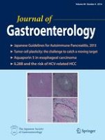
Obliterative phlebitis is a useful pathological finding for the diagnosis of lymphoplasmacytic sclerosing pancreatitis (LPSP), or type 1 autoimmune pancreatitis. The present study evaluated histological findings of obliterative phlebitis, including the significance of adding Elastica van Gieson stain (EVG) in comparison with other pancreatic conditions.
Methods
Specimens of LPSP (n = 18), chronic pancreatitis (CP; n = 24), and pancreatic ductal adenocarcinoma (PDA; n = 45) were enrolled. Obliterative venous lesions (OVLs), defined as the presence of inflammatory cells and/or fibrosis inside the tunica adventitia, were counted and compared between hematoxylin and eosin stain (H&E) and EVG. OVLs were classified into three types: OVL-1, lymphoplasmacytic infiltration and fibrosis against a loose textured background; OVL-2, dense fibrosis with minimal or no lymphoplasmacytic infiltration; and OVL-3, densely packed lymphoplasmacytic infiltration without fibrosis. OVL type and OVL size were compared between disease groups.
Results
OVL counts in LPSP, CP, and PDA were significantly higher with EVG than with H&E (p < 0.001). OVL-1 was most common in LPSP (H&E 92.4 %, EVG 79.8 %), and was identified in almost all cases of LPSP, but was less common in CP and PDA. Maximum diameter and OVL count in 1 cm2 of OVL-1 were high for LPSP. Maximum diameter of OVL-1 ≥150 μm was observed in 17 LPSP, 0 CP, and 1 PDA cases (sensitivity 94.4 %, specificity 98.6 %).
Conclusions
Additional EVG is useful for excluding conditions mimicking OVL-1 or detecting OVL in small specimens. The presence of OVL-1 with diameter ≥150 μm is highly diagnostic for LPSP.