There are several anatomical variations of the pulmonary vein which can cause serious complications in pulmonary lobectomy.
Case presentation
We inadvertently divided the left superior pulmonary vein during thoracoscopic left lower lobectomy in a lung cancer patient. Retrospective review of the preoperative computed tomography showed extra-pericardial common trunk of the left pulmonary venous system. Left superior pulmonary vein was reimplanted into stump of divided common trunk via thoracotomy.
Conclusions
Awareness of vascular anomalies will help thoracic surgeons to prevent potential morbidity and mortality from complications.
Abkürzungen
CT
computed tomography
LA
left atrium
LSPV
left superior pulmonary vein
Background
Pulmonary vascular variations are significant in thoracic surgery as such vascular anomalies can present a number of surgical complications [1]. Dividing a pulmonary vein that should be preserved can lead to potentially life-threatening complications [2]. In this report we describe a case where we inadvertently divided the left superior pulmonary vein (LSPV) during thoracoscopic left lower lobectomy in a lung cancer patient.
Case presentation
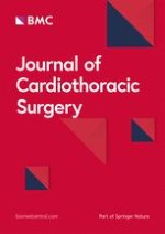
The chest computed tomography (CT) of a 74-year-old female showed a lobulated cancer in the lateral basal segment of the left lower lobe measuring 1.5 cm in diameter, for which the patient underwent thoracoscopic left lower lobectomy. During this operation, the pulmonary vein was initially divided using a 2.5-mm stapling device (Echelon Flex 60 Endopath Stapler, Ethicon Endo-Surgery, LLC, Guaynabo, Puerto Rico 00969 USA). Next, the common basal and the superior segmental arteries were divided simultaneously by employing the same device, then the left lower lobar bronchus was divided using a 4.1-mm stapling device (Echelon Flex 60 Endopath Stapler). During post-lobectomy exploration, the LSPV was not be visualized in the expected location. Retrospective review of the preoperative chest CT showed anatomical variation in the pulmonary venous system. In this case, the superior and inferior pulmonary veins had formed a common trunk outside the pericardium and drained into the left atrium (Fig. 1). After heparin injection, all staples on the LSPV and an half of the staples on the common trunk were removed under intra-pericardial partial clamping of left atrium (LA) via thoracotomy. The patient became bradycardic during first attempt of partial clamping of the LA. It was difficult to clamp the LA without causing bradycardia, and the operation only proceeded after the vital signs were stable for at least 5 min after clamping. The LSPV was reimplanted into the stump of the divided common trunk using continuous 4–0 Prolene sutures. The LSPV was reimplanted in the location of the left inferior pulmonary vein in the stump of the divided common trunk, rather than its original site. This was because the length of the original site of the LSPV was shortened due to the stapling direction at the time of pulmonary vein division, and the length for reimplantation could not be approximated. The patient recovered well from the procedure and was transferred to the general ward, and was discharged 6 days after the operation without any complications.
Fig. 1
Preoperative chest computed tomography showed that superior and inferior pulmonary veins (white arrow) had formed a extrapericardial common trunk (black arrow)
×
Anzeige
Discussion
Pulmonary vascular anomalies are significant in thoracic surgery [1]. Thoracic surgeons tend to focus more on variations of the pulmonary arteries than the pulmonary veins during preoperative evaluation because the majority of severe intra-operative complications during pulmonary lobectomy are related to the injury of major pulmonary arteries [3]. However, pulmonary venous anatomical variations are more common than those of pulmonary arterial branches [4]. Ligation of a pulmonary vein that should be preserved can lead to severe pulmonary edema, which may cause potentially life-threatening complications such as infection and respiratory distress [2]. Disruption of the pulmonary vein can also lead to complicated and longer procedures [5].
In consideration of the drainage pattern of the left pulmonary vein, the incidence of the common trunk forming one ostium in the LA is 14 % [6]. This can be divided into two types: the first type where the common trunk vein is less than 1 cm long, and the other where it is more than 1 cm long that drains into the LA. The incidence of having a common trunk that is longer than 1 cm is 3.5 % [6]. In this case, the superior and inferior pulmonary veins formed an extra-pericardial common trunk which was longer than 1 cm. The LSPV was reimplanted after lower lobectomy, because we had not confirmed the exact vascular anatomy and divided the common trunk consisting of the superior vein and the inferior vein, mistaking it as the inferior pulmonary vein. This is because of the surgeons’ tendency to only visualize the local surgical field rather than checking the general vascular anatomy around the operation site in thoracoscopic surgery. Retrospective review of the preoperative CT showed that the superior and inferior pulmonary veins joined to form the common trunk, which suggests that preoperative diagnosis of this variation is quite possible.
Conclusion
Awareness of vascular anomalies is very important in excising the pulmonary lobe for lung cancer. Especially closer attention is required in thoracoscopic procedures as the surgical view is more limited than in a thoracotomy. Keeping such vascular anomalies in mind will help thoracic surgeons to prevent potential morbidity and mortality from complications.
Authors’ contributions
All authors performed the operation. YS prepared the manuscript. EL edited manuscript. Both authors read and approved the final manuscript.
Anzeige
Competing interests
The authors declare that they have no competing interests.
Consent
Written informed consent was obtained from the patient for the publication of this report and any accompanying images.
Open AccessThis article is distributed under the terms of the Creative Commons Attribution 4.0 International License (http://creativecommons.org/licenses/by/4.0/), which permits unrestricted use, distribution, and reproduction in any medium, provided you give appropriate credit to the original author(s) and the source, provide a link to the Creative Commons license, and indicate if changes were made. The Creative Commons Public Domain Dedication waiver (http://creativecommons.org/publicdomain/zero/1.0/) applies to the data made available in this article, unless otherwise stated.
Nach der Katheterablation von Vorhofflimmern kommt es bei etwa einem Drittel der Patienten zu Rezidiven, meist binnen eines Jahres. Wie sich spätere Rückfälle auf die Erfolgschancen einer erneuten Ablation auswirken, haben Schweizer Kardiologen erforscht.
Schmerzen im Unterbauch, aber sonst nicht viel, was auf eine Appendizitis hindeutete: Ein junger Mann hatte Glück, dass trotzdem eine Laparoskopie mit Appendektomie durchgeführt und der Wurmfortsatz histologisch untersucht wurde.
Derzeit wird empfohlen, eine Therapie mit GLP-1-Rezeptoragonisten präoperativ zu unterbrechen. Eine neue Studie nährt jedoch Zweifel an der Notwendigkeit der Maßnahme.
Die Ureterstriktur ist eine relativ seltene Komplikation, trotzdem bedarf sie einer differenzierten Versorgung. In komplexen Fällen wird dies durch die roboterassistierte OP-Technik gewährleistet. Erste Resultate ermutigen.
Update Chirurgie
Bestellen Sie unseren Fach-Newsletterund bleiben Sie gut informiert.
Das Karpaltunnelsyndrom ist die häufigste Kompressionsneuropathie peripherer Nerven. Obwohl die Anamnese mit dem nächtlichen Einschlafen der Hand (Brachialgia parästhetica nocturna) sehr typisch ist, ist eine klinisch-neurologische Untersuchung und Elektroneurografie in manchen Fällen auch eine Neurosonografie erforderlich. Im Anfangsstadium sind konservative Maßnahmen (Handgelenksschiene, Ergotherapie) empfehlenswert. Bei nicht Ansprechen der konservativen Therapie oder Auftreten von neurologischen Ausfällen ist eine Dekompression des N. medianus am Karpaltunnel indiziert.
Das Webinar beschäftigt sich mit Fragen und Antworten zu Diagnostik und Klassifikation sowie Möglichkeiten des Ausschlusses von Zusatzverletzungen. Die Referenten erläutern, welche Frakturen konservativ behandelt werden können und wie. Das Webinar beantwortet die Frage nach aktuellen operativen Therapiekonzepten: Welcher Zugang, welches Osteosynthesematerial? Auf was muss bei der Nachbehandlung der distalen Radiusfraktur geachtet werden?
Inhalte des Webinars zur S1-Leitlinie „Empfehlungen zur Therapie der akuten Appendizitis bei Erwachsenen“ sind die Darstellung des Projektes und des Erstellungswegs zur S1-Leitlinie, die Erläuterung der klinischen Relevanz der Klassifikation EAES 2015, die wissenschaftliche Begründung der wichtigsten Empfehlungen und die Darstellung stadiengerechter Therapieoptionen.