The mechanistic study of glaucoma pathogenesis has shifted to seeking to understand the effects of immune responses on retinal ganglion cell damage and protection. Cytokines are the hormonal factors that mediate most of the biological effects in both the immune and nonimmune systems. CD4-expressing T helper cells are a major source of cytokine production and regulation. Type 1 helper T (Th1) cells are characterized by the production of proinflammatory cytokines such as interferon-gamma, interleukin (IL)-2, IL-12, IL-23, and tumor necrosis factor-alpha while type 2 helper T (Th2) cells are characterized by the production of IL-4, IL-5, IL-6, and IL-10. The balance of Th1/Th2 cytokine production influences many pathological processes and plays both causative and protective roles in neuronal damage. Growing evidence indicates that imbalances of Th1/Th2 cytokine production are involved in neural damage or protection in many neurological diseases. In this review, we discuss the possible roles of Th1/Th2 cytokine production and imbalance of Th1/Th2 cytokines in retina, especially glaucomatous optic neuropathy.
The following article was mistakenly run in volume 2, issue 2 (June 2009) instead of in the present special issue, for which it was intended. Please also see additional corrections to the dates originally listed in Table 3. The publisher regrets the error.
Introduction
Glaucoma is one of the leading causes of vision loss as a result of impairment of retina ganglion cells (RGCs). Although lowering of intraocular pressure (IOP) is currently the method of choice for the treatment of glaucoma, glaucomatous neuropathy may still develop and lead to loss of vision [1]. There are no other effective therapeutic and/or preventive interventions. Other mechanisms, besides the increase in IOP, have been associated with degeneration of RGCs. These include ischemia, glutamate excitotoxic stress, high reactive oxygen species production, and the loss of mitochondrial function. Recent mechanistic studies have focused on immunological changes during glaucomatous pathogenesis and possible preventive therapies [2]. The major targets of interest are cytokines and their functions in damage or protection of retinal ganglion cells. Recent advances in the studies of glaucoma or RGCs reveal that cytokines are a possible factor in the pathogenesis of glaucoma and may regulate RGCs survival or death. Here, we will review the roles of both type 1 helper T (Th1)-derived and type 2 helper T (Th2)-derived cytokines in the damage or protection of RGCs and the possible relationship of these cytokines with human glaucoma.
Cytokines from T helper cells and neuronal diseases
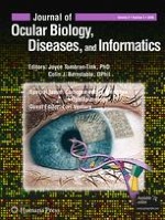
CD4-expressing T helper cells develop into two major subtypes of cells known as Th1 and Th2 cells [3, 4]. The two subtypes produce different sets of cytokines, which are involved in many physiological and pathological processes in humans. It has been considered that Th1 cells are characterized by the production of proinflammatory cytokines such as IFN-γ, IL-2, IL-12, IL-23, and TNF-alpha while Th2 cells are characterized by the production of IL-4, IL-5, IL-6, and IL-10 (Fig. 1). We now know that this classification is an oversimplification, and that the effects of cytokines are far more complicated in the tissue and cells in response to stress.
Fig. 1
Th1 and Th2 cytokine signaling and T helper cells. Th1 and Th2 cells can be induced from Th0 cells by IL-12 and IL-4, respectively. IL-2 promotes Th0 cell and also Th1 and Th2 cell proliferation. The major cytokines produced by Th1 or Th2 cells are listed
×
The functions of T helper cytokines have been extensively studied in a number of different conditions [5, 6], including neural damage and protection. In the autoimmune central nervous system disease multiple sclerosis and its animal model experimental autoimmune encephalomyelitis (EAE), T helper lymphocytes play a crucial role in inflammatory tissue damage to the neurons [7]. Cytokines, such as interferon-gamma, are also involved in neuron damage resulting from nonautoimmune conditions such as ischemia [8] and a number of neurodegenerative diseases [9]. In contrast, T helper cytokines can also play significant roles in protection against neural cell death [10], indicating two aspects of cytokine function, namely, in neural damage and protection.
Anzeige
Cytokines play essential roles in retina neural damage
The causes of RGC death have been studied in many model systems in vitro and in vivo, including glutamate or N-methyl-d-aspartate (NMDA) ocular injection and cytotoxicity, increase of intraocular pressure (IOP), ischemia/reperfusion (I/R), axotomy of optic nerves, optic nerve crush, experimental autoimmune uveoretinitis (EAU), EAE, and serum deprivation in cultured neural cells. Evidence over the past decade has revealed that cytokines produced from both Th1 and Th2 cells were involved in RGCs death in response to the insults listed above. Detailed information is listed in Table 1.
Table 1
Cytokines in retina neural damages
Cytokines
Systems
Methods
Cell types
Insults
Results
Author
Year
IFN-g
Animals
In vitro
Retina explants
Serum deprivation
RGC damage
Tura
2009
IL-1b
Animals
In vitro
RGC
Survival
RGC damage
Abcouwer
2008
Animals
In vivo
RGC
NMDA
RGC damage
Kitaoka
2007
Animals
In vivo
RGC
I/R
RGC damage
Zhang
2004
Animals
In vivo
RGC
I/R
RGC damage
Yoneda
2001
IL-2
Animals
In vivo
Retina
EAU
Retina damage
Amadi-Obi
2007
IL-6
Animals
In vitro
Retina explants
Serum deprivation
RGC damage
Tura
2009
Animals
In vivo
RGC
Glutamate toxicity
RGC damage
Fisher
2001
IL-17
Animals
In vivo
Retina
EAU
Retina damage
Amadi-Obi
2007
TNF-a
Animals
In vivo
Retina
EAU
Retina damage
Amadi-Obi
2007
Animals
In vitro
Retina
Serum deprivation
RGC damage
Tura
2009
Animals
In vitro
Retina explants
Culture
RGC damage
Hong
2009
Animals
In vivo
RGC
IOP
RGC damage
Nakazawa
2006
Animals
In vivo
RGC
TNF-a injection
RGC damage
Kitaoka
2006
Animals
In vitro
RGC
Survival
RGC damage
Tezel
2000
Human
In vivo
Optic nerve
Expression
Optic nerve damage
Yuan
2000
TNFR-1
Animals
In vivo
RGC
Optic nerve crush
RGC damage
Tezel
2004
Increased levels of Th1 cytokines, such as IFN-gamma [11], IL-1 [12], IL-2 [13], IL-17 [13], and TNF-alpha [11, 13‐18] were reported to be significantly associated with retina and RGC damage caused by distinct insults in vivo and in vitro, while IL-6 is the only Th2 cytokine that has been reported to show similar results [11, 19]. Most work has been focused on the actions of TNF-alpha. TNF-alpha was first found elevated in retina following increase of hydrostatic pressure. RGC apoptosis rates were attenuated approximately 66% by neutralizing antibody against TNF-alpha [14]. An increase of TNF-alpha expression has been associated with the most severely damaged optic nerve heads from human glaucomas [15], indicating that TNF-alpha contributes to the progression of optic nerve degeneration in this disease. Later work has confirmed that TNF-alpha is a critical cytokine in RGC damage caused by different insults, such as EAU [13], optic nerve crush [20], and serum withdrawal [11] in vitro. Direct ocular injection of TNF-alpha [16] in vivo also results in RGC death.
The Th2 cytokine IL-6 is a member of a large cytokine family that triggers the gp130 receptor and activates signal transducer and activator of transcription 1 (STAT1) and STAT3 in response to stress [21]. IL-6 has been considered a neurotrophic factor in protection of neurons from damage in many neural degenerative conditions [22, 23]. While IL-6 can play a neuroprotective role in retina, as described below, it has been shown to be involved in retina or RGC damage under a number of different conditions [11, 19]. In a study of Rho-kinase inhibition on retinal cell survival, the reduction of proinflammatory cytokines including TNF-alpha, IFN-gamma, and IL-6 likely contributed to the significantly lower toxicity on retina explants [11]. Other evidence showed that IL-6 deficiency could increase the rate of RGC survival during the first 24 h after optic nerve injury [19], indicating the complexity of IL-6 in both retina damage and protection.
IL-17 is produced by Th17 cells. This cell type can be stimulated to expand by IL-2 and IL-23. Besides IL-17 production, Th17 cells also secrete IL-6 and TNF-alpha and now are recognized as causative agents of several diseases previously attributed to Th1 cells, such as chronic inflammatory bowel disease [24]. IL-17 is an agent that contributes to retina damage in an EAU model [13]. Retinal damage in this model can be reduced by a neutralized by specific anti-IL-17 antibody.
Cytokines in retina neural protection
A significant number of studies on cytokine protection of neurons have been performed and these numbers are increasing year after year. As with the studies on neural damage, many kinds of insults have been used to test the roles of cytokines in protection of retinal neurons both in vivo and in vitro. In an EAU model, IL-27 and IFN-gamma have been shown to inhibit IL-2-induced expansion of IL-17, produced by Th17 cells, and to ameliorate retina damage by EAU [13]. This indicates that Th1 cytokines such as IFN-gamma could have protective roles on neurons under certain circumstances. In a similar case, IL-1, a mediator of neural injury, has a protective role by preventing neuronal cell death from glutamate neurotoxicity [25]. TNF-alpha, a clear mediator for neural damage, also can prevent secondary death of RGCs after axotomy of the optic nerve in vivo [26].
Th2 cytokines such as IL-4, IL-6, and IL-10 show neuroprotective functions in many different types of RGC injury such as optic nerve axotomy [27] and ischemia/reperfusion [28] in vivo and serum deprivation [29] in vitro (Table 2). Intraocular administration of adenoviral vectors encoding IL-10 and IL-4 may help prevent neurodegeneration caused by the activation of glial cells postaxotomy [27]. IL-6 is upregulated after retinal ischemia/reperfusion injury, and its expression by microglia/phagocytic cells may protect neurons in the RGC layer from this insult. Exogenously added IL-6 protects the inner retina after ischemia/reperfusion injury [28]. Our studies on STAT3 [30], the downstream effector of IL-6 and IL-10, have shown that STAT3 activation is essential for RGC survival, and persistent activation of STAT3 by neurotrophic factors and cytokines provides strong neuroprotection. This suggests that STAT3 activation will be an effective strategy in a number of chronic retinal diseases.
Table 2
Cytokines in retina neural protection
Cytokines
Systems
Methods
Cell types
Insults
Results
Author
Year
IFN-b1a
Animals
In vivo
Optic nerve
EAE
Optic nerve protection
Sättler
2006
IFN-b1b
Animals
In vivo
RGC
EAE
RGC protection
Maier
2006
IFN-g
Animals
In vivo/in vitro
Retina
EUA
Retina protection
Amadi-Obi
2007
IL-1
Animals
In vitro
Glail cell
Glutamate toxicity
RGC protection
Namekata
2008
IL-1b
Animals
In vivo
RGC
Axotomy
RGC protection
Diem
2003
IL-2
Animals
In vitro
RGC
Survival
RGC protection
Sholl-Franco
2001
IL-4
Animals
In vivo
RGC
Axotomy
RGC protection
Koeberle
2004
Animals
In vitro
RGC
Survival
RGC protection
Sholl-Franco
2001
IL-6
Animals
In vivo
RGC
I/R
RGC protection
Sanchez
2003
Animals
In vitro
RGC
Survival
RGC protection
Mendonça Torres
2001
IL-10
Animals
In vivo
RGC
Axotomy
RGC protection
Koeberle
2004
Animals
In vitro
RGC
Serum deprivation
RGC protection
Boyd
2003
IL-27
Animals
In vivo
Retina
EAU
Retina protection
Amadi-Obi
2007
STAT3
Animals
In vitro/in vivo
RGC
I/R and Glutamate toxicity
RGC protection
Zhang
2008
TNF-a
Animals
In vivo
RGC
Axotomy
RGC protection
Diem
2001
Leukocyte recruitment
Animals
In vivo
RGC
NMDA
RGC protection
Nakazawa
2007
T autoimmunity
Animals
In vivo
RGC
Optic nerve crush
RGC protection
Kipnis
2004
T autoimmunity
Animals
In vivo
RGC
Optic nerve crush
RGC protection
Yoles
2001
Cell-mediated immunity and inflammatory responses are also thought to be involved in neural damage and protection. There is evidence that inflammatory leukocyte recruitment can play a causative role in RGC cell death in NMDA-induced excitotoxicity. Anti-inflammatory agents improved RGC survival [31], suggesting that increases in inflammatory responses can lead to neural damage. On the other hand, an evoked T cell-dependent response has been shown to help neuron survival [32]. It was reported that using a single low dose of whole-body or lymphoid-organ gamma-irradiation significantly increased T cell-dependent responses, and this improved the spontaneous recovery of neurodegenerative conditions caused by injection of a toxic dose of intraocular glutamate [33]. Animals with severe immune deficiency or deprived of mature T cells were unable to benefit from this treatment [33], suggesting that this neuroprotection is immune mediated.
Cytokines in glaucomatous optic neuropathy
Alterations of cytokines in human glaucoma have been studied for over three decades. Fibroblasts from Tenon’s capsule are cellular components that contribute to unsuccessful glaucoma filtration surgery. Using the materials from Tenon’s capsule [34], many studies have been performed with examination of IFN signaling including IFN-alpha, IFN-beta, and IFN-gamma. As shown in Table 3, both IFN-alpha2b [35] and IFN-gamma [36] inhibit the fibroblast proliferation from Tenon’s capsule in vitro. More recently, IL-1 has been linked to an increase in extracellular matrix metalloproteinase-3 (MMP-3). IL-1alpha, and IL-1beta each individually can increase MMP-3 expression in the trabecular meshwork [37], and this affected the aqueous humor outflow facility. TNF-alpha, in combination with IL-1alpha or IL-1beta, produced intense synergistic increases in MMP-3 and MMP-12 but not in MMP-9. These results suggested a relationship between cytokines and glaucoma pathogenesis.
Table 3
Cytokines in human glaucoma studies
Cytokines
Systems
Methods
Cell types
Results
Author
Year
IFN-a2b
Human
In vitro
Tenon’s capsule fibroblasts
Inhibit cell proliferation
Gillies
1993
IFN-g
Human
In vitro
Tenon’s capsule fibroblasts
Inhibit collagen synthesis
Nguyen
1994
IFN-g
Human
In vivo
Blood serum
No change in glaucoma
Huang
unpublished
IL-1
Animals and Human
In vitro
Trabecular meshwork
Increase MMP-3 MMP-12
Kelly
2007
IL-1
Human
In vitro
Trabecular meshwork
ELAM-1
Wang
2001
IL-1
Human
In vitro
Trabecular meshwork
p38 or JNK activation
Zhang
2006
IL-2
Human
In vivo
Blood serum
No change in glaucoma
Yang
2001
IL-2
Human
In vivo
Blood serum
No change in glaucoma
Huang
unpublished
sIL-2R
Human
In vivo
Blood serum
High in glaucoma
Yang
2001
sIL-2R
Human
In vivo
Blood serum
No change in glaucoma
Huang
unpublished
IL-4
Human
In vivo
Blood serum
High in glaucoma
Huang
unpublished
IL-6
Human
In vivo
Blood serum
Low in glaucoma
Huang
unpublished
IL-10
Human
In vivo
Blood serum
High in glaucoma
Yang
2001
IL-12p40
Human
In vivo
Blood serum
High in glaucoma
Huang
unpublished
IL-12p70
Human
In vivo
Blood serum
No change in glaucoma
Huang
unpublished
IL-23
Human
In vivo
Blood serum
Low in glaucoma
Huang
unpublished
TNF-a
Animals and Human
In vivo
Trabecular meshwork
Increase MMP-3 MMP-12
Kelley
2007
TNF-a
Human
In vitro
Optic nerve
High in glaucoma
Yang
2000
TNF-a
Human
In vitro
Blood serum
Low in glaucoma
Huang
unpublished
Anzeige
Recent work has focused on the relationship between cytokine production and glaucomatous optic neuropathy. High levels of sIL-2R and IL-10 have been found in the serum of glaucoma patients compared with their normal controls [38, 39]. An increase in CD3(+)/CD8(+) lymphocytes was also associated with glaucoma, indicating that cellular immunity plays an important role in the initiation and/or progression of glaucomatous optic neuropathy. Recently, we compared human serum levels of Th1 and Th2 cytokines among two stages of primary open-angle glaucoma (POAG) and nonglaucomatous controls (Huang et al., unpublished data). The results showed that patients with POAG exhibited a significant elevation of IL-4 and a significant reduction of IL-6 compared to the control group, while no significant differences in IL-4 and IL-6 levels were observed between patients with severe optic neuropathy and patients with mild optic neuropathy. The level of IL-12p40 was significantly increased in patients with POAG compared to controls, while the average levels of IL-23 and TNF-alpha were significantly reduced in the POAG patients groups compared with controls. (Huang et al., unpublished data).
In conclusion
The interplay of Th1 and Th2 cytokine dynamics in retina neural damage and protection reflect the complexity of immune responses during glaucoma pathogenesis. Cytokines under different circumstances, or actions on different effectors such as RGC, astrocytes, microglial cells, and Müller glial cells, may result in distinct consequences. Although accumulated evidence has already sketched an outline of the roles of cytokines in glaucoma, further detailed studies will facilitate an understanding of the ways in which they influence the pathogenesis of glaucoma.
Open Access
This article is distributed under the terms of the Creative Commons Attribution Noncommercial License which permits any noncommercial use, distribution, and reproduction in any medium, provided the original author(s) and source are credited.
Open Access This is an open access article distributed under the terms of the Creative Commons Attribution Noncommercial License (
https://creativecommons.org/licenses/by-nc/2.0
), which permits any noncommercial use, distribution, and reproduction in any medium, provided the original author(s) and source are credited.
Die Verwendung von Ophthalmika in der Schwangerschaft und Stillzeit stellt immer eine Off-label-Anwendung dar. Ein Einsatz von Arzneimitteln muss daher besonders sorgfältig auf sein Risiko-Nutzen-Verhältnis bewertet werden. In der vorliegenden …
Die endogene Endophthalmitis ist eine hämatogen fortgeleitete, bakterielle oder fungale Infektion, die über choroidale oder retinale Gefäße in den Augapfel eingeschwemmt wird [ 1 – 3 ]. Von dort infiltrieren die Keime in die Netzhaut, den …
Eine endogene Endophthalmitis stellt einen ophthalmologischen Notfall dar, der umgehender Diagnostik und Therapie bedarf. Es sollte mit geeigneten Methoden, wie beispielsweise dem Freiburger Endophthalmitis-Set, ein Keimnachweis erfolgen. Bei der …
Die bestmögliche Wundheilung der Kornea, insbesondere ohne die Ausbildung von lichtstreuenden Narben, ist oberstes Gebot, um einer dauerhaften Schädigung der Hornhaut frühzeitig entgegenzuwirken und die Funktion des Auges zu erhalten.
Update Augenheilkunde
Bestellen Sie unseren Fach-Newsletterund bleiben Sie gut informiert.