Vitiligo is an acquired cutaneous depigmentation disorder which has a deleterious effect on the psychosexual function of many individuals; the genitalia are the common site for depigmentation. Here, the authors report two cases of focal vitiligo affecting the scrotum of the genital organs which were successfully treated by autologous cultured melanocyte transplantation. Autologous cultured melanocyte transplantation on the scrotum is shown to be a relative effective method of treatment for vitiligo.
The online version of this article (doi:10.1007/s13555-014-0050-5) contains supplementary material, which is available to authorized users.
Introduction
Vitiligo is an acquired cutaneous depigmentation disorder, which has a deleterious effect on the psychosexual function of many individuals; the genitalia are the common site for depigmentation [1]. There are few reports about treatment of genital vitiligo [2‐5]. Only one report discovered by the authors refers to depigmentation of the scrotum [5]. The importance of follicles as a reservoir of melanocytes is emphasized by the well-known fact that glabrous skin (i.e., non-hair-bearing skin), such as genitalia, rarely responds to therapy unless it has some residual pigment [6]. Therefore, the treatment of genital vitiligo is difficult. Here, the authors report two cases of focal vitiligo affecting the scrotum of the genital organs which were successfully treated by autologous cultured melanocyte transplantation.
Methods
Donor skin was obtained from the patients’ normally pigmented area of the abdomen or thighs. Specimens were washed with calcium-free Hanks’ solution, incubated in 0.25% trypsin solution for 10 min, followed by incubation with 0.02% ethylenediaminetetraacetic acid (EDTA) solution for 10 min at 37 °C. Cells were separated from the epidermal sheet under a stereomicroscope. The cell suspension was centrifuged, re-suspended with Hu16 medium (F12 medium with 20 ng/ml basic fibroblast growth factor, 20 μg/ml 3-isobutyl-1-methylxanthine, 10 ng/ml cholera toxin, 50 μg/ml gentamicin, and 10% fetal bovine serum) and seeded into a culture flask. Cells were incubated in a humidified 5% CO2 atmosphere at 37 °C. Geneticin (Sigma-Aldrich, Saint Louis, MO, USA) was added to the medium (100 μg/ml) 3 days later to eliminate contaminating cells. After primary cultures became confluent, the melanocytes were detached by 0.125% trypsin/0.01% EDTA solution, centrifuged, re-suspended, diluted at a ratio of 1:2–1:3, and seeded into culture flasks for subculture. After the cell number met the requirement for transplantation (approximately 80,000 melanocytes/cm2), the melanocytes were detached as described above, centrifuged, re-suspended in F12 medium, and transferred to the operating room for transplantation in a timely manner.
Anzeige
A topical anesthetic, lidocaine cream, was applied 2–4 h before transplantation. The recipient areas were cleaned with 70% alcohol and treated with UltraPulse® CO2 laser (Lumenis, Santa Clara, CA, USA; pulse rate 30–50 Hz at an energy level of 225 mJ per pulse) to remove the epidermis. The melanocyte suspension was applied to the laser-denuded area with a pipette at a density of 80,000 melanocytes/cm2. The recipient site was immediately covered with silicone gauze, followed by a gauze-soaked in F12 culture medium, and, finally, secured with Tegaderm (3 M Health Care Ltd, Laughborough, UK) and surgical tape. After the procedure, patients were instructed to recline flat for at least 1 h to allow successful attachment of the melanocytes to the recipient site. The patient was cautioned against any vigorous activities, which could displace the dressing. All dressings were removed 7–10 days later.
The Ethical Committee of the National Center for Vitiligo and Psoriasis approved the treatment. The study was approved by the institutional review Board of Third People’s Hospital of Hangzhou, and written informed consent was obtained from each patient for inclusion in this study and for publication of the photographs. All procedures followed were in accordance with the ethical standards of the Helsinki Declaration of 1975, as revised in 2000 and 2008.
Case Reports
Case 1
A 21-year-old male reported focal vitiligo of 5 years duration affecting the scrotum of the genital organ. The lesion was non-progressive for about 4 years. Physical examination revealed that the depigmentation patch affecting the scrotum had a total area of 20 cm2. The patient received the cultured autologous melanocytes transplantation in November 2011. The donor/recipient size ratio (DOT) of the melanocytes was 3.46, and the concentration of melanocytes applied to the recipient area was 0.197 ng/cell. Eighteen months post-transplantation, the vitiliginous area of the scrotum was almost completely repigmented (Fig. 1). The depigmented lesion had not received phototherapy before or after transplantation, and the patient was very satisfied with the cosmetic results.
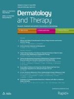
Fig. 1
Vitiligo lesion (20 cm2) in a 21-year-old male before (a) and 18 months after (b) the transplantation of autologous cultured melanocytes. A 90% repigmentation was achieved
×
Case 2
A 16-year-old male was first received in September 2012 complaining of a depigmented lesion on the scrotum with a duration of 3 years. The lesion had not shown any progress in the last 2 years. The area of the lesions was 15 cm2. The patient had no family history of vitiligo. He failed to respond to medical treatment administered to him (topical pimecrolimus used for 6 months, twice daily). The patient underwent transplantation in September 2012. The DOT of the melanocytes was 3.24, and the concentration of melanocytes applied to the recipient area was 0.186 ng/cell. In the latest follow-up in September 2013, the authors found the lesion 95% repigmentation was achieved, and the patient was very satisfied with the cosmetic results (Fig. 2).
Fig. 2
Vitiligo lesion (15 cm2) in a 16-year-old male before (a) and 12 months after (b) the transplantation of autologous cultured melanocytes. A 95% repigmentation was achieved
×
Anzeige
Discussion
The genital is a commonly affected site for vitiligo in men, and it is sometimes the only site affected [7]. The treatment of genital vitiligo is quite complex. There are very few reports in the literature for the treatment of genital vitiligo. Souza Leite et al. [4] reported one case of genital vitiligo successfully treated by pimecrolimus. However, this patient had a short duration of the disease considering the existence of residual melanocytes. Therefore, the use of medication was considered to be a better option. For vitiligo with longer duration or vitiligo without residual pigment in genitals, surgical intervention is recommended. Mulekar et al. [3] reported three cases of focal vitiligo affecting the glans and shaft of the penis successfully treated by antilogous non-cultured melanocyte–keratinocyte cell transplantation. Differing from their cases, the current transplantation site is located on the scrotum, and the scheme used was autologous cultured melanocyte transplantation. Mulekar et al. [3] needed to perform two rounds of surgery to achieve satisfying repigmentation, whereas the current authors achieved 90% repigmentation in case 1. Redondo et al. [5] used a graft of autologous melanocytes cultured on a denuded amniotic membrane to treat a patient with a scrotal lesion and achieved good regimentation. However, the current study did not use a denuded amniotic membrane, and achieved the same effect.
Patients with genital vitiligo often hide their lesions, and postpone their complaints until they miss the best time for treatment with medication. Therefore, the development of surgical interventions is particularly important for the patients with long duration vitiligo. The current study indicates that autologous cultured melanocyte cell transplantation not only provides an opportunity for patients with large areas of stable vitiligo, but can also be used as a very effective treatment for genital vitiligo, including the scrotum.
Acknowledgments
No funding or sponsorship was received for this study or publication of this article. All named authors meet the ICMJE criteria for authorship for this manuscript, take responsibility for the integrity of the work as a whole, and have given final approval for the version to be published.
Conflict of interest
Xiaowen Li, Weisong Hong, and Ai-E Xu have declared that they have no competing interests.
Compliance with ethics guidelines
The Ethical Committee of the National Center for Vitiligo and Psoriasis approved the treatment. The study was approved by the institutional review Board of Third People’s Hospital of Hangzhou, and written informed consent was obtained from each patient for inclusion in this study and for publication of the photographs. All procedures followed were in accordance with the ethical standards of the Helsinki Declaration of 1975, as revised in 2000 and 2008.
Open Access
This article is distributed under the terms of the Creative Commons Attribution Noncommercial License which permits any noncommercial use, distribution, and reproduction in any medium, provided the original author(s) and the source are credited.
Open AccessThis article is distributed under the terms of the Creative Commons Attribution 4.0 International License (https://creativecommons.org/licenses/by/4.0), which permits use, duplication, adaptation, distribution, and reproduction in any medium or format, as long as you give appropriate credit to the original author(s) and the source, provide a link to the Creative Commons license, and indicate if changes were made.
Die Therapie von Echinokokkosen sollte immer in spezialisierten Zentren erfolgen. Eine symptomlose Echinokokkose kann – egal ob von Hunde- oder Fuchsbandwurm ausgelöst – konservativ erfolgen. Wenn eine Op. nötig ist, kann es sinnvoll sein, vorher Zysten zu leeren und zu desinfizieren.
Seit November 2023 gibt es evidenzbasierte Empfehlungen zum perioperativen Management bei gastrointestinalen Tumoren (POMGAT) auf S3-Niveau. Vieles wird schon entsprechend der Empfehlungen durchgeführt. Wo es im Alltag noch hapert, zeigt eine Umfrage in einem Klinikverbund.
Mit dem demographischen Wandel versorgt auch die Chirurgie immer mehr betagte Menschen. Von Entwicklungen wie Fast-Track können auch ältere Menschen profitieren und bei proximaler Humerusfraktur können selbst manche 100-Jährige noch sicher operiert werden.
Worauf kommt es beim Management von Personen mit infektiöser Endokarditis an? Eine Kardiologin und ein Kardiologe fassen die zehn wichtigsten Punkte der neuen ESC-Leitlinie zusammen.
Update Innere Medizin
Bestellen Sie unseren Fach-Newsletter und bleiben Sie gut informiert.