Scleritis refers to a wide spectrum of ocular conditions ranging from mild to sight-threatening scleral inflammation that may compromise visual function and threaten the anatomical integrity of the ocular globe. Most aggressive forms like necrotizing or posterior scleritis are often difficult-to-treat cases, refractory to conventional treatment. The association with systemic diseases, namely rheumatoid arthritis, Sjögren syndrome, granulomatosis with polyangiitis, and relapsing polychondritis, may have prognostic implications as well. A better understanding of the pathogenesis of ocular inflammatory diseases have paved the way to more effective and targeted treatment approaches. In this regard, a growing body of evidence supports the potential role of biologic agents in the management of non-infectious scleral inflammation, either idiopathic or in a background of immune-mediated systemic disorders. Biologic agents such as anti-tumor necrosis factor agents, interleukin-1 and interleukin-6 inhibitors as well as CD20 blockade have displayed promising results. More specifically, several studies have reported their ability to control scleral inflammation, reduce the overall scleritis relapses, and allow a glucocorticoid-sparing effect while being generally well tolerated. Anecdotal reports have also been described with other biologic agents including abatacept, ustekinumab, daclizumab, and alemtuzumab as well as targeted small molecules such as tofacitinib. Further studies are warranted to fully elucidate the role of biologic agents in non-infectious scleritis and investigate specific areas with the aim to administer treatments in the context of personalized medicine. This review summarizes the available data regarding clinical trials, small pilot studies, and real-life experience of the last two decades reporting the use of biologic agents in the management of non-infectious scleritis.
Key Summary Points
This review summarizes the available evidence regarding the management of refractory non-infectious scleritis with biologic agents.
Non-infectious scleritis, particularly in its most aggressive forms such as necrotizing scleritis and posterior scleritis, may lead to visual impairment and severe sight-threatening ocular sequelae.
Biologic agents have shown to control scleral inflammation, reduce the overall scleritis relapses, and allow a glucorticoid-sparing effect.
Management of scleritis must take place in a multidisciplinary setting in order to maximize treatment benefits while minimizing safety concerns.
Introduction
The term scleritis includes a wide spectrum of clinical entities characterized by inflammation of the sclera. This clinical condition, particularly in its most aggressive forms such as necrotizing scleritis and posterior scleritis, may lead to visual impairment and severe sight-threatening ocular sequelae. Indeed, permanent scarring of the adjacent inflamed ocular structures may compromise visual function and result in anatomical damage as well. Severe and untreated cases may lead to perforation, which is among the most feared complications since it may be responsible for eye loss [1]. The overarching principles of its treatment consist in controlling the ocular inflammation and inducing long-term remission, thus preventing further sight-threatening complications and structural irreversible ocular impairment. Oral glucocorticoids (GCs) remain the mainstay of the short-term management of scleritis, while pulse intravenous steroids are reserved in sight-threatening emergencies requiring a rapid control of inflammation [2, 3]. Subconjunctival GC injections may be used as an adjunct therapy to systemic GCs to control acute exacerbation of unilateral non-infectious non-necrotizing scleral inflammation [4]. In recalcitrant and most aggressive cases of non-infectious scleritis, several biologic agents have been used to suppress ocular inflammation and reduce the burden of systemic GCs [5‐11]. Despite the revolution brought by biologic agents in many medical specialties, only an exiguous number of clinical trials, retrospective case series, or isolated case reports have been reported in literature regarding their employment in the treatment of non-infectious scleritis. Therefore, treatment guidelines or specific management protocols that include systemic immunosuppressive drugs are currently not available because of the lack of evidence-based data. We reviewed the current literature regarding the efficacy and safety of biologic agents in refractory non-infectious scleritis, with the aim to summarize the real-life experience in the management of this sight-threatening condition.
Anzeige
Research Strategy
A comprehensive literature research via Pubmed was performed and included articles published from June 2001 to June 2021. The following terms were searched in Medical Subjects Headings and/or as entrée words: “scleritis” and “biologic agents”. Thereafter, the name of each drug deemed relevant to the management of scleritis was searched together with the word “scleritis”. In particular, the following items were employed: “tumor necrosis factor inhibitors”, “adalimumab”, “infliximab”, “etanercept”, “golimumab”, “certolizumab”, “rituximab”, “ofatumumab”, “ocrelizumab”, “anakinra”, “canakinumab”, “tocilizumab”, “sarilumab”, “siltuximab”, “sirukumab”, “olokizumab”, “clazakizumab”, “abatacept”, “ustekinumab”, “secukinumab”, “ixekizumab”, “daclizumab”, “alemtuzumab”, “interferon”, “interferons” “baricitinib”, “tofacitinib”, “filgotinib”, and “upadacitinib”, each combined with “scleritis”. Case series, observational studies, and clinical trials were reviewed to find eligible papers according to the goal of our manuscript. Subsequently, relevant references cited in the eligible papers were searched by hand. This article is based on previously conducted studies and does not contain any new study with human participants or animals performed by any of the authors.
Tumor Necrosis Factor Inhibitors
Adalimumab
The first case reports and small case series of non-infectious scleritis successfully treated with adalimumab (ADA) [12‐16] were followed by retrospective studies assessing its efficacy in larger samples of refractory patients [7, 11, 17‐19]. Ragam et al. conducted a retrospective chart review of 17 patients with non-infectious, non-necrotizing scleritis and found that monoclonal anti-tumor necrosis factor alpha (TNFα) were able to reduce scleral inflammation and to concomitantly reduce the use of systemic GCs. More specifically, 10 patients were initially treated with infliximab (IFX) and seven with ADA. Control of active inflammation for at least 2 months was achieved in 15 out of 17 patients. The total rate of inflammatory control on ADA which included seven patients on first trial and two patients after IFX failure was 67% [7]. Moderate control of inflammation and a GC-sparing effect were also observed in nine patients with scleritis in a study investigating ADA efficacy. Almost half of the eyes achieved control of inflammation during follow-up. In addition, cystoid macular edema resolved in the majority of cases [17]. In their real-world prospective analysis, Sharma et al. found TNFα inhibitors ADA and IFX to be successful in several ocular inflammatory diseases. A sustained remission was noted in 91% of the cases, of whom four were diagnosed with non-infectious scleritis. Further detailed data on scleritis were not provided. These findings supports the long-term efficacy of TNF inhibitors and their GC-sparing effect [18]. We recently reported 19 patients diagnosed with non-infectious scleritis and treated with monoclonal TNFα blockers, mainly ADA (n = 13), followed by IFX (n = 5) and golimumab (GOL) (n = 1). Monoclonal TNFα inhibitors were shown to significantly reduce relapses as well as to control scleral inflammation both rapidly and with a long-lasting effect. Moreover, they allowed a GC-sparing effect and were associated with a good safety profile [11]. Successful results with TNFα inhibitors have also been reported in three patients affected by relapsing polychondritis (RP) [20]. Table 1 shows the retrieved studies regarding TNF inhibitors employed in the management of non-infectious scleritis and some of their respective findings.
Table 1
Anti-tumor necrosis factor therapy in the treatment of non-infectious scleritis
Successful control of inflammation. Improvement in visual acuity. Remission on 4 years. Mild relapse 6 months after IFX cessation responsive to ibuprofen 400 mg 3 times daily
IFX and ADA were effective in 1 and 2 patients, respectively. Two patients developed scleritis after IFX therapy and 1 of them was administered ADA and ETN without success. Finally CZP pegol was given, with no further relapses of scleritis
Control of active inflammation for at least 2 months was achieved in 88% of patients. Five (29%) patients ended up switching from one TNFα inhibitor to another
Stable visual acuity. No significant differences between ADA and IFX
Allergic reaction to a single infusion of IFX (n = 1). Transaminitis
After the third IFX infusion, the corneal ulceration stopped, and conjunctival hyperemia and ocular discomfort were relieved. Systemic immunosuppression tapered after 3 months
Favorable response in 90% of the patients, with 6 of them achieving remission and cessation of concomitant immunosuppression. Monthly infusions may be required to maintain remission
All patients achieved control of active scleritis within 14 weeks. One patient with GPA developed new-onset intraocular inflammation after 14 weeks. Clinical resolution in 4 out of 5
Ear infection with transient decreased hearing, UTI, lower RTI, and facial rash (n = 1)
UTI, diarrhea, upper RTI, nasal congestion and headache, mouth sores, head tremor, occasional numbness and tingling in extremities (n = 1)
IFX had a partial efficacy on ocular disease and no impact on intestinal symptoms. Two months after cyclophosphamide infusions the patient had a recurrence and was treated with ADA with no further relapses
Clinical relief in 10 days following the first IFX infusion. Reduction of scleral swelling on B-mode ultrasound examination and regression of papillitis at fundoscopy. Complete remission at 4 months
Nine out of 10 patient were classified as responders and 1 as partial responder requiring alkylating agent therapy. Five patients were able to reduce their concurrent immunosuppressive therapy. Three patients were able to remain relapse-free while not taking any medication
Dramatic response after three infusions of IFX. Improvement of visual acuity. Relapse upon reduction of prednisolone at 12.5 mg daily. Clinical resolution after increased dosages from 3 mg/kg to 5 mg/kg of IFX
Three out of 4 patients achieved remission. One patient showed a partial response but treatment was withdrawn. Repeated infusions were required to maintain remission
ETN proved to be effective in resolving scleritis and sterile corneal ulcerations by controlling inflammation, arresting tissue ulceration, and permitting in many cases tapering or cessation of immunosuppressive therapies
Development of scleritis 18 months after ETN treatment, treated with local and systemic GC, local cyclosporine and azathioprine. ETN was continued during disease course
Scleritis developed in 3 patients under ETN therapy, 1, 2, and 6 months respectively. In the remaining 3 patients, ocular disease was not influenced by TNF blockade (n = 1), remained quiescent (n = 1), or was minimally active (n = 1),
Paradoxical scleritis (n = 3)
–
ADA adalimumab, AE adverse event, AS anterior scleritis, AU anterior uveitis, BCVA best corrected visual acuity, BS Behçet’s syndrome, CD Crohn’s disease, CME cytoid macular edema, CR case report, CS case series, CT clinical trial, CZP certolizumab, ETN etanercept, GC glucocorticoid, GD Grave’s disease, GPA granulomatosis with polyangiitis, GOL golimumab, IFX infliximab, N° number of patients with scleritis, NAS necrotizing anterior scleritis, NeS necrotizing scleritis, NR not reported, pANCA perinuclear antineutrophil cytoplasmic antibody, PS posterior scleritis, PsA psoriatic arthritis, PU posterior uveitis, RP relapsing polychondritis, RS retrospective study, RTI respiratory tract infection, SpA spondyloarthritis, TNF tumor necrosis factor, UTI urinary tract infection
Infliximab
Similarly to ADA, IFX may be considered as a viable option in patients with refractory scleritis. The only prospective evidence regarding IFX employment in non-infectious scleritis dates back to 2009. In this prospective open-label pilot study, all patients met the primary outcome by achieving quiescence of their active scleritis by week 14 without associated conventional immunosuppressants. Additionally, four out of five patients achieved a sustained quiescence on treatment and 60% were able to successfully taper prednisone to daily doses lower than 10 mg [21]. These promising data were further corroborated by real-world experience in relatively large samples of non-infectious scleritis. Sobrin and colleagues found IFX therapy to be effective for the treatment of refractory ocular inflammation, with a low incidence of adverse events (AEs) prompting discontinuation. Nine out of 10 patient were classified as responders and one as a partial responder requiring alkylating agent therapy. Five patients were able to reduce their concurrent immunosuppressive therapy and three patients with scleritis were able to remain relapse-free despite having discontinued all medications [22]. Almost identical results were reported by another retrospective study demonstrating the efficacy of TNFα blockade with IFX in refractory scleritis. A favorable response was seen in 90% of the patients, with six of them achieving remission and cessation of concomitant immunosuppressants. Dosing intervals shorter than 8 weeks were found to be well tolerated and avoid inflammatory relapses that may occur during the last days of a standard interval before the next infusion [5]. In fact, increasing the dose or shortening the interval between doses is sometime required to control scleral inflammation [5, 22, 23].
To this end, patients should be closely monitored for potential AEs. Lupus-like reactions have been described in patients treated with IFX for severe scleritis [5, 22]. Patients treated with IFX may also develop new ocular inflammations defined as a paradoxical reaction. Possible mechanisms underlying such reactions may include interferon overproduction, Th17 cell expansion, and Th1 homing due to increased chemokine levels and receptors induced by interferon-α [24].
Anzeige
Murphy et al. reported seven IFX-treated patients with non-infectious ocular inflammatory disease refractory to conventional immunosuppression, of whom four were diagnosed with scleritis. Three out of four achieved remission, with the remaining patient showing a partial response but withdrawn from treatment because of an infusion-related reaction. Given its rapid action, the authors suggested IFX as a valid substitute for high-dose prednisolone as rescue therapy during ocular relapses [25]. A small series of three patients affected by idiopathic necrotizing anterior scleritis showed an excellent efficacy of IFX characterized by a complete clinical response as well as resolution of choroidal and retinal detachment [26]. Fifteen additional single-patient reports were retrieved [27‐41], and their respective findings are reported in Table 1. IFX has proven to be effective in the treatment of refractory scleritis also associated with rare disorders such as RP [33, 43], granulomatosis with polyangiitis (GPA) [29, 39], and surgically induced necrotizing scleritis [32] as well as in pediatric patients under appropriate circumstances [31, 39, 41].
Direct comparisons between ADA and IFX are currently limited. Ragam et al. found that the overall likelihood of achieving inflammation control on ADA and IFX was 66% and 77%, respectively, without any significant differences [7]. The lack of robust data does not allow one to establish the superiority of one agent over the other in the therapeutic approach of non-infectious scleritis. Moreover, in-class switching is a suitable solution in case loss of efficacy or safety concerns arise [7, 18].
Etanercept
Etanercept (ETN), either in association with conventional immunosuppressants or as monotherapy, has provided encouraging results in the early 2000s for the management of diffuse or necrotizing anterior scleritis as well as sterile peripheral ulcerative keratitis (PUK). Particularly, ETN was shown to control inflammation and arrest tissue ulceration while permitting a drug-sparing effect in 10 patients affected by non-infectious scleritis or PUK [42]. Its use, however, has lost popularity over time given the reported paradoxical effects on ocular inflammatory diseases. Indeed, several studies have disclosed controversial results of ETN in the management of such conditions. The first cases of paradoxical ocular inflammation were reported by Smith and colleagues. Three patients with RA developed bilateral scleritis for the first time, 1, 2, and 6 months after commencing ETN, respectively [43]. Another study performing direct comparisons between IFX and ETN reported two other patients with RA developing their first episode of anterior scleritis after ETN initiation. Furthermore, all ETN-treated patients in the study eventually required a change in medication to control ocular inflammation. The authors found IFX to be superior to ETN in the treatment of ocular inflammation and in decreasing the use of topical GCs [44]. Other single-patient reports and small case series of patients with RA have reported similar findings, raising the dilemma of whether to continue treatment with ETN despite an optimal control of extraocular manifestations [45‐48]. To the best of our knowledge, a total of 13 patients with ETN-related paradoxical scleritis, all affected by RA, have been described so far. The underlying mechanisms still need to be clarified, but a possible pathogenetic hypothesis may explain the tendency of TNF inhibition to precipitate an ocular inflammatory disease in predisposed individuals. First of all, TNF inhibition may interfere with apoptosis, which is believed to play a crucial role in the maintenance of ocular immune privilege [49]. Secondly, pharmacodynamic peculiarities may become relevant in specific situations. For instance, when TNFα levels are low, p75 TNF receptor, which possesses a fivefold higher affinity than p55, facilitates p55 activity possibly by a ligand-passing mechanism or as a result of intracellular kinase activation. Therefore, ETN, a soluble P75 TNF receptor, may interfere with immune homeostasis and influx of inflammatory cells in the eye [47]. Further data are required to draw firm conclusions in such a controversial topic.
Golimumab
Experience with golimumab (GOL) in treating non-infectious scleritis relies on rare single-patient reports [11, 50, 51]. GOL efficacy was described in a middle-aged woman affected by psoriatic arthritis and bilateral progressive necrotizing posterior scleritis associated with retinal vasculitis refractory to methotrexate, cyclosporine, and ADA treatment. Following the introduction of GOL, good control of the scleritis and arthritis without any relapse or progression of retinal lesions was described [50]. GOL proved to be rapidly effective in controlling scleral inflammation also in a patient affected by necrotizing scleritis and advanced glaucoma requiring Ahmed valve implant. Resolution of scleritis within 2 months allowed the insertion of an Ahmed valve in this patient, avoiding the high risk of globe perforation due to scleral thinning and a post-surgical relapse of scleritis [51].
Certolizumab Pegol
No comprehensive studies describing the treatment of scleritis with certolizumab have been reported so far, although a single case report described its efficacy in a case of rheumatoid arthritis (RA)-related bilateral scleritis refractory to ETN and following withdrawal of IFX because of AEs [52].
Anti-CD20 Therapy
Rituximab
Since the first reports of rituximab (RTX) efficacy in scleritis associated with systemic disorders [53, 54], a growing body of evidence in the management of refractory scleritis with RTX has emerged in the last 15 years. The only prospective, randomized, phase I/II clinical trial published so far disclosed an optimal RTX efficacy and safety profile in a large proportion of patients affected by refractory non-infectious scleritis, with nine out of 12 achieving a reduction of scleritis grading scale within 24 weeks. More than half of them required re-treatment to maintain inflammatory control. Interestingly, the authors did not find notable differences in terms of efficacy, safety, and B cell depletion between patients who received 500 mg and those treated with 1000 mg, suggesting that treatment regimens with lower dosages may be therapeutically equivalent to the standard protocol employed in many rheumatologic diseases [6]. However, the vast majority of the available data relies on real-life uncontrolled scenarios. Joshi and coworkers conducted a retrospective analysis of 37 patients with GPA treated with RTX, of whom 20 were diagnosed with scleritis. RTX was shown to induce complete and partial remission in 85% and 10% of patients, respectively, at 6 months [55]. Regarding cyclophosphamide use in GPA-related scleritis, RTX appeared to be superior in terms of safety and efficacy, with less need for treatment interruptions and dose adjustments, although both therapies proved to be effective and safe [56]. RTX was administered according to the rheumatologic protocol in eight patients with GPA-related refractory scleritis with optimal results [57]. A different posologic regimen may be required to maintain GC-free drug-induced remission in recalcitrant scleritis [8]. Yet, no firm conclusions can be drawn in this regard as the small case series reported do not allow head-to-head comparisons for the different protocols employed. A second cycle of RTX may be administered in case of relapse [6, 56, 57]. In fact, a considerable proportion of patients with GPA and scleritis have experienced relapses, thus advocating for a maintenance treatment instead of a single course of RTX that should be extended for at least 18 months in order to lower the relapse rate [58]. Morarji and colleagues described a unique and successful combination therapy of RTX with IFX. The patient affected by GPA received IFX during early stages whilst waiting for RTX to establish disease remission and, as a result, avoiding cyclophosphamide in the acute phase [59]. Potential usefulness of IFX in gaining rapid disease control and allowing time for RTX to take effect, at least in some patients with necrotizing scleritis, may be interesting to explore while controlling for AEs. A case of Pneumocystis jirovecii pneumonia after RTX therapy and prednisolone for posterior scleritis has been described [60]. Additionally, paradoxical ocular manifestations including cystoid macular edema following RTX administration were also reported [61].
Along with its efficacy in most patients with localized and generalized ocular GPA, RTX is an effective treatment modality for recalcitrant non-infectious scleritis either idiopathic or associated with different systemic inflammatory disorder. It may also provide long-term drug-free remission in some patients. In a relatively large cohort of patients, 14 out of 15 (93.3%) showed an initial improvement, and a subgroup analysis among those with longer follow-up disclosed a success rate of 61% [8]. Table 2 lists all studies published so far where RTX has been employed to treat non-infectious scleritis and details some data regarding ocular and systemic diagnosis, main findings, mean follow-up as well as safety profile if reported. Briefly, most patients with scleritis treated with RTX were affected by GPA (n = 68), followed by RA (n = 14) and idiopathic cases (n = 13). Most patients were diagnosed with diffuse anterior scleritis (n = 36), followed by necrotizing anterior scleritis (n = 22), posterior scleritis or panscleritis (n = 11), and nodular anterior scleritis (n = 5). However, the anatomical pattern was not specified in a considerable proportion of cases (n = 30). The first study of scleritis associated with RA and successfully treated with RTX dates back to 2009. The authors described three patients refractory or intolerant to conventional immunosuppressants or TNF inhibitors, suggesting a special consideration for RTX in inflammatory eye diseases secondary to RA or other rheumatologic disorders [62]. Other interesting case reports and small case series have also been reported [19, 63‐72]. RTX yielded a rapid benefit in an interesting case of severe and refractory scleritis associated with IgG4-related disease and initially misdiagnosed as idiopathic scleritis, advocating for a potential role of CD20 blockade as a therapeutic option in such cases [73]. Anecdotal evidence has been disclosed for RTX efficacy in scleritis diagnosed in the context of chronic lymphatic leukemia [74], lymphocytic hypophysitis [75], and surgically induced scleritis [76]. RTX appears to be a promising agent in the management of refractory scleritis in both idiopathic scleritis and scleritis associated with immune-mediated systemic disorders. Its administration, either as monotherapy or in combination with GCs or immunosuppressive agents, has shown a favorable ocular outcome. It would be tempting to test RTX efficacy as a first-line immunosuppressive therapy instead of a last resort option in the classic stepwise/stepladder approach in selected young patients with a particular severe form [67, 72].
Table 2
CD-20 blockade with rituximab in patients with non-infectious scleritis
Clinical resolution, improvement or stabilization of visual acuity (expressed in logMAR) in 79% of the eyes. One patient experienced relapse and was re-treated with a second cycle or RTX. Four eyes with NeS required scleral-patch grafting
All eyes achieved remission with RTX maintenance treatment. Reduction of the modified McCluskey scale. GC-sparing effect. Mild reduction of visual acuity expressed in logMAR. No differences in intraocular pressure between baseline and last follow-up visit
Herpes zoster (n = 1), cytopenia with bronchitis and bacterial pneumonia (n = 1)
Favorable response in 14 patients (93.3%) and significant improvement of McCluskey grading scale for scleritis activity score at 6 months. Steroid-free remission
Resolution of signs and symptoms on ophthalmological evaluation and GC-sparing effect
NR
7
AS anterior scleritis, AU anterior uveitis, BCVA best corrected visual acuity, CLL chronic lymphocytic leukemia, CME cystoid macular edema, GC glucocorticoid, GPA granulomatosis with polyangiitis, IFX infliximab, IgG immunoglobulin G, IOI idiopathic orbital inflammation, N° number of patients with scleritis treated with rituximab, NAS necrotizing anterior scleritis, NeS necrotizing scleritis, NR not reported, NS not specified, OG orbital granulomatosis, PS posterior scleritis, PUK peripheral ulcerative keratitis, RA rheumatoid arthritis, RTX rituximab, TFIL tumefactive fibroinflammatory lesion, UMCTD undifferentiated mixed connective tissue disorder, VA visual acuity
IL-1 Inhibitors
Interleukin (IL)-1 plays a key role in the pathogenesis of different inflammatory and degenerative eye diseases and its overexpression might be an initiating factor for many immunopathologic scenarios in the eye, as proven by the efficacy of the specific IL-1 blockade in different ocular conditions [77].
Anzeige
Anakinra
Botsios et al. described two patients affected by unilateral refractory non-infectious anterior scleritis related to RA and treated with anakinra (ANA) in combination with methotrexate 10 mg/week over a follow-up period of 3 years (patient 1) and 1 year (patient 2). Former IFX and ETN improved the articular domain, but did not ameliorate scleral inflammation. Both patients experienced a dramatic resolution of scleritis signs and symptoms within 6–8 weeks [78]. Treatment with the IL-1 receptor antagonist ANA was later employed in 10 consecutive patients affected by severe and refractory non-necrotizing scleritis. Seven patients presented bilateral anterior non-necrotizing diffuse scleritis while three patients manifested unilateral disease either with anterior or posterior involvement. Ninety percent of the patients were considered as complete responders at the end of follow-up. Remission, defined as resolution of scleral inflammation, edema, and pain, occurred after 1 month of ANA treatment in eight patients and after 2 months in one patient. Interestingly, a drug-sparing effect was recorded with a significant reduction of GC daily intake as well as discontinuation of conventional immunosuppressants in all but one. In addition to a rapid improvement of scleral inflammation within 1 month in almost all patients, the authors described a good safety profile with only four mild AEs that did not require ANA discontinuation [10]. We recently reported 14 patients with scleritis treated with biologic agents other than TNFα inhibitors. Three patients were administered ANA 100 mg/day subcutaneously. One of them has been receiving ANA for 44 months with resolution of scleral inflammation and scleritis relapses [79].
Gevokizumab
Blockade of IL-1β with gevokizumab has been shown to provide benefits in treating active, non-infectious, anterior scleritis. More extensively, gevokizumab has been used in a phase I/II clinical trial where seven out of eight patients with active non-infectious, non-necrotizing scleritis met the primary outcome. Seven eyes from seven patients exhibited at least a two-step reduction or reduction to grade 0 in scleral inflammation on a 0 to +4 scale according to a standardized photographic scleritis grading system within a median time of 2 weeks. Moreover, a good safety profile was reported [10].
Anti-IL-6 Therapy
Tociluzimab
Pathogenetic evidence linking IL-6 with intraocular inflammation has paved the way for newer treatment strategies. Indeed, manipulation of IL-6 in the inflammatory cascade of immune-driven uveitis has yielded encouraging results [80]. However, data on scleritis are still scarce and IL-6 inhibition in this sight-threatening disorder is still based on small case series and single-patient reports. Tode et al. [81] reported the first case of successful anti-IL6 treatment in refractory unilateral anterior necrotizing scleritis. The patient showed a reduction in scleritis grade and the necrotic process ceased. Scleritis remission was achieved after six combined steroid and tocilizumab (TCZ) infusions. Nearly a year later, a retrospective case series of 17 patients, six of whom were affected by recalcitrant non-infectious scleritis (anterior scleritis n = 5, panscleritis n = 1, eight eyes), was published. Five patients were diagnosed with an underlying systemic disease. At 6- and 9-month follow-up, 50% of patients with scleritis achieved control of inflammation as well as a steroid-sparing effect. Additionally, TCZ was able to control disease activity in one patient with scleritis and inflammatory bowel disease for almost 3 years before being discontinued because of secondary failure. Interestingly, in a subgroup analysis, time to inflammatory control of scleritis was found to be shorter than that of the patients affected by uveitis with a median time of 50 days and 140 days, respectively [82]. Since then, three other cases have been reported by different authors. Poelman et al. [83] described a case of a scleritis associated with biopsy-proven giant cell arteritis rapidly and persistently responsive to TCZ. We reported two patients treated with TCZ for 3 and 28 months, respectively, achieving resolution of the diffuse anterior scleritis in the affected eyes [79].
TCZ seems to be a promising treatment also for RP-related scleritis either as a first-line immunosuppressive treatment or in refractory patients unresponsive to conventional immunosuppressants and/or other biologic agents. A recent retrospective study of consecutive cases of scleritis and sclerouveitis showed that they optimally responded to TCZ after failing several conventional disease modifying anti-rheumatic drugs (cDMARDs) and biologic agents. Three patients clinically diagnosed with RP and recalcitrant anterior diffuse or nodular scleritis (five eyes) received monthly treatment with TCZ at a dose of 8 mg/kg. This approach resulted in complete and sustained ocular and extraocular remission up to 2 years, while allowing a steroid-sparing effect [84]. Another patient with RP-related scleritis and treated with TCZ has been reported. The report describes a case with bilateral scleritis and auricular chondritis refractory to GCs, methotrexate, and IFX that was successfully treated with TCZ [85]. Paradoxical reactions might also be possible. Anterior nodular scleritis following the second dose of TCZ therapy and responsive to orally administered prednisone was diagnosed in a patient with rheumatoid arthritis, pyoderma gangrenosum, and systemic lupus erythematosus. Hence, ocular manifestations following IL-6 inhibition, although rare, should be considered as potential AEs when treating rheumatic patients with inflammatory arthritis [86].
Anzeige
Other Biologics Agents and Targeted Small Molecules
Abatacept
Fabiani et al. employed co-stimulatory signal modulation with abatacept in patients with scleritis affected by RA. Two of three patients (four eyes) with active diffuse anterior scleritis achieved resolution of ocular inflammation at last follow-up visit [79]. Abatacept efficacy has also been reported in other ocular inflammatory conditions such as PUK [87] and in juvenile idiopathic arthritis-related uveitis also refractory to TNFα inhibitors [88]. Further prospective and well-designed studies are warranted to confirm these preliminary findings.
Ustekinumab
Ustekinumab is a human monoclonal antibody directed against the shared p40 subunit of IL-12 and IL-23 licensed for psoriasis, psoriatic arthritis, Crohn’s disease, and ulcerative colitis. Currently, no randomized clinical trials describing the effects of ustekinumab on scleritis have been reported. Its efficacy was described in a case of spondyloarthritis associated with Crohn’s disease where also extraintestinal manifestations, including scleritis, resolved shortly after the drug was initiated [89].
Daclizumab
Daclizumab is a humanized monoclonal antibody acting against the α-subunit-CD25 of the IL-2 receptor. In particular, it recognizes the high-affinity protein Tac-p55 of the IL-2 receptor and inhibits IL-2 signaling on activated T cells and thus has the potential to restore homeostasis of a dysregulated immune system. Daclizumab was shown to improve inflammation in one of two patients with scleritis in a retrospective case series analyzing its efficacy in various ophthalmologic inflammatory conditions refractory to standard therapy [90]. In March 2018, this drug was withdrawn from the market because of reports of autoimmune encephalitis.
Alemtuzumab
Kommaraju and colleagues reported a case of T cell prolymphocytic leukemia with central nervous system involvement that presented with bilateral diffuse anterior scleritis and anterior uveitis with secondary glaucoma. Systemic treatment with alemtuzumab and intrathecal therapies with alternating cytarabine/hydrocortisone and methotrexate/hydrocortisone improved the lymphadenopathy while resolving the pleural effusion and the ophthalmologic manifestations within 1 month. However, unresponsive central nervous system involvement was responsible for the fatal outcome 19 months thereafter [91].
Anzeige
Tofacitinib
Intracellular signaling pathways, such as those concerning Janus kinase (JAK), have emerged as a key axis in the cytokine network and, consequently, as important therapeutic targets. Tofacitinib is an oral reversible JAK inhibitor classified among targeted small molecules [92]. Its effectiveness in refractory scleritis has been suggested in single-patient reports [79, 93], including cases with necrotizing scleritis [94]. The underlying biologic rationale relies on the tight interplay between JAK/signal transducers and activators of transcription (STAT) intracellular pathways and inflammatory cytokines such as TNF and IL-6 which are involved in the pathogenesis of ocular inflammatory diseases. Paley et al. described two cases of recalcitrant uveitis and scleritis responsive to tofacitinib after failure or intolerance to multiple immunomodulatory regimes. One patient with scleritis responded within 3 weeks of tofacitinib treatment [93]. A similar time to response was also observed in an Indian patient affected by necrotizing scleritis [94]. Patients who are refractory to treatment with oral GCs and immunomodulatory drugs including biologic agents can benefit from this new class of small molecules that may therefore be employed as effective GC-sparing agents. However, further studies are warranted to confirm these anecdotal findings.
Table 3 provides all the available studies with some of their respective main findings regarding IL-1 inhibitors, TCZ, and other biologic agents as well as small molecules employed in non-infectious scleritis.
Table 3
Anti-IL-1 agents, tocilizumab, ustekinumab, abatacept, daclizumab, and alemtuzumab treatment in non-infectious scleritis
Clinical remission in 8 patients within 1 month and in one additional patient within 2 months, significant decrease in ocular relapse rate, GC-sparing effect
ABA abatacept, AE adverse event, ANA anakinra, AS anterior scleritis, FMF familial Mediterranean fever, GC glucocorticoid, GPA granulomatosis with polyangiitis, ISR injection-site reaction, NR not reported, NS not specified, PS posterior scleritis, PUK peripheral ulcerative keratitis, RA rheumatoid arthritis, RP relapsing polychondritis, SLE systemic lupus erythematosus, SS Sjögren’s syndrome, TCZ tocilizumab, TFC tofacitinib, VA visual acuity
Closing Remarks
The management of non-infectious scleritis may be challenging since its immunopathogenesis is not completely understood. Both innate and adaptive immunity may be advocated as the driving mechanisms leading to tissue damage, with a pivotal role played by inflammatory cytokines such as TNFα and IL-1, which in turn induce the secretion of matrix metalloproteinases from infiltrating inflammatory cells and stromal scleral fibroblasts [1]. Despite being hard to pinpoint where non-infectious scleritis is exactly situated in the immunological disease continuum [95], it is safe to assume that the pathophysiological mechanisms are largely dependent on the underlying systemic disease, if present. Indeed, many authors believe that non-infectious scleritis associated with systemic disorders represents an immune complex-mediated condition, whereas idiopathic scleritis may arise after a delayed hypersensitivity reaction [96]. The association with systemic diseases may have prognostic implications as well. Berkenstock and Carey conducted the largest retrospective review currently described in the literature to evaluate the association between scleritis and autoimmune entities. They reviewed 5.9 million charts of the electronic medical record and found 2702 patients affected by scleritis, of which approximately a third (31.4%) had an underlying autoimmune disease [97]. In this analysis, the most frequent associated autoimmune condition was RA (6.8%) followed by HLA-B27-related diseases (5.7%) and Sjögren/Sicca syndrome (4.5%), gout (3.5%), lupus (3.0%), and granulomatosis with polyangiitis (1.7%). Scleritis associated with systemic conditions, especially those affected by ANCA-associated vasculitis, appears to have a worse visual prognosis [98], making early diagnosis a crucial step for its successful management. The underlying systemic disease may also be considered as a driving factor in the choice of the more appropriate biologic for the management of scleritis. For instance, RTX may be particularly effective in the treatment of scleritis associated with systemic vasculitis. A better understanding of the pathogenesis of ocular inflammatory diseases and the development of new molecules have enriched the therapeutic armamentarium and created more effective treatment approaches. Anti-TNFα monoclonal antibodies, IL-1 and IL-6 inhibitors as well as anti-CD20 RTX were shown to control scleral inflammation and reduce the overall scleritis relapses, thus allowing a GC-sparing effect. Visual acuity is generally preserved and few adverse reactions have been reported [7, 8, 10, 11, 79]. Given the lack of robust evidence-based data, with the available literature reported so far and based on our personal experience, we suggest that TNFα inhibitors such as ADA or IFX should be trialed as first-line biologic agents unless the specific underlying systemic disease warrants otherwise. Non-responsive patients and/or those suffering from RA may benefit from IL-1 inhibitors or RTX. Other biologic agents including abatacept and ustekinumab or JAK inhibitors may be useful additions to the treatment armamentarium for refractory scleritis.
Another key aspect to consider is the potential difference between biologic monotherapy and combination therapy with cDMARDs. It is indeed still unclear whether concomitant treatment with cDMARDs may add clinical benefit and ultimately improve the overall prognosis of patients with scleritis. In a retrospective study of 17 patients with non-infectious, non-necrotizing scleritis treated with TNFα inhibitors, 11 patients were found to fail previous therapies with cDMARDs and 12 of them were receiving combination therapy while five were able to achieve clinical response with an TNFα inhibitor alone [7]. Sobrin et al. reported five out of 10 patients with scleritis treated with IFX being able to reduce or discontinue concurrent treatment with cDMARDs [22]. Other studies have reported different proportions of patients requiring concomitant cDMARDs, ranging from 10% [9] to more than 70% [11]. Cao et al. [8] described 13 out of 15 patients with scleritis achieving a scleritis grading score of 0 at 6 months during treatment with RTX: six patients using RTX in monotherapy and seven as combination therapy with another agent. Conversely, Joshi et al. found RTX to be effective in achieving remission in patients with GPA, but all of them required a maintenance treatment with cDMARDs [55]. Most of the patients receiving biologic agents or small molecules were previously treated with cDMARDs. Given the lack of response to monotherapy with cDMARDs, it can be safely assumed that the subsequent treatment with biologic agents was the one determining the improvement/resolution of scleritis. However, treatment should be ideally tailored according to the patient’s profile. Patients diagnosed with scleritis and an underlying associated systemic immune disease or those with a particularly active and severe disease may require an aggressive combination therapy to avoid long-term ocular and extraocular complications (Fig. 1). Studies specifically investigating this crucial endpoint are needed to fully explore the efficacy of monotherapy with biologic agents and their ability to decrease the immunosuppressive load.
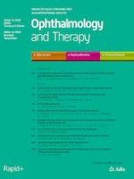
Fig. 1
Posterior and anterior unilateral (right eye) refractory scleritis in a patient affected by psoriatic arthritis undergoing subcutaneous treatment with monoclonal TNF inhibitor golimumab 50 mg every 28 days. Ocular ultrasonography (d) shows a circular acoustically hollow area called the ring sign corresponding to an edematous Tenon capsule (*), typical signs of an active posterior scleritis. Optical coherence tomography (OCT) scans show the presence of a concomitant subfoveal exudative neuroepithelium detachment (°) in the same eye (a) that decreased following 2 months of biologic treatment along with a short course of oral glucocorticoids (b), and totally disappeared after 4 months of treatment (c). Anterior segment photograph (e) shows the concomitant active anterior scleritis inflammation and its resolution after 2 months treatment (f)
×
Aggressive and early treatment is warranted to achieve rapid remission, prevent relapses visual acuity, and ultimately avoid long-term structural irreversible complications. Management of scleritis must take place in a multidisciplinary setting in order to maximize treatment benefits while minimizing safety issues. A future goal would be to tailor treatment strategies according to each patient’s needs and ideally associate patients’ ocular and systemic profiles with specific cytokine inhibitors. Biologic agents and small molecules represent a milestone for scleritis treatment but deserve further investigation in properly designed and powered studies to provide more robust evidence in terms of efficacy, optimal dosage regimen, and hopefully address some of the unmet needs in such a sight-threatening condition.
Acknowledgements
Funding
No funding or sponsorship was received for this study or publication of this article.
Authorship
All named authors meet the International Committee of Medical Journal Editors (ICMJE) criteria for authorship for this article, take responsibility for the integrity of the work as a whole, and have given their approval for this version to be published.
Authorship Contributions
Claudia Fabiani conceived the study design. Jurgen Sota and Matteo-Maria Girolamo performed the literature research. Claudia Fabiani, Jurgen Sota and Matto-Maria Girolamo retrieved the suitable articles and wrote the first draft. All authors critically revised the manuscript.
Compliance with Ethics Guidelines
This article does not include new studies with human participants conducted by any of the authors.
Disclosures
Jurgen Sota, Matteo-Maria Girolamo, Bruno Frediani, Gian Marco Tosi, Luca Cantarini and Claudia Fabiani have nothing to disclose.
Data Availability
Data sharing is not applicable to this article as no datasets were generated or analyzed during the current study.
Open Access This article is licensed under a Creative Commons Attribution-NonCommercial 4.0 International License, which permits any non-commercial use, sharing, adaptation, distribution and reproduction in any medium or format, as long as you give appropriate credit to the original author(s) and the source, provide a link to the Creative Commons licence, and indicate if changes were made. The images or other third party material in this article are included in the article's Creative Commons licence, unless indicated otherwise in a credit line to the material. If material is not included in the article's Creative Commons licence and your intended use is not permitted by statutory regulation or exceeds the permitted use, you will need to obtain permission directly from the copyright holder. To view a copy of this licence, visit http://creativecommons.org/licenses/by-nc/4.0/.
In der MONARCHE-3-Studie lebten Frauen mit fortgeschrittenem Hormonrezeptor-positivem, HER2-negativem Brustkrebs länger, wenn sie zusätzlich zu einem nicht steroidalen Aromatasehemmer mit Abemaciclib behandelt wurden; allerdings verfehlte der numerische Zugewinn die statistische Signifikanz.
Welchen Nutzen es trägt, wenn die Strahlentherapie nach radikaler Prostatektomie um eine Androgendeprivation ergänzt wird, hat die RADICALS-HD-Studie untersucht. Nun liegen die Ergebnisse vor. Sie sprechen für länger dauernden Hormonentzug.
Patienten mit metastasiertem hormonsensitivem Prostatakarzinom sollten nicht mehr mit einer alleinigen Androgendeprivationstherapie (ADT) behandelt werden, mahnt ein US-Team nach Sichtung der aktuellen Datenlage. Mit einer Tripeltherapie haben die Betroffenen offenbar die besten Überlebenschancen.
Das größte medizinische Problem bei Tattoos bleiben allergische Reaktionen. Melanome werden dadurch offensichtlich nicht gefördert, die Farbpigmente könnten aber andere Tumoren begünstigen.
Update Innere Medizin
Bestellen Sie unseren Fach-Newsletter und bleiben Sie gut informiert.