Aspergillus is a mold that may lead to different clinical pictures, from allergic to invasive disease, depending on the patient’s immune status and structural lung diseases. Chronic pulmonary aspergillosis is an infection with a locally invasive presentation, reported especially in patients with chronic pulmonary disease, while aspergilloma is typically found in patients with previously formed cavities in the lungs. Allergic bronchopulmonary aspergillosis is due to a hypersensitivity reaction to Aspergillus antigens and is more frequently described in patients with moderate-severe asthma or cystic fibrosis. Invasive pulmonary aspergillosis mainly occurs in patients with neutropenia or immunodeficiency, but has increasingly been recognized as an emerging disease of non-neutropenic patients. The significance of this infection has dramatically increased in recent years, considering the high number of patients with an impaired immune state associated with the management and treatment of neoplasm, solid or hematological transplantation, autoimmune diseases, and inflammatory conditions. Moreover, prolonged steroid treatment is recognized as an important risk factor, especially for invasive disease. In this setting, critically ill patients admitted to intensive care units and/or with chronic obstructive pulmonary disease could be at higher risk for invasive infection. This review provides an update on the clinical features and risk factors of pulmonary aspergillosis. Current approaches for the diagnosis, management, and treatment of these different forms of pulmonary aspergillosis are discussed.
ABPA, CPA, and IPA represent the three main categories of pulmonary aspergillosis.
ABPA should be suspected in patients with uncontrolled asthma or cystic fibrosis.
Treatment of CPA is important to prevent life-threatening hemoptysis.
Assessments of emerging risk factors for IPA and early diagnosis are crucial to improve outcome.
Adequate duration of antifungal therapy for IPA is an unresolved issue.
Introduction
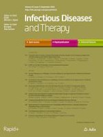
The clinical presentation of Aspergillus lung disease is determined by the interaction between the fungus and host. There are three main categories of pulmonary aspergillosis: allergic bronchopulmonary aspergillosis (ABPA), chronic pulmonary aspergillosis (CPA), and invasive pulmonary aspergillosis (IPA), as reported in Fig. 1.
Fig. 1
Categories of pulmonary aspergillosis based on underlying conditions; ABPA allergic bronchopulmonary aspergillosis, COPD chronic obstructive pulmonary disease, ICU intensive care unit
×
Anzeige
ABPA is due to a hypersensitivity reaction of the lung to Aspergillus inhalation, and it is a prerogative of patients with asthma or cystic fibrosis; CPA is a peculiar presentation of Aspergillus disease that is characterized by a local lung invasion mainly observed in patients with chronic pulmonary disease; and aspergilloma is a non-invasive form of pulmonary aspergillosis caused by a fungus ball that characteristically develops itself in a pre-existing cavity of the lung [1]. IPA is a severe acute/subacute disease and can be found not only in severely immunocompromised patients but also in non-neutropenic and/or critically ill patients, and those with chronic obstructive pulmonary disease (COPD) and/or Child–Pugh C liver cirrhosis. In non-neutropenic patients, a high suspicion of infection is reported for those without the classical risk factors of IPA, in whom, frequently, the clinical presentation is silent and nonspecific. Treatment is crucial for survival, and high rates of mortality are reported also in non-neutropenic patients, mainly due to delayed diagnosis [2, 3]. In this population, the non-specificity of clinical presentation and a lower sensitivity of diagnostic tests make it difficult to achieve a timely diagnosis of IPA compared to neutropenic patients.
The aim of this article is to present to clinicians a critical review on the risk factors, diagnosis, and therapy (as reported in Table 1) of the three main categories of pulmonary aspergillosis: ABPA, CPA, and IPA.
Table 1
Treatment of pulmonary aspergillosis entities
Aspergillus lung disease
First-line treatment
Duration of therapy
Alternative treatment
Comments
ABPA
Prednisolone 0.5 mg/kg/day for 4 weeks followed by 0.25 mg/kg/day for 4 weeks followed by 0.125 mg/kg/day for 4 weeks
Itraconazole 200 mg twice daily
3–5 months
Oral voriconazole
Posaconazole
Glucocorticoids are the first-line treatment for exacerbations
Antifungal therapy has a corticosteroid-sparing effect and could be considered in patients who fail to show improvement after steroid treatment
Aspergilloma
No therapy
Surgical resection when appropriate
–
Itraconazole
Voriconazole
Bronchial artery embolization
Anti-fungal therapy may be considered in cases of lung invasion or when there is the possibility of peri-operatively dissemination
CPA
Itraconazole 200 mg twice daily
Voriconazole 6 mg/Kg po/IVq12 h × 1 day followed by 4 mg/Kg po/IV q12 h
6 months
Posaconazole
Liposomal Amphotericin B
Caspofungin
Micafungin
Prolonged treatment may be necessary
Surgery has a limited role and may be associated with complications
Anti-fibrinolytic agent or bronchial artery embolization could be considered for management of hemoptysis
IPA
Voriconazole 6 mg/Kg po/IVq12 h × 1 day followed by 4 mg/Kg po/IV q12 h
Isavuconazole 372 mg po/IV q8 h × 6 doses followed by 372 mg po/IV daily
6–12 weeks
Liposomal Amphotericin B
Caspofungin
Combination therapy is not routinely recommended, but may be considered in selected refractory cases
Surgical resection is considered in selected situations
Empiric treatment could be considered in critically-ill patients with severe liver cirrhosis and/or end-stage chronic obstructive pulmonary disease and/or clinical worsening despite broad-spectrum antibiotics
ABPA allergic bronchopulmonary aspergillosis, CPA chronic pulmonary aspergillosis, IPA invasive pulmonary aspergillosis, po oral administration, IV intravenous
Methods
In May 2020, we performed a MEDLINE/PubMed search, employing various combinations of the following key words: Aspergillus, human, allergic bronchopulmonary aspergillosis, chronic pulmonary aspergillosis, and invasive pulmonary aspergillosis. The search period was from January 2000 to May 2020. Of the 1793 papers identified, 1348 were excluded by title, abstract screening, and the journal’s impact factor. The full texts of the remaining 445 papers and of pertinent references were then retrieved and discussed. The final decision on their inclusion in the present narrative review was based upon the subjective impression of the authors. This article is based on previously conducted studies and does not involve any studies on human or animal subjects performed by any of the authors.
Epidemiological Overview
Aspergillus species are ubiquitous in the environment, and the risk of infection is directly related to precipitation patterns, humidity, temperature, and wind conditions. The most common portal of entry in the lung is the inhalation of fungal spores; then, important efforts are made to decrease exposure to fungal spores, especially in immunocompromised patients, patients who have undergone solid organ transplantation (SOT), and burn patients. These special populations require the creation of a protected environment, and guidelines recommend the use of high-efficiency particulate air filtration and the maintenance of positive pressure rooms. However, most cases of pulmonary aspergillosis are sporadic, and outbreaks with onset of symptoms ≥ 7 days after hospital admission should be considered as hospital-acquired; however, in several cases, if it is not possible to identify an environmental source, it is not possible to distinguish community-acquired from hospital-acquired pulmonary aspergillosis [4].
Anzeige
Data about IPA reported worldwide have shown an incidence of almost 20% in SOT recipients, with a variable incidence of infection based on the organ transplanted: kidney (0.7–4%), liver (1–9.2%), pancreas (about 3%), and heart (from 1 to 14%). However, the incidence of invasive forms in general is related to patient-specific factors [4]. Overall mortality is about 22%.
Few data are reported about the incidence of ABPA and CPA. Finally, data on the prevalence of pulmonary aspergillosis have been systematically assessed in a few studies [5].
Allergic Bronchopulmonary Aspergillosis
ABPA is a lung inflammation characterized by pulmonary infiltrates and bronchiectasis [6], that is mainly observed in patients with asthma or cystic fibrosis (CF). In those patients, inhaled A. fumigatus may invade the lung ,evading the innate immune system and triggering a lymphocyte response, with activation of the inflammatory cytokines cascade resulting in sensitization [7]. The high IgE levels in serum for A. fumigatus antigens are the result of an immediate hypersensitivity to Aspergillus.
Symptoms of ABPA are often non-specific and reported in more common lung diseases [8], and the most common symptom is a chronic productive cough that could be associated with wheezing, hemoptysis, weight loss, and fever.
Generally, patients with controlled asthma may be asymptomatic for ABPA, and diagnosis is mainly based on routine testing; ABPA should be promptly suspected in patients with poorly controlled asthma or in patients affected by CF [9]. Screening tests are performed using the Aspergillus skin prick test or the A. fumigatus-specific IgE blood test which shows higher sensitivity [10]. If the screening is positive in the diagnostic tests for ABPA, IgE levels should be obtained in these patients. Then, ABPA is diagnosed based on these international criteria established in 2013 [6]:
1.
Presence of 1 of the following predisposing condition: asthma or CF.
2.
Main criteria:
Positive Aspergillus skin test or elevated IgE against A. fumigatus.
Total serum IgE > 1000 IU/mL.
3.
Adjunctive criteria (2 out of 3):
Serum precipitins or IgG against Aspergillus fumigatus;
radiological features suggestive for ABPA;
a blood eosinophil count > 500 cells/L in corticosteroid-naive patients.
Of interest, in 2013, Baxter and coworkers proposed three distinct classes of aspergillosis in CF using serologic, RT-PCR, and galactomannan (GM) data. This classification could improve phenotyping, studies on pathogenesis, and management of patients with CF and pulmonary aspergillosis [11]. The rationale of this specific classification is that patients with CF demonstrated a wide range of hypersensitivity responses to Aspergillus infection, beyond ABPA, which requires a different classification.
Few definitive data are available about the therapeutic approach. Therapy is mainly based on the use of glucocorticoids and antifungals [12]. In patients with acute ABPA, steroids are used alone, at an initial dose of 0.5 mg/kg for a total duration of 3–5 months [13]. Of interest, in a randomized trial, different dosages of prednisolone were compared: a medium-dose versus high-dose regimen in asthmatic patients with ABPA. Data reported an efficacy of both regimens with significantly fewer side effects in patients treated with a medium-dose regimen [14].
In patients with asthma and the development of ABPA plus bronchiectasis, in the absence of improvement after steroid treatment, antifungal therapy may be considered as an adjunctive therapy [15]. Itraconazole is mainly used as a second-line or adjunctive therapy (with or without steroids) to essentially maintain the disease’s remission for a longer period [16]. Of interest, the use of itraconazole was compared to prednisolone in a recent trial on patients with acute ABPA and asthma not previously treated. The authors reported, in patients of the prednisolone-group, after 6 weeks of therapy, a higher rate of efficacy compared to the itraconazole-group (100% vs. 88%; p = 0.007). However, the reduction of serum IgE levels and the rates of exacerbation/year and time to first exacerbation were similar in both groups [17]. Therefore, in selected cases, the combination therapy itraconazole-prednisolone could be considered, but definitive data on efficacy are necessary.
Anzeige
The assessment of therapy response is mainly based on a 25% decline in the total IgE level associated with a clinical and radiological improvement. In contrast, exacerbation is defined as at least a doubling in the baseline total IgE level plus clinical and/or radiological deterioration. Finally, remission is defined as the absence of exacerbations for at least 6 months after steroid therapy [18].
The overall prognosis of patients with ABPA is not well characterized [19]. However, early detection with prompt initiation of therapy generally leads to a good prognosis [20].
Chronic Pulmonary Aspergillosis
CPA involves a spectrum of diseases that affect immunocompetent patients with pre-existing structural pulmonary alteration [21]. These patients can show a clinical presentation from weight loss to the appearance of chronic productive cough, hemoptysis, and comparison of nodules and cavities at chest imaging. These clinical and radiological features should be present for at least 3 months at the time of diagnosis [4].
The most important progress of CPA is to chronic fibrosing pulmonary aspergillosis; aspergilloma represents a less severe form of CPA, consisting of Aspergillus hyphae with fibrin contained in a previously formed lung cavity [22, 23]. Its development is subsequent to colonization of the cavity by Aspergillus species: tubercular and nontubercular mycobacterial infections are the primary underlying lung conditions predisposing to the formation of aspergilloma [24]. Other less common predisposing conditions are ABPA, chronic obstructive pulmonary disease (COPD), lung transplantation, recurrent low respiratory tract infections, and sarcoidosis. Cough is the most common symptom, while life-threatening hemoptysis is reported in a high percentage of patients [25‐27].
Anzeige
Of importance, immunocompromised patients could develop a locally destructive CPA that tends to progress more rapidly, from 1 to 3 months. This subacute invasive aspergillosis is another subgroup of CPA that shows characteristics very similar to IPA.
CPA diagnosis is based on the presence of characteristic symptoms and radiologic features, present for at least 3 months, with microbiologic evidence of Aspergillus strains to confirm the diagnosis [4]. Symptomatic patients with cavities, aspergilloma, or nodular infiltrates at CT scan should be tested for the presence of serum A. fumigatus IgG; the presence of aspergilloma is associated with positivity of A. fumigatus IgG in serum. Alternatively, the when antibodies are negative, the positive Aspergillus cultures from the lower respiratory tract may support the diagnosis [28]. GM in bronchoalveolar lavage (BAL) showed a good diagnostic performance if compared to serum GM, and appears to be a valuable diagnostic assay [29]. Of interest, the combination of serum GM plus 1,3-beta-d glucan (BDG) could be help physicians to confirm or exclude Aspergillus infection, but their diagnostic values have not been well characterized [30, 31]. Finally, a biopsy from cavities showing the presence of Aspergillus hyphae is crucial to differentiate tissue invasion typical of subacute invasive aspergillosis from other forms of CPA, but the risks associated with the biopsy procedures should be carefully assessed in each patient [32].
The goal of CPA treatment is to prevent life-threatening hemoptysis and to improve symptoms and the patient’s quality of life. Oral itraconazole, at a dose of 200 mg twice daily, is considered the first-line therapy [22]. Voriconazole and posaconazole are second-line oral therapies [33, 34], and in selected cases the use of short-term courses of intravenous amphotericin B and echinocandins have also been successfully used, especially in patients with rapid progression of the infection, failure of therapy, or azole resistance of Aspergillus strains [35]. Of importance, therapeutic drug monitoring (TDM)-guided dosing has been shown to be clinically beneficial for voriconazole, especially in ICU patients. In critically ill patients treated with voriconazole, TDM should always be performed to assess adequate serum levels [36].
A 6-month duration of therapy is recommended, and asymptomatic patients can be reassessed every 3–6 months [37].
Anzeige
Finally, there is a strong recommendation to perform a surgical resection of a simple aspergilloma in symptomatic patients with low surgical risk, if important symptoms are reported and hemoptysis is persistent. Of importance, surgery should also be considered in patients with Aspergillus-localized CPA unresponsive to antifungal therapy [4].
Invasive Pulmonary Aspergillosis
IPA has been traditionally considered in the differential diagnosis of infection mainly occurring in patients with specific risk factors: neutropenia and hematologic malignancies, allogeneic bone marrow transplantation, SOT, neoplasm, or HIV patients [4]. Of importance, in recent years, an increasing number of studies have also reported the role Aspergillus spp. in non-neutropenic patients, including those with end-stage COPD requiring chronic high-dose steroid therapy, Child–Pugh C liver cirrhosis, and patients receiving immunosuppressive therapies (i.e., monoclonal agents) [38]. Moreover, patients admitted to the intensive care units (ICU) may also be susceptible to IPA, and recent important observations demonstrate the association between influenza, especially H1N1 virus, and IPA or other predisposing risk factors such as acute respiratory distress syndrome [3, 39‐41]. Of interest, a state of immunoparalysis is described in these categories of patients predisposing to the development of IPA [42‐44]. Moreover, recent data showed a possible association between COVID-19 caused by SARS-CoV-2 and the development of IPA in critically ill patients with moderate to severe ARDS [45]. Finally, environmental factors, including climatic variables, airborne mold concentration, geographic area, remodeling or construction work, and environmental quality of the air, may predispose to IPA [31].
Of importance, non-neutropenic patients show a non-specific symptomatology that makes clinical manifestations of IPA indistinguishable from other bacterial bronchopneumonia [46]. In this setting, the clinical diagnosis of IPA is a challenge, because diagnostic definitions [4, 47, 48] have been validated only for neutropenic patients and cannot also be used for those non-neutropenic.
Blot and coworkers proposed a clinical diagnostic algorithm aiming to discriminate colonization from probable IPA in ICU patients with Aspergillus-positive in bronchial cultures [49]. Fungal culture- and non-culture-based methods should be performed in all patients with relevant risk factors for IPA, and the development of pneumonia or the presence of a persistent pulmonary infection, despite broad-spectrum antibiotics, should drive physicians to further diagnostic exams to exclude or confirm IPA [50].
The clinical significance of Aspergillus from cultures of the lower respiratory tract remains a challenge for physicians, considering that Aspergillus spp. (especially in some specific populations like COPD patients) could be considered only a simple colonization [51]. The detection of fungus should be applied to the clinical characteristics of the patients. However, in a recent revision and update of the consensus definitions of invasive fungal disease, important aspects in the diagnosis of probable invasive pulmonary mold disease have been introduced, such as the use of Aspergillus PCR in diagnosis, as reported in Table 2 [48]. In recent years, the diagnosis of IPA has been improved using new markers based on the detection of fungal cell wall components or fungal DNA in blood or lung specimens; moreover, these markers showed the characteristic to differentiate colonization from infection. The detection of GM is currently the gold standard to early identification of IPA. Studies have reported that, in the BAL, a cutoff value of GM > 0.5 shows a sensitivity up to 100% and a specificity over 75% [52, 53]. The role of GM in hematological patients has been assessed, and the test may be used to obtain an early diagnosis and to monitor the treatment response. However, the efforts are now directed to also definitively assess the routine use of GM in non-neutropenic patients [54, 55].
Table 2
Criteria for diagnosis of probable IPA, which requires the presence of at least 1 host factor + a clinical feature + mycological evidence
Host factors
Clinical features
Mycological evidence
Recent history of neutropenia (< 500 neutrophils/mm3) for > 10 days or recognized hematologic malignancy
The presence of one of the following on CT scan:
Dense, well-circumscribed lesions with or without a halo sign
Air crescent sign
Cavity
Wedge-shaped and segmental or lobar consolidation
Aspergillus spp. detected in sputum, BAL, bronchial brush, or aspirate
Receipt of an allogeneic stem cell transplant or a solid organ transplant or acute graft-versus-host disease grade III or IV involving the gut, lungs, or liver that is refractory to first-line treatment with steroids
Tracheobronchial ulceration, nodule, pseudomembrane, plaque, or eschar seen on bronchoscopic analysis
Galactomannan antigen detected in plasma, serum, or BAL, any one of the following:
Single serum or plasma: ≥ 1.0
BAL fluid: ≥ 1.0
Single serum or plasma: ≥ 0.7 and BAL fluid ≥ 0.8
Prolonged use of corticosteroids for ≥ 3 weeks in the past 60 days or treatment with T-cell or B-cell immunosuppressants during the past 90 days
ICU admission
Aspergillus PCR, any one of the following:
Plasma, serum, or whole blood, 2 or more consecutive PCR tests positive
BAL fluid, 2 or more duplicate PCR tests positive
At least 1 PCR test positive in plasma, serum, or whole blood, and 1 PCR test positive in BAL fluid
Presence of a persistent pulmonary infection despite broad-spectrum antibiotic therapy
New tests under development:
Aspergillus species genes amplification
Lateral flow device
Detection of volatile organic compounds
Gliotoxin and bis(methylthio)gliotoxin
IPA invasive pulmonary aspergillosis, COPD chronic obstructive pulmonary disease, CT computed tomography, ICU intensive care unit, BAL bronchoalveolar lavage
Conversely, the 1-3-β-d-glucan assay is another important test that, in patients with hematological disease, showed a high sensitivity with a very low specificity for the diagnosis of fungal infection [56‐58]. In contrast, its negative predictive value of 80–90% could make 1-3-β-d-glucan potentially useful to rule out the diagnosis of IPA rather than to confirm it. However, the role of this marker in the diagnosis of IPA is still unknown, and future studies are necessary to definitively assess its use in clinical practice [59]. Few studies have evaluated the role of 1-3-β-d-glucan in BAL, also indicating a low specificity for IPA in immunocompromised patients [60].
Of importance, these tests, especially GM, could be affected by the high frequency of false-positive results based on the use of β-lactam antibiotics, human blood components, and hemodialysis [61]. New tests are actually under development and validation but not yet universally standardized [62, 63], and cannot be, to date, included as a criterion in the EORTC/MSG guidelines [4, 47]. The most important are: (1) Aspergillus species gene amplification in which the detection of genetic sequences, mainly represented by 18S rDNA, 28SrDNA, 5.8 SrDNA, and mithocondrial DNA, is obtained directly from fungal cultures and/or in direct clinical samples; Aspergillus PCR is processed in a few hours and, when these results are combined with other fungal biomarker (like GM or BDG) in serum or in BAL (mainly GM), the diagnostic sensitivity up to 100% further supports the introduction of this process in the new definitions of invasive fungal infection by the EORTC/MSG; (2) a lateral flow device (LFD) that detects a glycoprotein antigen in the serum and BAL of patients with IPA: this technique has been proposed as a new point-of care diagnostic approach for an early detection of IPA in non-neutropenic patients, but also in SOT or critically-ill patients in ICU; in a multicenter study evaluating the use of LFD devices in BAL of ICU patients showed a sensitivity of 80%, a specificity of 81%, positive and negative predictive values of 96% and 44%, respectively; however, further and larger studies are crucial to assess the use of LFD in clinical practice, despite these first promising results; (3) innovative technologies have recently been tested in the breath of patients infected with IPA: these technologies detect volatile organic compounds exhaled with a sensitivity ranging from 94 to 100% and a specificity from 83 to 93%; and (4) gliotoxin and bis(methylthio)gliotoxin have been applied in the diagnosis of IPA with significant and promising results [64‐68]. However, the diagnosis of IPA remains challenging considering that none of the available diagnostic tests, actually introduced in clinical practice, show high sensitivity and specificity if used alone. The rationale could be the use of diagnostic strategies, including cultures, surrogate biomarkers, and molecular tools in a simultaneous performance to achieve the best possible approach to patients with suspected IPA.
Despite the introduction in clinical practice of new antifungals and the use of supportive measures, the mortality in patients with IPA remains very high. In the IDSA guidelines, prophylaxis during prolonged neutropenia and immunosuppression is recommended [19]. Moreover, strong recommendations have been reported about the use of voriconazole or posaconazole for prophylaxis in large randomized clinical trials [69]. As second-line therapies also reported for prophylaxis are itraconazole, micafungin, and caspofungin, which may also be effective [70, 71]. Of interest, studies highlight the important role of non-pharmacologic prophylaxis measures to reduce exposure to fungal conidia. These strategies are based on placing severely immunocompromised patients in “protected environments”, with high-efficiency particulate air filtration and positive pressure, to avoid some activities that are associated with high exposure to Aspergillus spores, like moldy hay handling and construction, using personal protective equipment.
To date, despite the possibility of using many therapeutic options, the mortality rate of IPA remains high [72], and is reported to be higher in non-neutropenic patients than that reported in the neutropenic population. Probably, non-neutropenic patients at high risk of IPA for predisposing conditions like COPD, prolonged use of steroids and immunosuppressive therapy, Child–Pugh C liver cirrhosis, and ICU-related immunoparalysis should receive adequate antifungal therapy upon suspicion of the Aspergillus infection. The goal of IPA management is to obtain, as soon as possible, a CT scan, fungal cultures, and a combination of serological biomarkers represented by GM (especially in BAL), Aspergillus PCR, and 1-3-β-d-glucan assay. The antifungal treatment should be re-discussed and eventually discontinued if the diagnosis of IPA is not confirmed.
The antifungal agents approved as the first-line for the treatment of IPA are voriconazole, isavuconazole, and amphotericin B with its lipid formulation [19]. The selection of the best drug for the treatment of IPA is mainly based on different steps: the assessment of severity of the infection, clinical features, the presence of renal or hepatic insufficiency, possible drug–drug interactions (especially in patients undergoing particular treatments for underlying diseases), the need for therapeutic drug monitoring, and, no less important, the costs of antifungal drugs.
Of these, isavuconazole is a new drug of the triazole class that can be given once daily, and it shows a wider spectrum of antifungal activity compared to voriconazole. Isavuconazole activity also includes Mucorales infections and (as opposed to voriconazole) its intravenous formulation does not include cyclodextrin, which is a nephrotoxic and hepatotoxic compound typical of intravenous formulations of other triazoles, used to increase solubility. Also, compared to voriconazole, isavuconazole has fewer CYP enzyme-mediated drug interactions and shows linear and predictable pharmacokinetics, for which therapeutic drug monitoring is not necessary [73]. In an important randomized, double-blind trial, the non-inferiority of isavuconazole versus voriconazole has been demonstrated in terms of mortality. Isavuconazole has been used as a primary treatment for IPA or other filamentous fungi infections, also showing a superior safety profile [74]. Finally, all echinocandins have shown in vitro and in vivo activity against Aspergillus spp., but only caspofungin is licensed for the treatment of IPA, as second-line therapy. In specific cases or in refractory disease, the use of a combination therapy with echinocandin plus voriconazole or liposomal amphotericin B may be considered.
Adequate duration of antifungal therapy for IPA is an unresolved issue. IDSA guidelines recommend that the treatment of IPA should be continued for at least 6–12 weeks, considering the clinical condition of the patient and their response to therapy; moreover, serum biomarkers and radiological follow-up with a CT scan should be considered to monitor the therapeutic response to IPA.
Conclusion
Recently, an important evolution has occurred concerning the classification, diagnosis, and treatment of different forms of pulmonary aspergillosis. The semi-continuous spectrum of pulmonary aspergillosis starts from an allergic, noninvasive form to a more aggressive invasive disease. The different aspects of this infection represent a challenge for physicians, also considering its potential overlap with other common infectious and non-infectious pulmonary diseases. Clinicians should maintain a high index of suspicion for pulmonary aspergillosis because early diagnosis and treatment are associated with a favorable outcome, while a high rate of morbidity and mortality are reported in patients with a significant delay in diagnosis. For these reasons, all efforts should be guaranteed to identify diagnostic tools with a high sensitivity and specificity to reach early diagnosis, and more efficacious and better-tolerated therapies should be obtained.
IPA remains the most serious entity of this spectrum, with a high mortality rate despite optimal therapy. In these patients, an early diagnosis and identification of potential candidates for prophylaxis could be crucial to improve survival. The development of a standard set of definitions for invasive fungal diseases in non-neutropenic patients is the goal for improvement of the management of pulmonary aspergillosis. Finally, the duration of therapy and the frequency of follow-up should definitely be assessed. Recent data from FUNDICU project aimed to develop a standard set of definitions for invasive fungal infections in critically ill adult patients in ICU [75, 76]. In this project were summarized the available evidence on the diagnostic performance for IPA in non-hematological but also in non-SOT critically ill patients, such as classical immunocompromised or non-immunocompromised critically ill patients.
Acknowledgements
Funding
No funding or sponsorship was received for this study or the publication of this article.
Authorship
All named authors meet the International Committee of Medical Journal Editors (ICMJE) criteria for authorship for this manuscript, take responsibility for the integrity of the work as a whole, and have given final approval for the version to be published.
Disclosures
Alessandro Russo, Giusy Tiseo, Marco Falcone and Francesco Menichetti declare no conflicts of interest.
Compliance with Ethics Guidelines
This article is based on previously conducted studies and does not involve any studies on human or animal subjects performed by any of the authors.
Data Availability
Data sharing is not applicable to this article as no datasets were generated or analyzed during the current study.
Open AccessThis article is licensed under a Creative Commons Attribution-NonCommercial 4.0 International License, which permits any non-commercial use, sharing, adaptation, distribution and reproduction in any medium or format, as long as you give appropriate credit to the original author(s) and the source, provide a link to the Creative Commons licence, and indicate if changes were made. The images or other third party material in this article are included in the article's Creative Commons licence, unless indicated otherwise in a credit line to the material. If material is not included in the article's Creative Commons licence and your intended use is not permitted by statutory regulation or exceeds the permitted use, you will need to obtain permission directly from the copyright holder. To view a copy of this licence, visit http://creativecommons.org/licenses/by-nc/4.0/.
Fast ein Viertel der Personen mit mäßig dysplastischen Stimmlippenläsionen entwickelt einen Kehlkopftumor. Solche Personen benötigen daher eine besonders enge ärztliche Überwachung.
Bei Menschen mit Typ-2-Diabetes sind die Chancen, einen Myokardinfarkt zu überleben, in den letzten 15 Jahren deutlich gestiegen – nicht jedoch bei Betroffenen mit Typ 1.
Ob Patienten und Patientinnen mit neu diagnostiziertem Blasenkrebs ein Jahr später Bedauern über die Therapieentscheidung empfinden, wird einer Studie aus England zufolge von der Radikalität und dem Erfolg des Eingriffs beeinflusst.
„Kalte“ Tumoren werden heiß – CD28-kostimulatorische Antikörper sollen dies ermöglichen. Am besten könnten diese in Kombination mit BiTEs und Checkpointhemmern wirken. Erste klinische Studien laufen bereits.
Update Innere Medizin
Bestellen Sie unseren Fach-Newsletter und bleiben Sie gut informiert.