Much of our understanding of the molecular control of menstruation arises from laboratory models that experimentally recapitulate some, but not all, aspects of uterine bleeding observed in women. These models include: in vitro culture of endometrial explants or isolated endometrial cells, transplantation of human endometrial tissue into immunodeficient mice and the induction of endometrial breakdown in appropriately pretreated mice. Each of these models has contributed to our understanding of molecular and cellular mechanisms of menstruation, but nonhuman primates, especially macaques, are the animal model of choice for evaluating therapies for menstrual disorders. In this chapter we review some basic aspects of menstruation, with special emphasis on the macaque model and its relevance to the clinical issues of irregular and heavy menstrual bleeding (HMB).
1 Introduction
Much of our understanding of the molecular control of menstruation arises from laboratory models that experimentally recapitulate some, but not all, aspects of uterine bleeding observed in women. These models include: in vitro culture of endometrial explants or isolated endometrial cells [1‐3], transplantation of human endometrial tissue into immunodeficient mice [4], and the induction of endometrial breakdown in appropriately pretreated mice [5‐8]. Each of these models has contributed to our understanding of molecular and cellular mechanisms of menstruation, but nonhuman primates, especially macaques, are the animal model of choice for evaluating therapies for menstrual disorders. In this chapter we review some basic aspects of menstruation, with special emphasis on the macaque model and its relevance to the clinical issues of heavy and irregular menstrual bleeding.
Menstrual disorders including heavy menstrual bleeding (HMB), irregular uterine bleeding, and painful menstruation are common problems experienced by many women. HMB is considered as excessive menstrual blood loss, prolonged more than 8 days, which interferes with the woman’s physical, emotional, social and material quality of life, and which can occur alone or in combination with other symptoms [9]. Irregular menstruation is characterized by cycles that vary more than 8 days in length. HMB affects approximately one-third of reproductive aged women and although heavy bleeding can be associated with endometriosis and uterine fibroids, greater than 80 % of patients with HMB exhibit no clear underlying cause [10]. Idiopathic HMB is one of the most common gynecologic conditions requiring hospitalization, and 20–30 % of all hysterectomies are performed to stop abnormal bleeding. Expansion of medical therapies for HMB would greatly benefit the health of many women who cannot or do not wish to undergo surgery [11].
Anzeige
2 Phylogenetic considerations
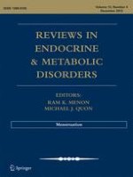
Periodic uterine bleeding is reported for a small number of non-primate mammals, including some species of tree shrew [12]. However, cyclic, hormonally regulated shedding of the endometrium, characteristic of true menstruation, is found only in a few species of bats [13, 14] and in primates, including Homo sapiens, the Great Apes (Hominidae), Lesser Apes (Hylobatidae) and Old World monkeys (Cercopithecidae) [15]. In these primate species, menstruation follows the fall in progesterone levels as the corpus luteum regresses at the end of the ovarian cycle, and cycle lengths are counted from the first day of menstrual bleeding. Menstrual cycle length varies slightly between primate species, from about 28 days in women and rhesus macaques to about 37 days in chimpanzees. Rhesus (Macaca mulatta), cynomolgus (Macaca fascicularis), and pigtailed macaques (Macaca nemestrina) display approximately 28 day menstrual cycles, similar to those of women [16‐18]. Rhesus macaques are the most widely used nonhuman primate model for menstrual bleeding studies [19‐21]. Macaques can be trained to allow daily vaginal swabs to detect menstrual bleeding [21] and can be fitted with tampons [22] for more quantitative analysis of menstrual blood loss. Naturally cycling macaques may display spontaneous menstrual disorders, including irregular bleeding and HMB (Fig. 1), making them ideal for clinically relevant studies. Further, macaques can be ovariectomized and treated with ovarian steroids to induce artificial menstrual cycles. Induced cycles reduce the variability inherent in natural cycles, and permit endometrial sampling during precisely defined premenstrual, menstrual and postmenstrual phases.
Fig. 1
Comparison of normal versus disordered and heavy menstrual bleeding (HMB) in rhesus macaques. Top panel: bleeding patterns of 8 animals with normal cycles. Bottom panel: bleeding patterns of 6 animals with abnormal cycles. Each row shows the days of bleeding of one animal followed for 110 days, aligned by the first day of bleeding. The lower panel shows the erratic bleeding patterns of monkeys with menstrual disorders including irregular bleeding and HMB
×
3 Anatomical considerations
The macaque uterus is morphologically similar to the human uterus, consisting of the fundus (dome-shaped top), corpus (body), and isthmus (neck) leading to the cervix. The endometrium lines the uterine cavity and is surrounded by the muscular wall or myometrium (Fig. 2a). In women [23] and macaques [24] the endometrium has 4 layers or “zones” extending from the luminal surface to the myometrial border. In that classification, the luminal epithelium and an underlying band of stromal cells is defined as Zone I. Slightly deeper, Zone II contains glands that run perpendicular to the surface. Zones I and II are sometimes referred to as the “compacta” in women and consist of densely packed stromal cells around the straight necks of the glands. Deeper still, Zone III, sometimes referred to as the spongiosa in women, contains glands that are branched. The deepest zone, Zone IV, the basalis, is adjacent to the myometrium, where the glands terminate (Fig. 3). The upper zones (e.g. the compacta and upper spongiosa) are also referred to as the functionalis, or functional layer, as opposed to the basalis, or basal layer. The functionalis, which undergoes secretory transformation under the influence of progesterone, derives its blood supply from specialized spiral arteries rather than the basal arteries that supply the deepest, basal zone [25]. The spiral arteries are unique endometrial vessels in menstruating primates that undergo hormonally driven regeneration after menstruation in each cycle. In the secretory phase, progesterone stimulates spiral artery hypertrophy, and during a fertile cycle these arteries play an important role in embryo implantation. Abnormal spiral arteriogenesis can contribute to pregnancy failure and may lead to breakthrough bleeding and HMB [26].
Fig. 2
Comparison of rhesus uterus in proliferative versus menstrual phase. Freshly removed uteri were cut in half along the fundal-cervical axis and photographed with macro lenses. a a dark line marks the endometrial-myometrial border and another line delineates the plane along which sections of the endometrium were cut. This specimen was taken on day 14 of the induced proliferative phase. Endo = endometrium. Myo = myometrium. b on day 2 of the menstrual cycle, bleeding is restricted to the upper third of the endometrium. The basalis and the lowest part of the functionalis does not bleed or slough. Scale bar = 1 cm; applies to both images
Fig. 3
Histology of the rhesus endometrium in the proliferative, secretory and menstrual phases. Histological sections of the endometrium were cut on a plane running from the luminal to the myometrial border. Straight versus tortuous glands are evident in (a versus b). Sections taken during the menstrual phase (c–e) show that menstrual breakdown is only evident in the functionalis (c–d) not the basalis (e). Scale bar for a–c = 1 mm; scale bar for d–e = 100 μm
×
×
While the macaque uterus is strikingly similar to that of most other Old World nonhuman primates, the anatomy of the cervix varies greatly between primate species [27]. In baboons [28] ( as in women) the cervical canal is relatively free from obstruction, which facilitates hysteroscopy for the assessment of endometrial bleeding and collection of samples by curettage [29‐31]. However, in macaques there is a prominent colliculum (Fig. 4) that obstructs passage of instruments into the uterine cavity [32]. Estrogen stimulation induces increased curvature that further restricts access to the cervical canal.
Fig. 4
The rhesus macaque cervix. A cervix was freshly removed from a rhesus macaque at midcycle and cut longitudinally along the axis from the External os to Internal os. A projection of the cervical wall (colliculum) forces the cervical canal into a Z-shaped path. Scale bar = 1 cm
×
4 The macaque menstrual cycle
Central regulation of female sexual maturation, follicular development, ovulation, and ovarian hormone secretion in primates involves interplay between the hypothalamus, pituitary and ovary, relationships that have been reviewed extensively [33‐35]. In primates, ovarian cyclicity is not dependent on endocrine feedback from the uterus. Estradiol (E) and progesterone are the only ovarian factors required for induction of menstruation [36‐38]. Briefly, the menstrual cycle can be divided into three phases: the menstrual phase, the follicular (or proliferative) phase, and the luteal (or secretory) phase. Estradiol secreted by the ovary during the follicular phase stimulates cell proliferation in both the endometrial glands and stroma [39]. In macaques the length of the proliferative phase of the natural cycle is normally 10–14 days. After ovulation, progesterone levels rise during the luteal phase [39] of the cycle, which usually lasts for 12 to 14 days. If pregnancy does not occur, the corpus luteum regresses and the decline of progesterone triggers menstruation .
Anzeige
In various studies, we induced cycles in ovariectomized macaques by treating the animals with Silastic capsules that release E and progesterone [40]. Briefly, animals are treated first with E alone, which stimulates an artificial proliferative phase, characterized by tubular endometrial glands and abundant proliferating cells in the glandular epithelium. After 14 days of E priming, addition of a progesterone capsule induces an artificial secretory phase, which becomes evident by day 3 of progesterone treatment. Progesterone acts to gradually suppress cell proliferation in the glandular epithelium, which thickens, and the glands become saw-toothed and sacculated in appearance, with shrunken nuclei and jagged apical surfaces. In the macaque, the early secretory phase is marked by a striking up regulation of cell proliferation in the glands of the basalis zone [41, 42]. Withdrawal of the progesterone capsule (with E left in place) completes the cycle and induces menses on days 2–5 of the next cycle. We refer to this 2–5 days interval as the luteal-follicular transition (LFT) [43, 44].
The effects of ovarian steroid hormones on estrogen and progesterone receptors have been extensively reviewed [45‐47]. Briefly, during the proliferative phase, E stimulates expression of estrogen receptor-1 (ESR-1) and progesterone receptor (PGR) in the endometrial glandular epithelium and stromal fibroblasts. During the secretory phase, progesterone suppresses ESR-1 in glands and stroma of the functionalis zone, while suppressing PGR only in the glands, not the stroma. Thus, progesterone can act through PR in the stromal fibroblasts to control expression of tissue degrading enzymes, growth factors, and extracellular matrix molecules. The basalis zone is unique in retaining both ESR-1 and PGR in both the glands and stroma during the secretory phase. The vascular endothelium in all zones fails to expresses ESR-1 or PGR but does express ESR-2 (ER-beta [45, 48]).
Endometrial angiogenesis, vascular growth, and vasoconstriction [49, 50] have been extensively studied in macaques [51, 52] because the vessels of the upper functionalis are shed and regenerated after menstruation. Endothelial cell proliferation peaks in small vessels during the proliferative phase [51, 53]. After ovulation, the endometrial spiral arteries hypertrophy [42], and provide increased blood flow [54] to the upper zones where embryo implantation and early placentation occur [55]. The effects of progesterone on the spiral arteries are probably mediated by factors arising from PGR-positive stromal cells or through novel non-nuclear receptors [56]. At the end of a nonfertile cycle the fall in progesterone triggers waves of vasoconstriction [57] of the spiral arteries typically followed 4 to 24 h later by menstruation.
5 Histological aspects of menstrual breakdown
On day 28 of an induced cycle, the endometrium appears thickened with stromal cell hypertrophy, tissue edema and tortuous glands in the functionalis zone. Twenty four hours after progesterone withdrawal the endometrium shrinks dramatically and the stroma becomes highly compacted. By day 2 the uppermost third of the endometrium undergoes extensive fragmentation accompanied by hemorrhage and sloughing (Fig. 2b). The deeper functionalis and basalis zones show no signs of fragmentation, but the glandular epithelial cells in the basalis undergo extensive apoptotic cell death during days 2–6.
Overt menstrual bleeding typically ends on day 4–6, and around day 5 mitotic activity begins in the necks of the surviving glands [44]. By day 8 there is increased endometrial growth of the glands as well as maximal endothelial cell proliferation [51]. Structural regrowth of the endometrium in the follicular phase appears complete by day 10–14, at which time the endometrium consists of straight, nonsacculated tubular glands encompassed within a moderately loose stroma.
6 Effects of progesterone withdrawal on endometrial matrix metalloproteinases
The decline of progesterone at the end of the cycle is followed by increased expression of tissue degrading matrix metalloproteinase (MMP) enzymes [19, 58]. The MMPs have the enzymatic capability to mediate tissue dissolution in the upper functionalis zones and are considered primary effectors of menstrual sloughing [59, 60]. MMPs are a multigene family of enzymes that require zinc for their activation [61] . Secreted as proenzymes, the MMPs undergo activation to a catalytically active form in the endometrium. Enzyme activation is regulated by specific endogenous tissue inhibitors of metalloproteinases (TIMPs) [61] that are also expressed in the endometrium [62, 63]. In vitro studies on endometrial stromal cells in culture demonstrate that progesterone suppresses expression of several MMPs [59, 64]. Paradoxically, progesterone receptor consensus sequences have not been identified on the MMPs associated with menstruation. Moreover, in the macaque, endometrial expression of several MMPs declines rapidly after menstruation, well before progesterone levels increase [19]. Therefore, progesterone mediated regulation of MMPs appears to be either indirect through paracrine/autocrine factors, or through non-traditional mechanisms. Because of the focal nature of menstrual breakdown and the restriction of MMP expression to the mid and upper functionalis zones, it has been suggested that local factors such as specific cytokines can regulate endometrial MMPs. For instance, TGF-beta has been identified as a mediator of MMP-7 suppression by progesterone [65]. Also, endometrial bleeding associated factor (ebaf; lefty-A) has been identified as an inhibitor of TGF-beta [66], and lefty-A can induce expression of several MMPs in human endometrial stromal cells. Because progesterone can still suppress MMPs upregulated by lefty-A in vitro [67], the mechanism through which progesterone regulates endometrial MMPs remains unclear.
In earlier studies we documented the timing of menstrual sloughing and the expression of various MMP enzymes in rhesus and pigtailed macaques [19, 40]. For these studies the animals were treated with E and progesterone to induce artificial menstrual cycles, and the uterus was removed from the animals on days 1,2,3,4,5,6,8,10,14,21 and 28 of the cycle. In this design, day 0 represents the day of progesterone implant removal. In other animals we removed both the E and the progesterone releasing capsules to determine whether the absence of E would affect MMP expression. These latter animals were referred to as either spayed or hormone-deprived. In each case, samples of endometrium were prepared for immunocytochemistry and analysis of MMP mRNA expression. In general, the expression of endometrial MMPs in vivo during the menstrual cycle of the rhesus macaque was very similar to that reported for women. Most of the MMPs were expressed at relatively high levels just before and during menstrual breakdown (Fig. 5). Immunolocalization studies revealed strong staining for MMP-1, MMP-2, and MMP-3, localized primarily to the stromal cells of the fragmenting zones, with intense staining in the cytoplasm of stromal cells at the basement membrane of regressing spiral arteries. Immnocytochemical staining patterns of MMP-2 are illustrated in Fig. 6. MMP-7 was initially expressed by the glands immediately below the fragmenting zone, and later in the upper glands as the endometrium entered the repair phase. MMP-7 localization became stronger as MMP-2 and MMP-3 staining waned in the early follicular phase. Tissue inhibitor of metalloproteinases-1 (TIMP-1) also increased, at both the protein and gene expression level, by cycle day 2. This increase was especially apparent in the smooth muscle and perivascular stroma of the spiral arteries and in large numbers of stromal cells in the fragmenting and sloughing endometrial regions. All these effects were similar in the presence and absence of E.
Fig. 5
mRNA expression of various MMPs in the rhesus menstrual cycle. Plots of northern hybridization data reveal two patterns of MMP mRNA expression. Top Row: Certain MMPs were strongly expressed during the menstrual phase and declined to minimal levels by days 5–6. Bottom Row: Other MMPs peaked similarly during menstruation but declined much more slowly
Fig. 6
Immunohistochemistry of MMP-2 during the proliferative phase. Endometrial sections were stained for MMP-2 on days (D) 0,1,2,3,4 and 14 after P withdrawal. The black line marks the myometrial border. Strong dark brown staining for MMP-2 protein was confined to the upper functionalis zone during the menstrual phase (D1-4). The staining became nondetectable by day 14. Scale bar = 1 mm; applies to all images
×
×
Several studies have proposed that induction of menstruation occurs in two phases [21, 68, 69]. In the first phase, progesterone withdrawal initiates up regulation of specific paracrine regulators, especially those secreted by the perivascular cells of the spiral arteries. Once these regulators are fully expressed, a second phase dominated by tissue degrading enzymes develops. A corollary of this hypothesis is that the first, but not the second phase can be interrupted by re-elevation of progesterone [21]. This suggests that progesterone withdrawal is followed by a “critical period” after which tissue breakdown, sloughing and bleeding become inevitable. To further define the critical period in macaques, we induced cycles as described above and, at the end of the luteal phase, withdrew progesterone at time 0 and then either replaced progesterone (or not) 12, 24, or 48 h later. Uteri from experimental (progesterone-replaced) and control (not replaced) animals were collected 36, 48 and 72 h after progesterone withdrawal (n = 3/interval). Strong immunocytochemical staining for MMP-1 and MMP-2 was evident in controls at 36, 48 and 72 h. Progesterone replacement at 12–24 h after P withdrawal (the critical period), blocked menses and blocked expression of both MMPs. This confirms a role for both MMP-1 and MMP-2 in the initiation of menstruation. However, progesterone replacement after the critical period failed to suppress menses and failed to suppress MMP2, though MMP-1 was suppressed. Therefore, some MMPs may be more important for initiating menses, and others for sustaining menstrual breakdown. Our results confirm and extend the view that there is a physiologically significant critical period underlying the menstrual process. The particular role that each MMP plays in the menstrual cascade remains to be determined.
Anzeige
7 Vascular events associated with menstruation
Much of our understanding of menstruation arises from classical studies by Markee [49]. Markee transplanted rhesus macaque endometrium to the anterior chamber of the eye and visualized the events of menstruation directly. He observed pulses of vasoconstriction in the spiral arteries after progesterone withdrawal that could lead to localized ischemic hypoxia in the superficial zones. Vascular endothelial growth factor (VEGF) is reported to be upregulated by hypoxia in the human endometrium [70], and in our macaque studies, stromal and glandular VEGF were dramatically up regulated premenstrually. VEGF may interact with several receptors, including VEGFR-1 and VEGFR-2 which are located on the membranes of endothelial cells [71, 72]. However, VEGFR-2 expression was also upregulated by progesterone withdrawal in the stromal cells of the functionalis zone. Whether VEGF interacts with VEGFR-2 in these stromal cells after progesterone withdrawal remains to be determined.
We also examined the expression and localization of hypoxia inducible factor 1 (HIF-1α) in the rhesus macaque endometrium during the menstrual-early proliferative stage of an induced cycle. HIF-1α is a nuclear protein that mediates the effects of hypoxia on gene expression. Artificial cycles were induced in 26 ovariectomized rhesus macaques. After 14 days the P implant was removed and the uterus was collected from 2 animals each on days 0, 1, 2, 3, 4, 5, 6, 8 and 14 (P withdrawn). In another group, the P and E implants were both withdrawn on day 0 and the uteri were collected on days 1, 2, 3, and 14 (hormone deprived). Menses occurred in both groups on days 2–5. Endometrial samples were analyzed for HIF-1 mRNA by Northern blot and in situ hybridization . Northern blots revealed that HIF-1α mRNA levels were low on day 0, increased strikingly on days 1–3 of the cycle and then declined. Similarly, in the hormone-deprived group, the HIF-1α mRNA increased on days 1 and 2 and then declined. These increases occurred primarily in the glands and the small blood vessels of the upper functionalis zone. Grain counts of the ISH preparations showed a 6.6 fold increase in the blood vessels and a 1 .4 fold increase in the glands over baseline during days 1 and 2 followed by a decline. Immunocytochemical staining of the HIF-1α nuclear protein showed a similar pattern of expression (Fig. 7). In sum, HIF-1α was strikingly elevated in endometrial vessels and glands in the functionalis on days 1–3 after P withdrawal whether or not E was present. These data support a role for hypoxia, mediated by HIF-1α, in the early phases of the menstrual cascade.
Fig. 7
Immunohistochemistry of hypoxia inducible factor in the macaque endometrium. Endometrial sections were stained for hypoxia inducible factor protein on days 0, 2,3, and 8 after P withdrawal. A mouse monoclonal antibody against HIF-1α at a concentration of 1:1000 (Novus Biologicals) was used. Nuclear HIF-1α staining was low on day 0, increased strikingly on days 1–3 of the cycle and then declined to undetectable by day 8. These increases occurred primarily in the glands and the small blood vessels of the upper functionalis zone, presumably in response to local hypoxia. Scale bar = 50 um; applies to all images
×
8 Endometrial repair
Much more is known about the destructive sloughing of the menstrual process than the reconstructive processes involved in menstrual repair. These include re-epithelialization, surface healing and cessation of bleeding. Immediately after menstruation the endometrial surface is ragged and torn with multiple gland openings surrounded by denuded stromal elements [73]. Healing of this denuded stromal surface begins with the transformation of the uppermost gland cells into a migratory phenotype. Cells move from the necks of the glands, spread out over the raw suface, meet migrating cells from other glands and combine with them to form a new luminal surface. By day 5, estrogen driven mitotic activity begins in the necks of the glands [44].
The underlying cellular and molecular mechanisms that support this form of endometrial “wound healing” are not understood. In other wounded tissues, particularly skin [74], a variety of molecules including fibronectin, various collagens, laminin and vitronectin are involved in the healing process . Fibronectin is a large fibrillar glycoprotein, secreted usually as a homodimer, composed of two ~250 kDa monomers linked together by a pair of disulphide bonds. Interactions between fibronectin and specific integrins are known to enhance cell adhesion and migration during wound healing [75‐77]. In a recent study, [78], we analyzed samples of rhesus macaque endometrium with pathway-focused arrays of human genes associated with the extracellular matrix and found that fibronectin and integrin β1 transcripts were dramatically increased during menstruation and repair, compared to the late secretory phase of the cycle. The results showed that fibronectin and specific fibronectin receptors increased dramatically during menstruation and repair and that these molecules were only expressed in the uppermost endometrial zones. Additional research on the related factors in the extracellular matrix is needed to further define endometrial “wound healing”, a process that is highly relevant to the clinical problem of uncontrolled endometrial bleeding.
Anzeige
9 Studies of menstrual blood loss
Recently we utilized a rigorous method for measuring menstrual blood loss in rhesus macaques. Macaque-size tampons were devised and used to collect the menstrual flow. Tampons were removed daily, vacuum dried and mixed with 5 % sodium hydroxide. The resulting alkaline haematin extract was quantified spectrophotometrically at 564 nm on Elisa plates. Average total blood loss during the menstrual cycle in 11 animals was 5.87 +/− 0.6 ml (over 6 days). Removal of the tampons daily revealed that menstrual blood loss peaked in all the animals on day 3 after progesterone withdrawal. To assess the effects of ovarian hormones on menstrual blood loss, we manipulated both the serum level of progesterone and the length of the secretory phase. Short menstrual cycles were created by treating the animals with progesterone (5–6 ng/ml) for 7 days. Normal menstrual cycles had similar levels of progesterone for 14 days. Extended 35 day menstrual cycles had 2 progesterone implants that produced 11–13 ng/ml for 21 days. Interestingly, treatment with short cycles had no significant effect on total menstrual blood loss compared to normal length cycles (Fig. 8). In contrast, treatment with extended cycles with elevated progesterone resulted in significantly greater menstrual blood loss (Fig. 8). We also treated animals with two anti-fibrinolytic therapies, tranexamic acid (TXA) or ε-aminocaproic acid (EACA;) beginning on cycle day 0 for 5 days. Both significantly reduced menstrual blood loss. We will evaluate alternative therapies for ameliorating menstrual blood loss in rhesus macaques with a similar quantitative approach.
Fig. 8
Mean menstrual blood loss over 6 days in artificially cycled macaques. Menstruation peaked in all animals on day 3 after progesterone withdrawal. Short (21 days) menstrual cycles had no significant effect on total menstrual blood loss compared to normal length (28 days) cycles, but treatment with artificially amplified cycles (35 days) in which the secretory phase had elevated and prolonged exposure to progesterone resulted in increased bleeding (P < 0.01). Treatment with antifibrinolytics including tranexamic acid (TXA; 75 mg/kg/day) or ε-aminocaproic acid (EACA; 100 mg/kg/day) significantly reduced menstrual blood loss (P < 0.05). Values were compared statistically by Analysis of Variance and means were compared by Fisher’s protected LSD
×
10 Conclusion
In sum, the rhesus macaque has contributed significantly to our understanding of both the control and the nature of menstruation. Many novel target molecules associated with menstruation, such as the MMPs, VEGF, TIMPs and components of the extracellular matrix, have been identified. Exploration of these and other target molecules should lead to a deeper understanding of severe menstrual blood loss, and may lead to new suppressive therapies for HMB. Our laboratory remains committed to this goal.
Acknowledgments
This work has been supported by the following grants from the National Institutes of Health:HD43209, HD18185 and RR00163. We wish to thank Kunie Mah for excellent technical assistance, and Christopher S. Keator, Ph.D. for assistance with the research on HIF-1alpha.
Open Access
This article is distributed under the terms of the Creative Commons Attribution License which permits any use, distribution, and reproduction in any medium, provided the original author(s) and the source are credited.
Anzeige
Open AccessThis article is distributed under the terms of the Creative Commons Attribution 2.0 International License (https://creativecommons.org/licenses/by/2.0), which permits unrestricted use, distribution, and reproduction in any medium, provided the original work is properly cited.
Mit e.Med Innere Medizin erhalten Sie Zugang zu CME-Fortbildungen des Fachgebietes Innere Medizin, den Premium-Inhalten der internistischen Fachzeitschriften, inklusive einer gedruckten internistischen Zeitschrift Ihrer Wahl.
Ob bei einer Notfalloperation nach Schenkelhalsfraktur eine Hemiarthroplastik oder eine totale Endoprothese (TEP) eingebaut wird, sollte nicht allein vom Alter der Patientinnen und Patienten abhängen. Auch über 90-Jährige können von der TEP profitieren.
Wenn unter einer medikamentösen Hochdrucktherapie der diastolische Blutdruck in den Keller geht, steigt das Risiko für schwere kardiovaskuläre Ereignisse: Darauf deutet eine Sekundäranalyse der SPRINT-Studie hin.
Insektenstiche sind bei Erwachsenen die häufigsten Auslöser einer Anaphylaxie. Einen wirksamen Schutz vor schweren anaphylaktischen Reaktionen bietet die allergenspezifische Immuntherapie. Jedoch kommt sie noch viel zu selten zum Einsatz.
Beginnen ältere Männer im Pflegeheim eine Antihypertensiva-Therapie, dann ist die Frakturrate in den folgenden 30 Tagen mehr als verdoppelt. Besonders häufig stürzen Demenzkranke und Männer, die erstmals Blutdrucksenker nehmen. Dafür spricht eine Analyse unter US-Veteranen.
Update Innere Medizin
Bestellen Sie unseren Fach-Newsletter und bleiben Sie gut informiert.