Erschienen in: Journal of Cardiothoracic Surgery 1/2009

Open Access 01.12.2009 | Case report

Giant right coronary artery aneurysm- case report and literature review

verfasst von: Neerod K. Jha, Husam Z. Ouda, Javed A. Khan, Gregory P. Eising, Norbert Augustin

Erschienen in: Journal of Cardiothoracic Surgery | Ausgabe 1/2009

download
DOWNLOAD
print
DRUCKEN
insite
SUCHEN
Hinweise

Electronic supplementary material

The online version of this article (doi:10.​1186/​1749-8090-4-18) contains supplementary material, which is available to authorized users.

Competing interests

The authors declare that they have no competing interests.

Authors' contributions

NJ collected the data and has written the manuscript. HO is the referring and treating cardiologist. JK is co-author and assisted the surgery. NA is consultant in-charge surgeon who operated upon the patient. GE is also a senior author who also managed this case.

Introduction

Although the coronary artery aneurysms (CAA) are not uncommon, giant aneurysms are rare. Clinical presentation, prognosis and management of giant CAA are not well defined due to limited experience. Recently, there are increasing reports suggesting the occurrence of CAA as a complication of drug eluting stent implantation and angioplasty. Therefore, there is a need to report present case and review the available literature to remind, update and discuss this anomaly for better awareness, understanding and management especially in view of the expected increase in their incidence.

Case presentation

A 54 years-old-hypertensive male patient was presented to us with history of recent inferior wall myocardial infarction (MI) which was managed with medical treatment in the referring hospital. On clinical examination, there was a 3/6 ejection systolic murmur along the left lower sternal border. Electrocardiogram was consistent with inferior MI. The 2-D echocardiogram revealed a large cystic mass adjacent to the right atrium. Coronary angiography revealed significant coronary artery disease in the proximal left anterior descending artery (LAD) and a giant aneurysm of middle segment of right coronary artery (RCA). There was a mild ectatic segment in the proximal circumflex coronary artery, as well.
Patient underwent successful resection of giant aneurysm of RCA under cardiopulmonary bypass (CPB) via median sternotomy. Proximal and distal communications of RCA were ligated from within the aneurysmal sac and then coronary artery bypass graft surgery (CABG) was performed using right internal mammary artery to the distal RCA and left internal mammary artery to the LAD.
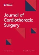
The aneurysmal sac was found to be 12 × 9 × 1 cms in dimension, occupying the entire right atrioventricular groove and displacing the right atrium (Figure 1 &2). There was no luminal thrombus or calcification. Histopathology of excised aneurysm had shown widespread myxoid degeneration in the media, focal necrosis, atherosclerosis and fibrosis of the medial muscles.

Discussion

Coronary artery aneurysm is defined as a localized dilatation exceeding the diameter of adjacent normal segment by 50% and occurs in approximately 1.5–5% of patient undergoing coronary angiography [1]. However, a coronary artery with a diameter more than 2 cm is termed as "giant aneurysm " and only a few cases have been described in the literature [223] (Table 1).
Table 1
Reported cases of giant coronary artery aneurysm in adults
Author
Year
Number
(patients)
Site
Presentation
Etiology
Management
Nobrega[2]'et al'
1996
1
RCA, LAD
MI
SLE
Medical
Channon 'et al'[3]
1998
1
RCA
Medistinal mass
Atherosclerotic
Medical
Yu 'etal'[4]
2001
2
RCA, LAD
LV fistula
Congenital
Surgical
Konen 'et al'[5]
2001
1
RCA
Medistinal mass
Atherosclerotic
Surgical
Hao 'et al'[6]
2003
1
RCA
Angina
Atherosclerotic
Surgical
Banerjee 'et al'[7]
2004
1
RCA
Medistinal mass
Atherosclerotic
Medical
Grandmougin 'et al'[8]
2005
1
RCA
Cardiac tumour
Atherosclerotic
Surgical
McGlinchey 'et al'[9]
2005
1
RCA
Cardiac compression
Atherosclerotic
Surgical
Dianyuan 'et al'[10]
2005
6
3RCA,2LAD,1Diagonal
LV fistula
Congenital, Atherosclerotic
Surgical
Shakir 'et al'[11]
2005
1
RCA, LAD
CHF
Hypercholesterolemia
Surgical
Augustin 'et al'[12]
2006
1
RCA
Cardiac compression
Atherosclerotic
Surgical
Kumar 'et al'[13]
2006
1
RCA
SVC syndrome
Atherosclerotic
Surgical
Manghat 'et al'[14]
2006
1
LAD
Chest pain
Kawasaki disease
Medical
Jindal 'et al'[15]
2007
1
LAD
Angina
Stent implantation
Surgical
Takano 'et al'[16]
2007
1
RCA
MI
Atherosclerotic
Surgical
Blank 'et al'[17]
2007
1
RCA
Medistinal mass
Atherosclerotic
Surgical
Malero 'et al'[18]
2008
1
RCA
Intracardiac mass
Atherosclerotic
Surgical
Vlachou 'et al'[19]
2008
1
RCA
Medistinal mass, MI
Atherosclerotic
Surgical
Eshtehardi'et al'[20]
2008
1
LAD
Angina
Atherosclerotic
Stenting
Matsubayashi 'et al'[21]
2008
1
LM
Angina
Atherosclerotic
Surgical
Kanaan 'et al'[22]
2008
1
RCA, LAD
Angina
Takayasu disease
Surgical
Sharma 'et al'[23]
2009
1
LAD
Angina
Stent implantation
Medical
RCA-right coronary artery, LAD-left anterior descending artery, LM-left main coronary artery, MI-myocardial infarction, CHF-congestive heart failure, LV-left ventricle, SVC-superior vena cava, SLE-systemic lupus erythematosus
In adults, CAA is predominantly atherosclerotic in origin however, other causes include Kawasaki disease, autoimmune disease, trauma, infection, dissection, congenital malformation and angioplasty [1, 24]. Recently, with the advent of implantation of drug eluting stents there are increasing reports suggesting stents causing coronary aneurysm months or years after the procedure [1, 14, 22, 24]. The proposed pathogenesis of stent-related aneurysm formation is multi-factorial. The drug-eluting stent contains immunosuppressant such as Sirolimus, which inhibits inflammation, or chemotherapeutic agents like Paclitaxel, which is an anti-inflammatory agent and inhibits cell proliferation. In due course of time, once drug is eluted, the polymer in which the drug is embedded may elicit a hypersensitivity reaction and vasculitis and results in weakening of vessel wall and subsequent dilatation [1, 14, 22, 24]. Mechanical damage to the arterial wall during balloon angioplasty and stent placement or turbulent blood flow may be an added factor for the development of an aneurysm [1].
The majority of the patients with CAA are asymptomatic but they may present with angina pectoris, myocardial infarction, sudden death or complications such as thrombus formation, embolisation, fistula formation, rupture, hemo-pericardium, tamponade, compression of surrounding structure or congestive heart failure [1, 3, 4, 912]. Coronary artery aneurysms are small, thick-walled structures with a relative low-risk of rupture but usually associated with myocardial ischemia [5] However, giant CAA are associated with advance age, tendency for complications including rupture and may present as mediastinal, intracardiac mass or superior vena cava syndrome in addition to ischemic symptoms [3, 5, 7, 13, 1719].
Clinical presentation of giant CAA may mimic aneurysm of ascending aorta, pulmonary trunk, cardiac tumour, pericardial tumour or thymoma [1]. Giant CAA may be detected by non-invasive tools like echocardiography, computed tomography, magnetic resonance imaging but coronary angiography remains the gold standard which provides information about size, shape, location and co-existing anomalies such as coronary artery disease [24]. Since, our patient presented with MI, a diagnostic coronary angiography was done straightaway.
Due to rarity and non-availability of controlled trials, there is no optimal management strategy for patients with giant CAA. However, depending on the symptoms, etiology and associated lesions medical treatment (anti-platelet agent, anti-coagulation), stent implantation or surgical exclusion of the aneurysm using resection or ligation technique have been described [1, 10]. A review of literature suggests that surgery is the preferred approach for Giant CAA in order to achieve excision of the aneurysmal sac, manage associated surgical condition and prevent complications [223].
The surgical management requires median sternotomy, cardiopulmonary bypass and myocardial revascularization (CABG). Occasionally femoral artery is cannulated for CPB to decompress the aneurysm and the ventricle before opening the chest for safety [10]. Prognosis of CAA is controversial but overall 5-year survival is reported to be 71% [1, 24].
Therefore, Giant CAA is an uncommon lesion with varied clinical presentation and should be considered in the differential diagnosis of other conditions mimicking similar symptoms and need proper use of imaging technology to diagnose this rare anomaly and associated lesions for successful management. Surgical management need to be planned carefully and requires appropriate techniques for a better outcome.
Written informed consent was obtained from the patient for publication of this case report including pictures for review.

Acknowledgements

Authors appreciate Mr Eberhard Vestweber-Wilmes for photography.
This article is published under license to BioMed Central Ltd. This is an Open Access article distributed under the terms of the Creative Commons Attribution License (http://​creativecommons.​org/​licenses/​by/​2.​0), which permits unrestricted use, distribution, and reproduction in any medium, provided the original work is properly cited.

Competing interests

The authors declare that they have no competing interests.

Authors' contributions

NJ collected the data and has written the manuscript. HO is the referring and treating cardiologist. JK is co-author and assisted the surgery. NA is consultant in-charge surgeon who operated upon the patient. GE is also a senior author who also managed this case.
Anhänge

Authors’ original submitted files for images

Below are the links to the authors’ original submitted files for images.
Literatur
1.
Zurück zum Zitat Nichols L, Lagana S, Parwani A: Coronary artery aneurysm: a review and hypothesis regarding etiology. Arch Pathol Lab Med. 2008, 132: 823-28.PubMed Nichols L, Lagana S, Parwani A: Coronary artery aneurysm: a review and hypothesis regarding etiology. Arch Pathol Lab Med. 2008, 132: 823-28.PubMed
2.
Zurück zum Zitat Nobrega TP, Klodas E, Breen JF, Liggett SP, Higano ST, Reeder GS: Giant coronary artery aneurysms and myocardial infarction in a patient with systemic lupus erythematosus. Catheterization and Cardiovascular Diagnosis. 1998, 39 (1): 75-79. 10.1002/(SICI)1097-0304(199609)39:1<75::AID-CCD16>3.0.CO;2-N.CrossRef Nobrega TP, Klodas E, Breen JF, Liggett SP, Higano ST, Reeder GS: Giant coronary artery aneurysms and myocardial infarction in a patient with systemic lupus erythematosus. Catheterization and Cardiovascular Diagnosis. 1998, 39 (1): 75-79. 10.1002/(SICI)1097-0304(199609)39:1<75::AID-CCD16>3.0.CO;2-N.CrossRef
3.
Zurück zum Zitat Channon KM, Wadsworth S, Bashir Y: Giant coronary aneurysm presenting as a mediatinal mass. Am J Cardiol. 1998, 82 (10): 1307-8. 10.1016/S0002-9149(98)00626-2.CrossRefPubMed Channon KM, Wadsworth S, Bashir Y: Giant coronary aneurysm presenting as a mediatinal mass. Am J Cardiol. 1998, 82 (10): 1307-8. 10.1016/S0002-9149(98)00626-2.CrossRefPubMed
4.
Zurück zum Zitat Yu WQ, Shou HS, Tao QS: Surgical treatment of giant coronary artery aneurysm. Asian Cardiovasc Thorac Ann. 2001, 9: 215-7.CrossRef Yu WQ, Shou HS, Tao QS: Surgical treatment of giant coronary artery aneurysm. Asian Cardiovasc Thorac Ann. 2001, 9: 215-7.CrossRef
5.
Zurück zum Zitat Konen E, Feinberg MS, Morag B, Guetta V, Shinfeld A, Smolinsky A, Rozenman J: Giant right coronary aneurysm-CT angiographic and echocardiographic findings. AJR. 2001, 177: 689-91.CrossRefPubMed Konen E, Feinberg MS, Morag B, Guetta V, Shinfeld A, Smolinsky A, Rozenman J: Giant right coronary aneurysm-CT angiographic and echocardiographic findings. AJR. 2001, 177: 689-91.CrossRefPubMed
6.
Zurück zum Zitat Hao WR, Chen FC, Kao PF, Hsieh MH, Chen YJ, Chan P: Adult giant coronary artery aneurysm-a case report and literature review. Acta Cardiol Sin. 2004, 20: 187-90. Hao WR, Chen FC, Kao PF, Hsieh MH, Chen YJ, Chan P: Adult giant coronary artery aneurysm-a case report and literature review. Acta Cardiol Sin. 2004, 20: 187-90.
7.
Zurück zum Zitat Banerjee P, Houghton T, Walters M, Kaye GC: Giant right coronary artery aneurysm presenting as a mediastinal mass. Heart. 2004, 90: e50-10.1136/hrt.2002.002519.CrossRefPubMedPubMedCentral Banerjee P, Houghton T, Walters M, Kaye GC: Giant right coronary artery aneurysm presenting as a mediastinal mass. Heart. 2004, 90: e50-10.1136/hrt.2002.002519.CrossRefPubMedPubMedCentral
8.
Zurück zum Zitat Grandmougin D, Croisille P, Robin C, Pacoch M, Barral X: Giant coronary aneurysm mimicking a compressive cardiac tumor: imaging features and operative strategy. Cardiovasc Pathol. 2005, 14 (5): 272-5. 10.1016/j.carpath.2005.04.001.CrossRefPubMed Grandmougin D, Croisille P, Robin C, Pacoch M, Barral X: Giant coronary aneurysm mimicking a compressive cardiac tumor: imaging features and operative strategy. Cardiovasc Pathol. 2005, 14 (5): 272-5. 10.1016/j.carpath.2005.04.001.CrossRefPubMed
9.
Zurück zum Zitat McGlinchey PG, Maynard SJ, Graham AN, Roberts MJD, Khan MM: Giant aneurysm of the right coronary artery compressing the right heart. Circulation. 2005, 112: e66-e67. 10.1161/CIRCULATIONAHA.104.496224.CrossRefPubMed McGlinchey PG, Maynard SJ, Graham AN, Roberts MJD, Khan MM: Giant aneurysm of the right coronary artery compressing the right heart. Circulation. 2005, 112: e66-e67. 10.1161/CIRCULATIONAHA.104.496224.CrossRefPubMed
10.
Zurück zum Zitat Dianyuan L, Wu Q, Sun L, Song Y, Wang W, Pan S, Luo G, Liu Y, Qi Z, Tao T, Sun JZ, Hu S: Surgical treatment of giant coronary artery aneurysm. J Thorac Cardiovasc Surg. 2005, 130: 817-21. 10.1016/j.jtcvs.2005.04.004.CrossRef Dianyuan L, Wu Q, Sun L, Song Y, Wang W, Pan S, Luo G, Liu Y, Qi Z, Tao T, Sun JZ, Hu S: Surgical treatment of giant coronary artery aneurysm. J Thorac Cardiovasc Surg. 2005, 130: 817-21. 10.1016/j.jtcvs.2005.04.004.CrossRef
11.
Zurück zum Zitat Shakir D, Carr C, Suwaidi J: Giant coronary artery aneurysm and severe mitral regurgitation in patients with familial hypercholesterolemia-case report and review of the literature. Int J Angiology. 2005, 14 (4): 249-52. 10.1007/s00547-005-2000-7.CrossRef Shakir D, Carr C, Suwaidi J: Giant coronary artery aneurysm and severe mitral regurgitation in patients with familial hypercholesterolemia-case report and review of the literature. Int J Angiology. 2005, 14 (4): 249-52. 10.1007/s00547-005-2000-7.CrossRef
12.
Zurück zum Zitat Augustin N, Wessely R, Porner M, Schomig A, Lange R: Giant coronary aneurysm obstructing the right heart. Lancet. 2006, 368 (9533): 386. 10.1016/S0140-6736(06)69112-5.CrossRefPubMed Augustin N, Wessely R, Porner M, Schomig A, Lange R: Giant coronary aneurysm obstructing the right heart. Lancet. 2006, 368 (9533): 386. 10.1016/S0140-6736(06)69112-5.CrossRefPubMed
13.
Zurück zum Zitat Kumar G, Karon BL, Edwards WD, Puga FJ, Klarich KW: Giant coronary artery aneurysm causing superior vena cava syndrome and congestive heart failure. Am J Cardiol. 2006, 98: 986-88. 10.1016/j.amjcard.2006.04.047.CrossRefPubMed Kumar G, Karon BL, Edwards WD, Puga FJ, Klarich KW: Giant coronary artery aneurysm causing superior vena cava syndrome and congestive heart failure. Am J Cardiol. 2006, 98: 986-88. 10.1016/j.amjcard.2006.04.047.CrossRefPubMed
14.
Zurück zum Zitat Manghat NE, Hughes GJM, Cox ID, Roobottom CA: Giant coronary artery aneurysm secondary to Kawasaki disease: diagnosis in an adult by multi-detector row CT coronary angiography. BJR. 2006, 79: e133-136. 10.1259/bjr/16077689.CrossRefPubMed Manghat NE, Hughes GJM, Cox ID, Roobottom CA: Giant coronary artery aneurysm secondary to Kawasaki disease: diagnosis in an adult by multi-detector row CT coronary angiography. BJR. 2006, 79: e133-136. 10.1259/bjr/16077689.CrossRefPubMed
15.
Zurück zum Zitat Jindal RK, George R, Singh B: Giant coronary aneurysm following drug-eluting stent implantation presenting as fever of unknown origin. J Invasive Cardiol. 2007, 19 (7): 313-314.PubMed Jindal RK, George R, Singh B: Giant coronary aneurysm following drug-eluting stent implantation presenting as fever of unknown origin. J Invasive Cardiol. 2007, 19 (7): 313-314.PubMed
16.
Zurück zum Zitat Takano MI, Oikawa M, Yamaki T, Yamaguchi O, Nakazato K, Ohsugi T, Kobayashi A, Watanabe M, Yaoita H, Yukio M: A case of recurrent myocardial infarction caused by a giant right coronary artery aneurysm. J Am Soc Echocardiogr. 2007, 20 (11): 1318.e5-1318.e8. 10.1016/j.echo.2007.04.008.CrossRef Takano MI, Oikawa M, Yamaki T, Yamaguchi O, Nakazato K, Ohsugi T, Kobayashi A, Watanabe M, Yaoita H, Yukio M: A case of recurrent myocardial infarction caused by a giant right coronary artery aneurysm. J Am Soc Echocardiogr. 2007, 20 (11): 1318.e5-1318.e8. 10.1016/j.echo.2007.04.008.CrossRef
17.
Zurück zum Zitat Blank R, Haager PK, Maeder M, Genoni M, Rickli H: Giant right coronary artery aneurysm. Ann Thorac Surg. 2007, 84: 1740-42. 10.1016/j.athoracsur.2007.05.006.CrossRefPubMed Blank R, Haager PK, Maeder M, Genoni M, Rickli H: Giant right coronary artery aneurysm. Ann Thorac Surg. 2007, 84: 1740-42. 10.1016/j.athoracsur.2007.05.006.CrossRefPubMed
18.
Zurück zum Zitat Malero EA, Martin YD, Janelle GM, Peng YG: An unusual giant right coronary artery aneurysm resembles an intracardiac mass. Anesth Analg. 2008, 107: 1161-62. 10.1213/ane.0b013e318181f74f.CrossRef Malero EA, Martin YD, Janelle GM, Peng YG: An unusual giant right coronary artery aneurysm resembles an intracardiac mass. Anesth Analg. 2008, 107: 1161-62. 10.1213/ane.0b013e318181f74f.CrossRef
19.
Zurück zum Zitat Vlachou PA, Mulcahy K, Adair W: Giant coronary artery aneurysm: an unusual cause of a mediastinal mass. Eur Radiol. 2008, 18 (12): 3007-3009. 10.1007/s00330-008-1010-1.CrossRefPubMed Vlachou PA, Mulcahy K, Adair W: Giant coronary artery aneurysm: an unusual cause of a mediastinal mass. Eur Radiol. 2008, 18 (12): 3007-3009. 10.1007/s00330-008-1010-1.CrossRefPubMed
20.
Zurück zum Zitat Eshtehardi P, Cook S, Moarof I, Triller HJ, Windecker S: Giant coronary artery aneurysm. Circulation: Cardiovascular Interventions. 2008, 1: 85-86. 10.1161/CIRCINTERVENTIONS.107.763656. Eshtehardi P, Cook S, Moarof I, Triller HJ, Windecker S: Giant coronary artery aneurysm. Circulation: Cardiovascular Interventions. 2008, 1: 85-86. 10.1161/CIRCINTERVENTIONS.107.763656.
21.
Zurück zum Zitat Matsubayashi K, Asai T, Nishimura O, Kinoshita T, Ikegami H, Kambara A, Suzuki T: Giant coronary artery aneurysm in the left main coronary artery: a novel surgical procedure. Ann Thorac Surg. 2008, 85 (6): 2130-32. 10.1016/j.athoracsur.2007.12.052.CrossRefPubMed Matsubayashi K, Asai T, Nishimura O, Kinoshita T, Ikegami H, Kambara A, Suzuki T: Giant coronary artery aneurysm in the left main coronary artery: a novel surgical procedure. Ann Thorac Surg. 2008, 85 (6): 2130-32. 10.1016/j.athoracsur.2007.12.052.CrossRefPubMed
22.
Zurück zum Zitat Kanaan S, Baker C, Starnes V: resection of giant coronary artery aneurysm in a Takayasu's arteritis patient. Ann Thorac Surg. 2008, 85 (5): 1795-96. 10.1016/j.athoracsur.2007.10.053.CrossRefPubMed Kanaan S, Baker C, Starnes V: resection of giant coronary artery aneurysm in a Takayasu's arteritis patient. Ann Thorac Surg. 2008, 85 (5): 1795-96. 10.1016/j.athoracsur.2007.10.053.CrossRefPubMed
23.
Zurück zum Zitat Sharma J, Kanei Y, Kwan TW: A case of giant coronary artery aneurysm after placement of a heparin-coated stent. J Invasive Cardiol. 2009, 21 (2): e22-23.PubMed Sharma J, Kanei Y, Kwan TW: A case of giant coronary artery aneurysm after placement of a heparin-coated stent. J Invasive Cardiol. 2009, 21 (2): e22-23.PubMed
24.
Zurück zum Zitat Pahlavan PS, Niroomand F: Coronary artery aneurysm: a review. Clin Cardiol. 2006, 29: 439-43. 10.1002/clc.4960291005.CrossRefPubMed Pahlavan PS, Niroomand F: Coronary artery aneurysm: a review. Clin Cardiol. 2006, 29: 439-43. 10.1002/clc.4960291005.CrossRefPubMed
Metadaten
Titel
Giant right coronary artery aneurysm- case report and literature review
verfasst von
Neerod K. Jha
Husam Z. Ouda
Javed A. Khan
Gregory P. Eising
Norbert Augustin
Publikationsdatum
01.12.2009
Verlag
BioMed Central
Erschienen in
Journal of Cardiothoracic Surgery / Ausgabe 1/2009
Elektronische ISSN: 1749-8090
DOI
https://doi.org/10.1186/1749-8090-4-18

Weitere Artikel der Ausgabe 1/2009

Journal of Cardiothoracic Surgery 1/2009 Zur Ausgabe

Deutlich weniger Infektionen: Wundprotektoren schützen!

08.05.2024 Postoperative Wundinfektion Nachrichten

Der Einsatz von Wundprotektoren bei offenen Eingriffen am unteren Gastrointestinaltrakt schützt vor Infektionen im Op.-Gebiet – und dient darüber hinaus der besseren Sicht. Das bestätigt mit großer Robustheit eine randomisierte Studie im Fachblatt JAMA Surgery.

Chirurginnen und Chirurgen sind stark suizidgefährdet

07.05.2024 Suizid Nachrichten

Der belastende Arbeitsalltag wirkt sich negativ auf die psychische Gesundheit der Angehörigen ärztlicher Berufsgruppen aus. Chirurginnen und Chirurgen bilden da keine Ausnahme, im Gegenteil.

Ein Drittel der jungen Ärztinnen und Ärzte erwägt abzuwandern

07.05.2024 Medizinstudium Nachrichten

Extreme Arbeitsverdichtung und kaum Supervision: Dr. Andrea Martini, Sprecherin des Bündnisses Junge Ärztinnen und Ärzte (BJÄ) über den Frust des ärztlichen Nachwuchses und die Vorteile des Rucksack-Modells.

Echinokokkose medikamentös behandeln oder operieren?

06.05.2024 DCK 2024 Kongressbericht

Die Therapie von Echinokokkosen sollte immer in spezialisierten Zentren erfolgen. Eine symptomlose Echinokokkose kann – egal ob von Hunde- oder Fuchsbandwurm ausgelöst – konservativ erfolgen. Wenn eine Op. nötig ist, kann es sinnvoll sein, vorher Zysten zu leeren und zu desinfizieren. 

Update Chirurgie

Bestellen Sie unseren Fach-Newsletter und bleiben Sie gut informiert.

S3-Leitlinie „Diagnostik und Therapie des Karpaltunnelsyndroms“

Karpaltunnelsyndrom BDC Leitlinien Webinare
CME: 2 Punkte

Das Karpaltunnelsyndrom ist die häufigste Kompressionsneuropathie peripherer Nerven. Obwohl die Anamnese mit dem nächtlichen Einschlafen der Hand (Brachialgia parästhetica nocturna) sehr typisch ist, ist eine klinisch-neurologische Untersuchung und Elektroneurografie in manchen Fällen auch eine Neurosonografie erforderlich. Im Anfangsstadium sind konservative Maßnahmen (Handgelenksschiene, Ergotherapie) empfehlenswert. Bei nicht Ansprechen der konservativen Therapie oder Auftreten von neurologischen Ausfällen ist eine Dekompression des N. medianus am Karpaltunnel indiziert.

Prof. Dr. med. Gregor Antoniadis
Berufsverband der Deutschen Chirurgie e.V.

S2e-Leitlinie „Distale Radiusfraktur“

Radiusfraktur BDC Leitlinien Webinare
CME: 2 Punkte

Das Webinar beschäftigt sich mit Fragen und Antworten zu Diagnostik und Klassifikation sowie Möglichkeiten des Ausschlusses von Zusatzverletzungen. Die Referenten erläutern, welche Frakturen konservativ behandelt werden können und wie. Das Webinar beantwortet die Frage nach aktuellen operativen Therapiekonzepten: Welcher Zugang, welches Osteosynthesematerial? Auf was muss bei der Nachbehandlung der distalen Radiusfraktur geachtet werden?

PD Dr. med. Oliver Pieske
Dr. med. Benjamin Meyknecht
Berufsverband der Deutschen Chirurgie e.V.

S1-Leitlinie „Empfehlungen zur Therapie der akuten Appendizitis bei Erwachsenen“

Appendizitis BDC Leitlinien Webinare
CME: 2 Punkte

Inhalte des Webinars zur S1-Leitlinie „Empfehlungen zur Therapie der akuten Appendizitis bei Erwachsenen“ sind die Darstellung des Projektes und des Erstellungswegs zur S1-Leitlinie, die Erläuterung der klinischen Relevanz der Klassifikation EAES 2015, die wissenschaftliche Begründung der wichtigsten Empfehlungen und die Darstellung stadiengerechter Therapieoptionen.

Dr. med. Mihailo Andric
Berufsverband der Deutschen Chirurgie e.V.