The air embolism in this case was likely to have been caused by positioning the patient in a prone position, which was associated with the lesion to be biopsied being at a maximum height over the left atrium. Due to the resulting negative pressure, air entered through a fistula that formed between the airspace and the pulmonary vein. The air could have been trapped in the left atrium by positioning the patient in left lateral position. The event itself could have been prevented by positioning the patient in an ipsilateral dependent position during the biopsy. In addition to hyperbaric oxygen therapy, the preferred treatment options are positioning maneuvers, administration of pure oxygen, and heparinization.
Hinweise
Competing interests
The authors declare that they have no competing interests.
Authors’ contributions
RR and BG are the authors responsible for this letter. RR and BG conducted the literature research, the writing and revision of the draft, the production of the figure and the final approval of the letter. FJW contributed to the writing and revision of the draft, especially concerning the intensive care therapy of acute air embolism. AL, AEG and TD contributed to the writing and critical revision of the manuscript, with special regard to neurological and radiological considerations. All authors read and approved the final manuscript.
Correspondence
Dear Sir,
We read with interest the report by Hung et al. [1] describing the case of a 63-year-old patient who suffered an acute stroke of the left middle cerebral artery and a non-ST elevation myocardial infarction due to an air embolism after a biopsy of a tumor in the lower lobe of the left lung. Although a very large amount of air had entered, filling the aorta to almost 40 % (Fig. 1b [1]), the patient fortunately survived the event and according to the report, was discharged to care at home 7 days later with residual hemiplegia on the right side and presumably persisting global aphasia.
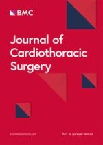
Fig. 1
Pulmonary venous pressures depending on the position of the lesion in relation to the left atrium. If the lesion is located below the level of the atrium, the pulmonary venous pressure at this location is the existing pressure in the left ventricle plus the hydrostatic pressure over the lesion to be biopsied (“T”), defined as the distance from the center of the left atrium to the lesion, marked “A” in the figure. If a pulmonary vein is injured, bleeding occurs here. If the lesion is located above the level of the atrium, the pulmonary venous pressure at this location is the existing pressure in the left atrium minus the hydrostatic pressure below the lesion to be biopsied, defined as the distance from the center of the left atrium to the lesion, marked “B” in the figure. If a pulmonary vein is injured, air enters the pulmonary vein due to the lower viscosity of air compared with blood. LV: Left ventricle; RV: right ventricle; RA: right atrium
×
A few remarks are necessary, both regarding the risk of this event and its treatment.
Anzeige
The biopsy was performed in prone position [1], one of the main risk factors for an air embolism [2]. As Fig. 1a shows, the lesion was in the posterior basal segment of the left lower lobe, at the time of the biopsy far above the level of the left atrium, not “under the level of the left atrium“, as the authors describe [1]. In this case, the prone position of the patient placed the lesion as far as possible above the left atrium. This is associated with negative intrapulmonary venous pressure that allows air to enter if an alveolar to pulmonary venous fistula, a bronchial to pulmonary venous fistula, or a direct connection between the tip of the needle and the pulmonary vein occurs. Figure 1 illustrates how the “position of the lesion above or below the level of the left atrium” should be understood. If the patient had been positioned in “ipsilateral dependent position” [3] i.e. in a supine position with the right side elevated somewhat, this complication may not have occurred.
From Fig. 1c [1], which like 1a and 1b is shown laterally reversed, but correctly labeled, it can be seen that a considerable amount of air was in the left atrium of the heart. This air could have been trapped in the left atrium by positioning the patient on the left side [4]. As can be seen in Fig. 1d [1], the patient was presumably turned over his right side onto his back after the biopsy, so the stroke occurred in the left medial cerebral artery. As both the desired and initially existing Trendelenburg positioning of the patient and the left lateral position that would have been protective in this case were abandoned, immediate transport to a pressure chamber should have been considered.
We agree in principle with the risk factors described [1, 2], but would like to note that Hiraki et al. [5] described no increased risk associated with the biopsy of a rather “centrally located lesion” [1] and that in 1990, CT fluoroscopy was not available to Worth et al. [6]. Of course the needle should not be placed directly in a central pulmonary vein and of course it is extremely important to monitor the needle position in real time if possible. However, in order to avoid mid-sized or small pulmonary veins, we recommend setting a thinner collimation than the 5 mm presented in this case [1].
Conservative treatment with aspirin is understandable considering its indication to prevent the development of microthrombi via the tiny arterial air emboli. In this case, however, considering the risk of hemorrhagic transformation of the middle cerebral artery stroke, it would have been preferable to administer heparin because, unlike aspirin, it can be antagonized. In general, administration of 100 % oxygen, not 50 %, is recommended, on the one hand to minimize the size of the gas bubbles by eliminating nitrogen from them and on the other hand to ensure the greatest possible oxygenation of the tissue [7]. Under this treatment, it would have been possible to await the physical resorption of the air in the Trendelenburg position or in left lateral position.
Anzeige
Open AccessThis article is distributed under the terms of the Creative Commons Attribution 4.0 International License (http://creativecommons.org/licenses/by/4.0/), which permits unrestricted use, distribution, and reproduction in any medium, provided you give appropriate credit to the original author(s) and the source, provide a link to the Creative Commons license, and indicate if changes were made. The Creative Commons Public Domain Dedication waiver (http://creativecommons.org/publicdomain/zero/1.0/) applies to the data made available in this article, unless otherwise stated.
Competing interests
The authors declare that they have no competing interests.
Authors’ contributions
RR and BG are the authors responsible for this letter. RR and BG conducted the literature research, the writing and revision of the draft, the production of the figure and the final approval of the letter. FJW contributed to the writing and revision of the draft, especially concerning the intensive care therapy of acute air embolism. AL, AEG and TD contributed to the writing and critical revision of the manuscript, with special regard to neurological and radiological considerations. All authors read and approved the final manuscript.
Die Therapie von Echinokokkosen sollte immer in spezialisierten Zentren erfolgen. Eine symptomlose Echinokokkose kann – egal ob von Hunde- oder Fuchsbandwurm ausgelöst – konservativ erfolgen. Wenn eine Op. nötig ist, kann es sinnvoll sein, vorher Zysten zu leeren und zu desinfizieren.
Der OP in der Zukunft wird mit weniger Personal auskommen – nicht, weil die Technik das medizinische Fachpersonal verdrängt, sondern weil der Personalmangel es nötig macht.
Seit November 2023 gibt es evidenzbasierte Empfehlungen zum perioperativen Management bei gastrointestinalen Tumoren (POMGAT) auf S3-Niveau. Vieles wird schon entsprechend der Empfehlungen durchgeführt. Wo es im Alltag noch hapert, zeigt eine Umfrage in einem Klinikverbund.
Auch wenn sich Krankenhäuser nachhaltig und grün geben – sie tragen aktuell erheblich zu den CO2-Emissionen bei und produzieren jede Menge Müll. Ein Pilotprojekt aus Bonn zeigt, dass viele Op.-Abfälle wiederverwertet werden können.
Update Chirurgie
Bestellen Sie unseren Fach-Newsletterund bleiben Sie gut informiert.
Das Karpaltunnelsyndrom ist die häufigste Kompressionsneuropathie peripherer Nerven. Obwohl die Anamnese mit dem nächtlichen Einschlafen der Hand (Brachialgia parästhetica nocturna) sehr typisch ist, ist eine klinisch-neurologische Untersuchung und Elektroneurografie in manchen Fällen auch eine Neurosonografie erforderlich. Im Anfangsstadium sind konservative Maßnahmen (Handgelenksschiene, Ergotherapie) empfehlenswert. Bei nicht Ansprechen der konservativen Therapie oder Auftreten von neurologischen Ausfällen ist eine Dekompression des N. medianus am Karpaltunnel indiziert.
Das Webinar beschäftigt sich mit Fragen und Antworten zu Diagnostik und Klassifikation sowie Möglichkeiten des Ausschlusses von Zusatzverletzungen. Die Referenten erläutern, welche Frakturen konservativ behandelt werden können und wie. Das Webinar beantwortet die Frage nach aktuellen operativen Therapiekonzepten: Welcher Zugang, welches Osteosynthesematerial? Auf was muss bei der Nachbehandlung der distalen Radiusfraktur geachtet werden?
Inhalte des Webinars zur S1-Leitlinie „Empfehlungen zur Therapie der akuten Appendizitis bei Erwachsenen“ sind die Darstellung des Projektes und des Erstellungswegs zur S1-Leitlinie, die Erläuterung der klinischen Relevanz der Klassifikation EAES 2015, die wissenschaftliche Begründung der wichtigsten Empfehlungen und die Darstellung stadiengerechter Therapieoptionen.