Erschienen in: Journal of NeuroEngineering and Rehabilitation 1/2023

Open Access 01.12.2023 | Review

Electrical stimulation methods and protocols for the treatment of traumatic brain injury: a critical review of preclinical research

verfasst von: D. Ziesel, M. Nowakowska, S. Scheruebel, K. Kornmueller, U. Schäfer, R. Schindl, C. Baumgartner, M. Üçal, T. Rienmüller

Erschienen in: Journal of NeuroEngineering and Rehabilitation | Ausgabe 1/2023

Abstract

Background

Traumatic brain injury (TBI) is a leading cause of disabilities resulting from cognitive and neurological deficits, as well as psychological disorders. Only recently, preclinical research on electrical stimulation methods as a potential treatment of TBI sequelae has gained more traction. However, the underlying mechanisms of the anticipated improvements induced by these methods are still not fully understood. It remains unclear in which stage after TBI they are best applied to optimize the therapeutic outcome, preferably with persisting effects. Studies with animal models address these questions and investigate beneficial long- and short-term changes mediated by these novel modalities.

Methods

In this review, we present the state-of-the-art in preclinical research on electrical stimulation methods used to treat TBI sequelae. We analyze publications on the most commonly used electrical stimulation methods, namely transcranial magnetic stimulation (TMS), transcranial direct current stimulation (tDCS), deep brain stimulation (DBS) and vagus nerve stimulation (VNS), that aim to treat disabilities caused by TBI. We discuss applied stimulation parameters, such as the amplitude, frequency, and length of stimulation, as well as stimulation time frames, specifically the onset of stimulation, how often stimulation sessions were repeated and the total length of the treatment. These parameters are then analyzed in the context of injury severity, the disability under investigation and the stimulated location, and the resulting therapeutic effects are compared. We provide a comprehensive and critical review and discuss directions for future research.

Results and conclusion

We find that the parameters used in studies on each of these stimulation methods vary widely, making it difficult to draw direct comparisons between stimulation protocols and therapeutic outcome. Persisting beneficial effects and adverse consequences of electrical simulation are rarely investigated, leaving many questions about their suitability for clinical applications. Nevertheless, we conclude that the stimulation methods discussed here show promising results that could be further supported by additional research in this field.
Hinweise

Publisher's Note

Springer Nature remains neutral with regard to jurisdictional claims in published maps and institutional affiliations.
Abkürzungen
BDNF
Brain-derived neurotrophic factor
CaMKII
Calcium/calmodulin-dependent protein kinase 2
cAMP
Cyclic adenosine monophosphate
CCI
Controlled cortical impact
CLOCK
Circadian locomotor output cycles protein kaput
CMG
Cystometrogram
CREB
CAMP response element-binding protein
CRY2
Cryptochrome 2
CST
Corticospinal tract
CT
Computed tomography
DBS
Deep brain stimulation
DoC
Disorders of consciousness
ECS
Electrical cortical stimulation
EE
Environmental enrichment
EEG
Electroencephalogram
EES
Epidural electrical stimulation
ELISA
Enzyme-linked immunoassay
ES
Electrical stimulation
EUS-EMG
External urethral sphincter electromyography
fMRI
Functional magnetic resonance imaging
FPI
Fluid percussion injury
GABA
Gamma-aminobutyric acid
GABABR
Gamma-aminobutyric acid beta receptor
GAD
Glutamic acid decarboxylase
GAP43
Growth associated protein 43
GFAP
Glial fibrillary acidic protein
IL-10
Interleukin-10
IL-1β
Interleukin-1 beta
LCN
Lateral cerebellar nucleus
LFMS
Low-field magnetic stimulation
LFP
Local field potential
LHA
Lateral hypothalamic area
LSCI
Laser speckle contrast imaging
LSM
Laser scanning microscopy
LTD
Long-term depression
LTP
Long-term potentiation
M1
Primary motor cortex
mCBF
Microvascular cerebral blood flow
MEP
Motor-evoked potential
MeSH
Medical subject headings
mNSS
Modified Neurological Severity Score
MR
Midbrain raphe
MRI
Magnetic resonance imaging
mRNA
Messenger RNA
MRS
Magnetic resonance spectroscopy
MSN
Medial septal nucleus
mTBI
Mild traumatic brain injury
MUA
Multi-unit activity
NADH
Nicotinamide adenine dinucleotide
NeuN
Neuronal nuclei
NMDA
N-Methyl-d-aspartate
NSS
Neurological severity screen
OX1R
Orexins receptor type 1
P-CREB
Phosphorylated CREB
p75NTR
P75 neurotrophin receptor
PCR
Polymerase chain reaction
PET
Positron emission tomography
PnO
Rostral pontine reticular nucleus
PPTg
Pedunculopontine tegmental nucleus
PrPc
Prion protein
PSD-95
Postsynaptic density protein 95
RMT
Resting motor threshold
rmTBI
Repetitive mild traumatic brain injury
RNA
Ribonucleic acid
RRT
Rotarod test
rTMS
Repetitive transcranial magnetic stimulation
RT-PCR
Reverse transcription PCR
SEP
Somatosensory-evoked potential
SPRT
Single-pellet reaching task
STDP
Spike timing-dependent plasticity
SVZ
Subventricular zone
SYN
Synaptophysin
TBI
Traumatic brain injury
tDCS
Transcranial direct current stimulation
TEM
Transmission electron microscopy
TMS
Transcranial magnetic stimulation
TNF-α
Tumor necrosis factor alpha
TrkB
Tropomyosin receptor kinase B
VNS
Vagus nerve stimulation
α1-AR
Alpha-1 adrenoceptor

Background

Most recent epidemiological surveillance reports indicate around 223,000 traumatic brain injury related hospitalizations in 2019 and more than 64,000 TBI-related deaths in 2020 in the USA alone [1]. Recent analysis of data from the European Union in 2017 shows a much higher number of TBI-related hospitalizations, although there are less TBI-related deaths [2]. Despite substantial differences among countries, TBI remains a leading cause of mortality and morbidity, particularly amongst the younger population. Decades-long accumulation of clinical and experimental data has set the path to considerable achievements in the clinical management of TBI, which brought a remarkable and gradual reduction in mortality due to head injury [3, 4]. Nevertheless, neurological deficits, cognitive and motor impairments, psychiatric disorders or other morbidities remain among the major sequelae of TBI [5]. Whilst these disabilities render TBI survivors dependent on assistance for daily activities, they also cause severe psychological and economic burden on families due to lifelong patient care.
A modest list of major pathophysiological changes after TBI includes dysregulated cerebral blood flow [6] and impaired cerebral oxygenation leading to ischemic insult [7], glutamate excitotoxicity [8, 9], blood brain barrier breakdown [10], cerebral edema [11, 12], oxidative and nitrosative stress [13, 14], cerebral inflammation [15, 16], hypo- and hyper perfusion [17], mitochondrial dysfunction [18], hemorrhage [19] and hyperemia [20]. The cascade of these pathophysiological changes starts within minutes to hours and days following the primary injury, and may directly or indirectly induce secondary damage to brain tissue, resulting in impaired connectivity and a delayed loss of neuronal cells. Moreover, chronic microglial activation and axonal damage may persist over much longer periods, leading to connectivity loss even years after trauma [21]. Based on the order of appearance of those pathologies, the post-TBI period can be roughly divided into the acute phase lasting minutes to hours after trauma, the subacute phase that lasts several days and is connected to the beginning of the secondary injury, and the chronic phase covering the weeks, months or even years following TBI [22, 23]. Decades of immense clinical and preclinical research were dedicated to deciphering the mechanisms of secondary damage and cell loss. Nevertheless, the continuously increasing knowledge in this field has not yet yielded the desired clinical applications for targeted pharmacological therapies to prevent or attenuate these mechanisms and stop further progression of tissue damage.
Neuromodulation by means of electrical and magnetic stimulation has been used to promote neuroplasticity and connectivity. Although the limited capabilities of nervous tissue to self-repair hinders complete regeneration of damaged brain tissue, processes involved in neuroplasticity can at least partially restore neuronal connectivity. Promising results observed in preclinical and clinical studies with electrical stimulation provide a good basis for the exploitation of neuroplasticity for functional restoration to alleviate trauma-induced disabilities [24, 25]. Hypo- or hyper-excitability, for instance, provide suitable targets for neuromodulatory interventions such as transcranial magnetic stimulation and deep brain stimulation [26, 27]. Supportive treatment of post-traumatic depression using electrical stimulation has also been subject to an immense interest [28, 29]. Preclinical studies, however, which are required to corroborate findings on underlying mechanisms of electrical stimulation and reveal neurobiological correlates of these stimulation techniques, are disproportionately sparse and appear to have attracted increased interest only over the last decade.
In the first part of this article, we give an overview on what is known about the effects of stimulation on neuronal cells and the state-of-the-art of the most commonly used electrical stimulation methods for therapeutic applications. In the second part we present a critical review of the available literature on preclinical studies using electrical stimulation in animal models of traumatic brain injury. The aims are (1) to assess the efficacy of these stimulation methods as post-TBI treatments in preclinical research across several selected studies, (2) to critically compare stimulation protocols as well as treatment time after traumatic insult and (3) to infer the translational value of the reported outcomes for clinical applications.

State-of-the-art

Effects of electrical stimulation on neurons

The excitability of neuronal cells facilitates modulation of their firing activity using external stimulation to enhance or suppress endogenous activity [30]. This modulation can be utilized for therapeutic or diagnostic purposes in several neurological diseases or injuries to nervous tissue [3134]. To better understand the advantages of therapeutic electrical stimulation following TBI, it is necessary to gain extensive insights into how and to which extent stimulation influences neuronal physiology and morphology.
Artificial electrical stimulation may change the electrical potential of the surrounding extracellular region through anodic as well as cathodic protocols [3537]. In cathodic stimulation, a negative current pulse is delivered to the extracellular area, which in turn depolarizes the cellular membrane with the aim to elicit an action potential. Anodic stimulation instead hyperpolarizes the region near the site of interest and thus decreases the membrane potential [38]. This results in a flux of positive ions towards the stimulation site from surrounding areas, which leads to a depolarization of the cell membrane further away from the site of stimulation, possibly triggering an action potential at the nearest Ranvier node [39, 40].
The effect of electrical stimulation on the brain depends on the inherent characteristics of the tissue. At the cellular level, it is easier to excite an axon than a soma, and myelinated axons are the most excitable part of the cell [41, 42]. Induced voltages differ between nodes and internodes due to the drastic differences in voltage-gated ion channel density [43, 44]. Activated axons progress the signal antidromically to the soma [45, 46] and orthodromically to the synaptic terminals [47, 48]. Bending, branching and significant changes in the diameter of an axon determine the effective site and threshold of the stimulation [43]. Generally, it is easier to elicit action potentials with negative currents in almost all cell compartments, except for some types of dendrites that are more prone to stimulation with positive currents [43, 49, 50].

Long-term potentiation (LTP), long-term depression (LTD) and plasticity

Electrical stimulation deeply influences brain electrophysiology through modulation of neuronal signaling not only in the short-term, but also in facilitating or attenuating long-term modifications on a cellular level [51, 52]. Activity-dependent synaptic plasticity can either strengthen or weaken the development of synapses [53, 54], which is crucial for post-traumatic regeneration and recuperation of high-level cognitive abilities like learning and memory formation, loss of which is a typical outcome of TBI [55, 56]. Long-term potentiation (LTP) and long-term depression (LTD) are highly complex and pivotal processes of synaptic plasticity, which may be heavily modified as a consequence of TBI, possibly leading to severe cognitive impairments [56].
LTP is a form of synaptic enhancement resulting in a lasting facilitation of signal transduction. Classically, LTP is elicited through brief high frequency stimulation [57], although it may also be triggered successfully with theta-burst stimulation protocols [58] or chemical compounds [59]. Initiation of LTP requires the activation of postsynaptic N-methyl-d-aspartate (NMDA) receptors with glutamate. Subsequent rapid increase of calcium concentration within the cell initiates multiple metabolic cascades and the modulation of gene transcription, resulting in long-term changes to receptor expression, synaptic vesicle transport and other cytoskeletal interactions [55, 60]. LTD is a process analogous to LTP, but leads to reduction of synaptic efficacy. It is usually induced by low frequency stimulation, leading from low to moderate influx of calcium into the neuron mostly through voltage-gated calcium channels and, to a lesser extent, through the activation of NMDA receptors [60, 61].
During a head trauma, mechanical forces applied to nervous tissue disturb ionic fluxes and the concomitant depolarization [62]. This leads to excessive glutamate release from presynaptic axon terminals in the acute phase of the injury that may result in neuronal hyperexcitability and changes in synaptic plasticity. In general, TBI attenuates synaptic LTP responses, while its effect on LTD may vary [56]. LTP deficits and overall increased neuronal excitability were observed soon after injury in in vivo and ex vivo TBI models [63, 64], while the ability to induce LTD was left unchanged [64]. In a controlled cortical impact model in rats, LTD was enhanced as long as 2 days after the initial injury [65]. Considering all of the above, pertinent electrical stimulation protocols to effectively modulate LTP and LTD could be advantageous in the recovery of physiological neuroplasticity mechanisms and the recuperation of impeded motor and cognitive functions following TBI.

Spike timing-dependent plasticity (STDP)

Timing of the activation of presynaptic and postsynaptic cells plays a pivotal role in synaptic plasticity [66, 67]. Spike timing-dependent plasticity (STDP) is considered a biologically plausible model for synaptic modifications occurring in vivo [68, 69] and its occurrence has been reported in several brain regions, such as the corticostriatal pathway [7072], the barrel cortex [73, 74] and the visual cortex [75, 76]. It is determined by the temporal order of action potential initiations and the narrow time between subsequent action potentials. In general, activation of the presynaptic cell immediately before activation of the postsynaptic cell leads to timing-dependent LTP, while activating the presynaptic neuron immediately after the postsynaptic cell elicits timing-dependent LTD [68, 69]. The time window between these activations needs to be in the order of milliseconds, is specific for each synapse and depends on receptor kinetics, current densities and the release of retrograde messengers such as endocannabinoids [69]. Spontaneous spiking as well as changes in the spike frequency can further modulate the strength of plasticity, e.g. higher frequency stimulation has been described to increase the effect of timing-dependent LTP [69]. STDP was observed in both excitatory and inhibitory neurons and could be further modified by cholinergic, dopaminergic and adrenergic signaling [68], enabling prospective pharmacological modulation. It offers an alternative to frequency-dependent stimulation in clinical settings and has already been deployed in human studies to successfully modulate the force of the long-latency stretch reflex in healthy volunteers [77], while overall lower limb motor output was improved in patients with spinal cord injury [78].

Electrical stimulation methods

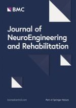
The most prevalent electrical stimulation methods used in post-TBI treatment studies, which are in the focus of this review, are transcranial magnetic stimulation (TMS), transcranial direct current stimulation (tDCS), deep brain stimulation (DBS) and vagus nerve stimulation (VNS) [25, 79]. TMS and tDCS are amongst the most commonly used non-invasive brain stimulation techniques [80, 81]. They are effective in the treatment of a wide variety of neurologic impairments, but their efficiency and precision is limited by the distance of the stimulator to the target region. Invasive stimulation methods, such as DBS or VNS, may achieve higher precision and efficiency by bringing the stimulation electrodes closer to the desired area. A schematic overview of these four stimulation methods and their preclinical usage is depicted in Fig. 1.

Transcranial magnetic stimulation

TMS is a non-invasive method that utilizes magnetic fields to inhibit or enhance the electrical activity of brain tissue with the aim to improve various neurologic disabilities [82, 83]. This technique utilizes the physical principle of electromagnetic induction by running a high alternating current through a magnetic coil positioned tangentially to the skull of a subject, leading to the formation of a magnetic field that is able to penetrate the skull. When stimulation is applied in the form of pulses, the rapid changes in the magnetic field create electrical currents in the brain, which in turn leads to excitation or inhibition of electrical activity, depending on the frequency of stimulation [84].
The main limitation of this method is that the electromagnetic field created by the coil rapidly decreases in strength with increasing distance. Thus, TMS is mainly used to stimulate cortical areas near the surface of the brain; however, functionally connected regions deeper in the brain can be stimulated indirectly through projecting axons [24]. The depth that the magnetic field penetrates into the brain as well as the size of the stimulated area can be adapted to specific requirements by selecting different coil types with various geometries, materials and designs. Circular coils, for example, can be used to uniformly stimulate a larger volume of neuronal tissue, resulting in greater penetration depth. Figure-of-eight shaped designs, comprising two circular coils positioned next to each other, allow for more selective stimulation at the cost of penetration depth [85]. The area where the two electromagnetic fields produced by this arrangement overlap is characterized by an increased current density compared to the surrounding regions [84]. TMS can be applied in a wide variety of different protocols, most commonly in the form of repetitive pulses, which is usually referred to as repetitive TMS (rTMS) [86].
The therapeutic potential of rTMS is widely recognized, particularly in the field of psychiatry, and it is applied as a treatment option for depression [87, 88] and obsessive–compulsive disorder [89, 90]. Its efficacy was further tested as a treatment for a number of different neurological conditions, such as neuropathic pain [91, 92], epilepsy [93], stroke [94], multiple sclerosis [95] and post-traumatic stress disorder [96], as well as Parkinsonian movement disorders [97, 98].

Transcranial direct current stimulation

In contrast to other stimulation methods that employ pulsed protocols for neurostimulation, tDCS uses direct current to influence the cell membranes of neurons in the desired cortical area [99, 100]. A current of several milliamperes is applied via a pad electrode, called the active electrode, attached to the pericranium near the area of interest, which leads to changes in cortical excitability and neuronal activity [101, 102]. A second, larger reference electrode is usually placed further away from the stimulation site. During anodal tDCS, a positive current is applied between the two electrodes, leading to a hyperpolarization of the area near the active electrode, whilst cathodal tDCS depolarizes the tissue with the use of negative currents. The resulting excitation or inhibition of neurons may lead to neuromodulation in affected areas [99, 103]. The current density is crucial for the efficacy and propagation depth of the stimulus [104].
This method is painless, non-invasive and used as a treatment for depression and a variety of cognitive dysfunctions [105, 106]. However, lack of precision is a limiting factor in cases where targeted neurostimulation would be necessary, such as post-traumatic tremor [107].

Deep brain stimulation

DBS is an invasive approach that requires the implantation of a stimulation electrode directly into the targeted brain area [108, 109]. The stimulation setup comprises an implanted stimulation electrode and a connected subcutaneous wire that forwards signals from an external stimulating device. Stimulation electrodes are often implanted bilaterally and commonly have multiple metal contacts, which can be used both as anodes and as cathodes [110]. In bipolar configurations, an electrical field is generated between two adjacent contacts, allowing for a concentrated electric field and thus a higher precision [110]. The optimal electrode position is usually determined beforehand with the help of neuroimaging via computed tomography (CT) or magnetic resonance imaging (MRI), which can also be used to guide the surgeon during implantation. Throughout the procedure, electrical activity is continuously measured to ensure correct electrode placement. Afterwards, the efficacy of the implanted device is verified by applying initial stimulation pulses [111].
This method is approved for the symptomatic treatment of Parkinson’s disease, essential tremor, obsessive compulsive disorder and some cases of severe epilepsy in humans [112, 113]. Thanks to its versatility and high spatial resolution, DBS has potential use in the treatment of higher-order cognitive dysfunction and disorders of consciousness in patients with TBI [114].

Vagus nerve stimulation

VNS is an indirect brain stimulation method that excites the afferents of the vagus nerve to modulate activity of the central nervous system. While vagal afferents provide sensory information to the brain stem from multiple internal organs, efferents mediate the parasympathetic control of various bodily functions. Thus, VNS results in a wide range of different effects caused by the stimulation of medulla and brainstem including the modulation of neurotransmitters: notably epinephrine, serotonin and gamma-aminobutyric acid [115]. Other potential modes of action include changes in blood flow in several brain regions [116118], upregulation of neurotrophin production [119], reduction of damage to the blood brain barrier [120122] and anti-inflammatory effects [123, 124]. VNS systems are approved for treatment of drug-resistant epilepsy [125] and severe, recurrent unipolar and bipolar depression [126], both of which are common disorders developing as a consequence of TBI [127129].
Most commonly, VNS is used as an invasive modality, employing helical cuff electrodes in monopolar, bipolar or tripolar configurations. These electrodes are usually wrapped around the left cervical vagus nerve [130] to indirectly stimulate distant brain regions. Stimulation of the right vagus nerve might lead to severe bradycardia and is therefore generally avoided [130]. Monopolar electrodes are comparatively cheap, but require an additional ground electrode. Bipolar configurations allow the induced current to flow between two electrodes, enabling a much greater control of the current path. Tripolar electrodes are more expensive, but have the advantage of preventing leakage currents to the surrounding tissue since the stimulating electrode is positioned between two common counter-electrodes.

Stimulation waveforms and protocols

The selection of suitable protocols is an important factor for efficacious stimulation, but also for the prevention of damage to the stimulating electrodes and the surrounding tissue [35]. This is particularly relevant for invasive approaches, such as DBS and VNS, where implanted electrodes need to last for longer periods of time and are in direct contact with neural tissue [131]. Unwanted electrochemical reactions at the electrode-tissue interface include corrosion and oxygen reduction reactions, which can be minimized by selecting appropriate stimulation protocols and waveforms [132, 133]. While monophasic pulses are more efficacious for stimulation than biphasic pulses, they potentially result in greater tissue damage, since all injected charge creates electrochemical reaction products and result in greater negative overpotentials over time [35, 134]. Biphasic stimulation, on the other hand, has the potential to reverse electrochemical processes at the electrode-tissue interface, but may also reverse some of the desired effects necessary for efficacious charge induction. Introducing a short interphase delay reduces the suppressing effect of the reversal phase, as long as the delay is short enough to prevent excessive accumulation of electrochemical reaction products [35].
Another important part of the stimulation protocol is the timing of the treatment application after injury, which depends on the selected treatment modality, the severity of the trauma and the goal of the treatment [135]. The onset of stimulation in preclinical studies varies from immediately to several weeks after trauma [25]. In clinical settings, these techniques are usually applied at later stages as a support to traditional rehabilitation methods for treating disabilities that persist after TBI [114, 136].

Additional stimulation methods

In addition to the methods mentioned above, there are several other promising electrical stimulation modalities that may be effective in the treatment of TBI sequelae. Electrical cortical stimulation, an invasive method where electrodes are implanted near the cortical surface, can be used to modulate brain plasticity to treat sensorimotor and cognitive deficits in rats [137]. Similarly, epidural electrical stimulation utilizes pulsed stimulation protocols applied to electrodes implanted in the epidural or subdural space to enhance motor recovery and brain activity [138140]. Promising non-invasive TBI treatment methods include electroconvulsive therapy, which finds use as the treatment for mood disorders such as depression [141], but has not yet been investigated in preclinical TBI models.
Temporal interference stimulation is another novel treatment modality that can be used to stimulate deep brain regions non-invasively, exploiting a well-known acoustic phenomenon [142]. By applying two sinusoidal stimuli in the kilohertz-range with slightly differing frequencies through electrode pairs placed on the head of a patient, interference patterns can be generated inside the brain [143]. The effect of stimuli in the kilohertz range on the underlying tissue is only small due to the filtering properties of cellular membranes [144, 145], and the amplitude of the individual signals is comparably low. Constructive interference of these two signals in the target area leads to an electric field oscillating with an envelope frequency equal to the difference between the two individual signal frequencies. This method has successfully been applied to mouse motor cortex, leading to the elicitation of movements [146].
It is also possible to implant passive components in the brain that convert an external impulse from a source outside the skull into an electrical stimulus. An example for this would be photocapacitors [147149], which charge up when they are irradiated by light pulses, creating an electric field at their surface, leading to the depolarization of adjacent neural cells. These photocapacitive devices can also be used in combination with temporal interference stimulation protocols [150].

Systematic literature review

To gain further insight into the methods and protocols used for TBI therapy in preclinical studies, an extensive systematic literature search was conducted. The articles included in this survey were found in PubMed and Web of Science. The scientific integrity of the review was ensured by closely following the PRISMA 2020 guidelines [151]. A flow diagram detailing the literature assessment process is given in Fig. 2.

Search terms: literature identification

To cover the most commonly used variations that describe the stimulation methods selected for this review as well as TBI, the search query consisted of the following MeSH terms:
(“transcranial magnetic stimulation” OR “transcranial direct current stimulation” OR “deep brain stimulation” OR “vagus nerve stimulation” OR "vagal nerve stimulation") AND (“traumatic brain injury” OR “tbi” OR “concussion”)
The search was conducted in the PubMed and Web of Science databases. To obtain as many relevant records as possible, the query was searched in all fields of the respective databases, which includes titles, abstracts and keywords of publications, among other information. The list of search results was last updated on the 8th of September 2022 and the search yielded 358 results in PubMed and 524 in Web of Science, amounting to a total of 583 different records after removing duplicates. The results were sorted by publication date from oldest to most recent and the titles, authors and publication years of these records were exported from the respective databases and collected in a Microsoft Excel spreadsheet to organize further screening.

Inclusion criteria: literature screening

The abstracts of all 583 individual search results were screened by one investigator for five different criteria of interest to this review. This was done manually without the use of any advanced automation tools except for a simple text search function. First, the abstract needed to mention TBI as the underlying cause of the disability under investigation. Next, an electrical stimulation method had to be utilized in the study and third it had to be used for a therapeutic purpose or as a treatment, as opposed to a diagnostic application. The record also needed to consist of original research, which excluded other review articles and excerpts from larger studies, such as meeting abstracts and conference papers. Finally, only preclinical studies were included, where an animal model was utilized to investigate certain parameters of interest.
These five criteria were assessed in the order described above, and when an article did not contain that criterion, it was immediately excluded from the review. A total of 543 records were excluded, 93 of which did not investigate TBI sequelae, 60 were not about electrical stimulation, 97 used these methods for an application other than therapy, 162 were not original research and 131 of the remaining articles were not preclinical studies. This abstract screening resulted in 40 articles for the following full-text assessment step.

Eligibility: full-text assessment

Out of the 40 articles selected for full-text assessment, another six were excluded. Four of the excluded articles used electrical stimulation not for the therapy but for the assessment of stimulation effects on healthy animals. One study was not original research, which was not immediately apparent in the abstract, and another article did not utilize electrical stimulation altogether. Ultimately, literature screening led to a total of 34 articles that were reviewed in this study. Eight separate studies used TMS and VNS respectively, seven employed tDCS, and eleven utilized DBS for the treatment of TBI sequelae.

Results

During full-text assessment, multiple parameters were collected from the 34 selected articles for further analysis and comparison. The first two columns list general information about the respective study, such as its main focus and the impairment under investigation. The next column describes the animal model used in each study, which includes the number and type of animals, the applied TBI model, and if animals were anesthetized during stimulation. After that, the technical aspects of the applied stimulation are summarized, such as the stimulation protocol that was used, the time frame of the stimulation, and the location that was stimulated. The last set of parameters focuses on the assessment of the study results, namely the tests that were conducted with the animals, the parameters that were studied, if they observed any long-term effects of stimulation, and a short summary of the main findings of the paper. All this information was collected in four individual tables, one each for TMS, tDCS, DBS and VNS, which are displayed below (Tables 1, 2, 3, 4).
Table 1
Overview of preclinical transcranial magnetic stimulation (TMS) studies
References
Main focus
Impairment
Animal model
Stimulation protocol
Stimulation time frame
Yoon YS et al. [138]
Effects of rTMS and EES on TBI
Motor function
51 male Sprague–Dawley rats (21 died from TBI), Marmarou’s weight drop (450 g from 1 m, diffuse, mild TBI, medial impact), awake and immobilized during TMS
90% of max. device output, 10 Hz, 3 s stim and 6 s pause, for 10 min
Twice per day, day 1–14 post-injury
Yoon KJ et al. [152]
rTMS for behavioral recovery
Motor function, brain metabolism, cell death
20 adult male Sprague–Dawley rats, lateral FPI (3.5–4 atm pressure, severe TBI), awake and immobilized during TMS
80% of RMT, 10 Hz, 15 trains of 2 s, 1 s inter-train interval
10 sessions over 2 weeks, beginning on 4th day post-injury
Lu H et al. [153]
rTMS for pediatric TBI
Motor function
26 juvenile Sprague–Dawley rats, CCI over left primary somatosensory cortex (severity unclear), TMS under 2% isoflurane
25% of max. device output, 20 Hz, 9 trains of 100 pulses, 55 s inter-train interval, for 9 min
Twice per week, starting 9 post-injury, for 4 weeks
Lu X et al. [156]
rTMS for neuromodulation and neurogenesis
Loss of brain parenchyma, reduced brain metabolism, neurological impairment
38 adult Sprague–Dawley rats, Feeney’s weight drop (moderate TBI, right hemisphere), awake and immobilized during TMS
60% of max. device output, 5 Hz, 36 trains of 25 pulses, 15 s inter-train interval, 900 pulses/day, figure-of-eight coil
From 2 days post-injury until 1 day before sacrificed (7/14/28 days after TBI)
Verdugo-Diaz et al. [157]
Treatment with intermediate frequency rTMS
Mortality, general behavioral changes
97 male Wistar rats, Marmarou’s weight drop (motor cortex, severe TBI), awake and immobilized during TMS (animals trained for immobilization)
50% of max. device output (120% of RMT), 2 Hz, 15 min per day, figure-of-eight coil
Starting 1 day post-injury, for 7 consecutive days
Shin et al. [154]
Therapy with rTMS and environmental enrichment
motor function
97 male Sprague–Dawley rats, CCI (4 m/s, moderate TBI, right hemisphere), MEP assessment under isoflurane, electrophysiological recordings under urethane, fMRI under sedation, rTMS under 2% isoflurane
10 Hz, 7 cycles of 4 s, 26 s between cycles, figure-of-eight coil, (stim. intensity unclear)
Starting 1 day post-injury, daily, for 6 days
Sekar et al. [155]
Low-field magnetic stimulation (LFMS, rTMS variant) treatment after TBI
Cognitive and motor functions
48 male C57BL/6 mice, weight drop (60 g from 1 m, closed head trauma, repetitive TBI, once daily for 3 consecutive days, severity unclear), awake and immobilized during TMS
40 Hz, 6 ms pulses, 80 trains of 2 s, 8 s pause, magn. field changes between uniform and linear gradient every 2 min, for 20 min
Once per day, following recovery from rightening reflex after TBI, for 3 days and once on day 4
Qian et al. [158]
Investigation of cellular mechanisms caused by rTMS treatment
General overview
45 male Sprague–Dawley rats, Feeney's weight drop (20 g × 30 cm impact force, moderate TBI), awake and immobilized during rTMS
30% of motor threshold, 40 Hz, 40 trains of 1 s, in 15 s intervals
Starting 4 days post-injury, once daily, for 2 weeks, five times per week
References
Stimulus location
Tests
Acquired parameters
Persistent effects
Main findings
Yoon YS et al. [138]
Center of the coil placed above injury site
Limb placement test, SPRT, RRT, immunohistochemistry
Limb placement changes, SPRT success rate, RRT performance time rate, c-Fos expression
Not investigated
TMS and EES resulted in significant improvement in SPRT and accelerated improvement in RRT, with particularly robust effects of EES
Yoon KJ et al. [152]
Area with largest MEP amplitude at the weaker biceps femoris after suprathreshold stim., side not stated (probably ipsilateral)
Rotarod and beam balance tests, brain MRI, magnetic resonance spectroscopy, western blot, immunohistochemistry
Motor coordination, balance ability, intact and lesioned hemispheric volume, brain metabolism, apoptotic signaling
Not investigated
rTMS did not have beneficial effects on motor recovery, enhancement of anti-apoptotic response in perilesional area
Lu H et al. [153]
Contralateral primary sensory region
Extracellular electrophysiological recordings, fMRI, open field test, forelimb and hindlimb reflex test, immunostaining
CaMKII expression (LTP), MUA responses, LFP magnitude, evoked fMRI cortical responses, behavioral tests (physiology and hyperactivity)
Long-lasting increase of excitability in non-injured cortex after 4 weeks of TMS therapy
Significant increases in evoked-fMRI cortical response, evoked synaptic activity, evoked neuronal firing and expression of neuroplasticity markers, decreased hyperactivity in behavioral tests
Lu X et al. [156]
Whole brain influenced by magnetic field (max. stim. over the center of the brain)
Behavioral tests (mNSS evaluation), hematoxylin and eosin staining, immunohistochemistry, PET examination
Behavioral recovery, relative brain parenchyma loss, cell proliferation and neurogenesis, neuron protection, cell apoptosis, metabolic activity
Not investigated
High-frequency rTMS may decrease mortality, mature neuron loss, apoptosis, improve behavioral recovery, cell proliferation and neurogenesis in the SVZ, metabolic activity in the contralateral site was not affected
Verdugo-Diaz et al. [157]
Injury site
Hunter’s 21-point behavioral-neurological scale, histology
Body weight, food intake, post-TBI bleeding and mortality, neurobehavioral score, cellular morphological changes, disruptions in hippocampal tissue architecture
Not investigated
Movement restriction prevents damage caused by TBI, intermediate-frequency rTMS slightly promotes behavioral and histologic recovery after TBI
Shin et al. [154]
Midpoint between lambda and bregma, medial located
Beam walk and challenge ladder tests, electrophysiology, evoked LFP, MEP assessment, fMRI in the contralateral cortex
Beam traversal latency, mean speed and slips from ladder, MEP amplitude, LFP magnitude, fMRI activation maps
Combination of EE and TMS led to benefits in sensorimotor function lasting up to 6 weeks
Combined therapy with TMS and EE after TBI leads to functional improvements, possibly via cortical excitability and reorganization, long-term effects probably due to EE rather than TMS
Sekar et al. [155]
Cortical and subcortical areas
RRT, open field test, novel location recognition test, immunohistochemistry, western blot
Time on rotarod, locomotor activity, cognitive function, PrPc level in plasma, GFAP, NeuN and PrPc protein levels, CLOCK and CRY2 levels
Not investigated
LFMS treatment improved motor and cognitive function in mice after repetitive TBI, restored PrPc level, decreased proteins associated with circadian rhythm, decreased GFAP levels, increased NeuN levels, and showed neuroprotective effects
Qian et al. [158]
Coil placed above ipsilateral side, close to the scalp
mNSS assessment, TEM, immunohistochemistry, western blot, RT-PCR detection
Injury severity, synaptic ultrastructure, protein expression (BDNF, TrkB, NMDAR1, P-CREB, SYN), mRNA expression levels
Not investigated
rTMS may promote recovery of neurological functions in TBI rats through enhanced SYN protein levels to promote synaptic reconstruction and affecting the expression of proteins related to LTP occurrence
Table 2
Overview of preclinical transcranial direct current stimulation (tDCS) studies
References
Main focus
Impairment
Animal model
Stimulation protocol
Stimulation time frame
Yoon et al. [159]
Effects of anodal tDCS on behavioral and spatial memory in early stage TBI
Behavioral and spatial memory
36 male Sprague–Dawley rats, lateral FPI (moderate TBI), anesthetized during tDCS
Anodal tDCS, 0.2 mA, (2.82 mA/cm2 current density), for 20 min
Once per day, for 5 days, starting 1 or 2 weeks post-injury
Kim and Han [160]
Effects of anodal tDCS on neuroplasticity
Motor and sensory cortical excitability
31 male Sprague–Dawley rats (postnatal day 42), weight drop (175 g from 30 cm, 3 consecutive times, repetitive mild TBI), anesthetized during all procedures and evaluations
Anodal tDCS, 0.2 mA (0.255 mA/cm2 current density), for 30 min
Once, directly after TBI
Bragina et al. [161]
Perfusion and tissue oxygenation after anodal tDCS, motor and cognitive neurologic outcome
mCBF and tissue oxygenation, motor function
40 mice, CCI (5 m/s, 2 mm from cortical surface, mild to moderate TBI), awake during tDCS
Repetitive anodal tDCS, 0.1 mA, for 15 min
Over 4 weeks, for 4 consecutive days at 3-day intervals, starting 1 or 3 weeks post-injury
Yu et al. [162]
Effects of tDCS and ECS on motor and cognitive recovery, brain plasticity, spatial learning and memory
Motor and cognitive function
30 male Sprague–Dawley rats, weight drop (moderate TBI), awake during tDCS
Anodal tDCS, 0.1 mA, 50 Hz, 200 µs pulses, for 30 min
Once per day from days 3 to 28 after electrode positioning
Martens et al. [165]
Cathodal tDCS in the treatment of psychiatric-like symptoms after TBI
Impulsivity and attention
20 male Long-Evans rats, bilateral, frontal CCI (severe TBI), anesthetized during tDCS
Cathodal tDCS, 800 µA (0.708 mA/cm2), 10 min
Once per day for 7 days (2 h before testing), starting 6 weeks post-injury
Bragina et al. [164]
Effects of anodal tDCS on cerebrovascular reactivity and mCBF regulation
Cerebrovascular reactivity and mCBF
20 mice, CCI (5 m/s, 2 mm from cortical surface, mild to moderate TBI), awake during tDCS
Anodal tDCS, 0.1 mA, for 15 min
Once, 3 weeks post-injury
Park et al. [163]
Anodal tDCS to improve motor function after repetitive mild TBI
Motor function
65 male Sprague–Dawley rats, weight drop (175 g from 30 cm, once daily for 3 days, repetitive mTBI), anesthetized during tDCS
Anodal tDCS, 0.2 mA (0.255 mA/cm2), for 30 min
Once, 24 h after last induction of mTBI
References
Stimulus location
Tests
Acquired parameters
Persistent effects
Main findings
Yoon et al. [159]
Anode over perilesional area, cathode on chest
RRT, Barnes maze test, brain MRI, MRS, immunohistochemical analysis
Behavioral ability, spatial memory, lesion volume, brain edema, metabolites, BDNF expression
Beneficial effects visible 1 weeks after stimulation, no sustained effects after 3 weeks
tDCS increases recovery of spatial and memory functions when applied 2 weeks post injury, only improves spatial memory when applied 1 week post-injury
Kim and Han [160]
Anode around left motor cortex, counter electrode on thorax
MEP and SEP test, brain MRI, immunohistochemical analysis
Recovery of righting reflex, MEP latency and amplitude, SEP latency and amplitude, brain volumetric changes, GFAP expression
Immunohistochemistry performed 12 days after stimulation, showed no significant improvements
Single anodal tDCS after rmTBI induces early recovery of consciousness, increases modulation of cortical excitability and promotes transient motor recovery
Bragina et al. [161]
Anode near craniotomy, counter electrode on thorax
Custom-made LSCI, two-photon LSM, RRT, passive avoidance test, Y-maze test, Nissl staining
Regional and microvascular cerebral blood flow, motor deficits, learning, spatial and working memory
Preserved improvement in learning and motor abilities 1 week after stimulation was ended
Anodal tDCS increases brain microvascular blood flow and tissue oxygenation in TBI and sham mouse brain and could contribute to neurologic improvement
Yu et al. [162]
Anode above lesion, cathode at trunk
Rehabilitation training (SPRT, RRT, Y-maze), neurological examination, histology, immunohistochemistry
Success rate of SPRT and Y-maze tests, average rates of RRT, lesion assessments, c-Fos expression
Not investigated
ES with rehabilitation training for TBI rats is effective for motor recovery and brain plasticity, ECS induces faster behavioral and cognitive improvements than tDCS
Martens et al. [165]
Cathode near bregma, anode between scapulae
Five-choice serial reaction time task, analysis of brain slices to verify injury severity
Motor impulsivity, attention, relationship between magnitude of impairment and recovery
No lingering effects observed, disappeared after stimulation stopped
Relationship between magnitude of impulsive deficit and degree of tDCS-recovery, the most severely impaired subjects benefit the most from neuromodulation
Bragina et al. [164]
Anode near craniotomy, cathode on thorax
Two-photon LSM (before and after stimulation), cerebrovascular reactivity test (hypercapnia)
mCBF (arteriolar diameter), brain tissue oxygen flow (NADH autofluorescence)
Not investigated
Anodal tDCS restores cerebrovascular reactivity of parenchymal arterioles and regulation of mCBF, could contribute to neurologic improvement
Park et al. [163]
Anode over left M1 area, cathode on trunk
Brain MRI, histology, MEP evaluation (via TMS and needle electrodes), foot-fault test, rotarod test
Damage evaluation after repetitive mTBI, MEP amplitude and latency, motor coordination, sensorimotor function, balance alterations
Not investigated
Anodal tDCS at the M1 area after repetitive mTBI could improve MEP amplitude, balance control, postural orientation and motor endurance by activating the CST
Table 3
Overview of preclinical deep brain stimulation (DBS) studies
References
Main focus
Impairment
Animal model
Stimulation protocol
Stimulation time frame
Lee et al. [166]
Theta frequency DBS to improve spatial memory
Cognitive deficits
56 adult male Sprague–Dawley rats, lateral FPI (moderate TBI), awake during DBS
80 µA, 7.7 Hz, 1 ms pulses, for 1 min in exp. 1 and for 15 min in exp. 2
From post-injury days 5 to 7, directly before Barnes maze experiment
Gonzalez et al. [167]
Behavioral and anatomical recovery after TBI
Cognitive deficits
79 adult male Sprague–Dawley rats, FPI (moderate TBI), awake during DBS
30 µA, 8 or 24 Hz, 1 ms pulses, 5 min alternated with 5 min break, over 12 daylight hours
Starting 4–6 h post-injury (or after 7 days in one group), for 8 weeks
Tabansky et al. [175]
Temporally-patterned DBS after multiple TBI
Decreased arousal
25 C57BL/6J mice (6–9 weeks old), weight drop (20 g from 25 cm, up to 5 times, moderate TBI), awake during DBS
150 µA, 200 µs biphasic pulses, 125 Hz, for 10 min every 4 h over 1 day, diff. temporal patterns (varying interpulse intervals)
Starting 4–6 h post-injury, over the course of 1 day
Lee et al. [168]
DBS to improve cognition after TBI
Cognitive deficits
136 adult male Harlan Sprague–Dawley rats, lateral FPI (moderate TBI), awake during DBS
20/80/200 µA, 7.7/100 Hz, 1 ms pulses
exp. 1: for 15/30/60 s; exp. 2 and 3: starting 1 min before task, for 6 min
Exp. 1: 4 and 5 days post-injury, 2x/day;
exp. 2 and 3: 5–7 days post-injury, 2x/day
Chan et al. [171]
Motor recovery with DBS
Motor deficits
32 male Long Evans Rats (7 were withdrawn), FPI in motor cortex contralateral to dominant forelimb (severity unclear), awake during DBS
80% of individual motor threshold, 30 Hz, 400 µs pulses, 12 h per day
Starting 4 weeks post-injury, for 4 weeks
Jen et al. [172]
DBS to modulate bladder function in TBI animals
Bladder dysfunction
22 female Sprague–Dawley rats, weight drop (450 g from 2 m, severe TBI), anesthetized during DBS and cystometry
1.5 V, 50 Hz, 182 µs pulses
One session, 1 week post-injury, during cystometry, triggered by EUS-EMG
Praveen Rajneesh et al. [173]
DBS to treat bladder dysfunction after TBI
Bladder dysfunction
49 male Sprague–Dawley rats, weight drop (450 g from 0.5, 1, 1.5, 2 and 2.25 m, severity unclear), anesthetized during DBS and cystometry
1/1.5/2/2.5 V, 50 Hz, 182 µs biphasic pulses, for 10 s
One session, 1 week post-injury, during cystometry when bladder pressure exceeded threshold
Praveen Rajneesh et al. [174]
DBS to improve bladder function after TBI
Bladder dysfunction
28 male Sprague–Dawley rats, weight drop (450 g from 2 m, severe TBI), anesthetized during DBS and cystometry
1/1.5/2/2.5 V (randomized sequence), 50 Hz, 182 µs pulses, for 10 s
One session, 1 week post-injury, during cystometry when bladder pressure exceeded threshold
Dong et al. [176]
DBS to promote wakefulness after TBI
DoC
55 Sprague–Dawley rats (28 male, 27 female), weight drop (400 g dropped from 40 to 44 cm, severity unclear), comatose but without anesthesia during DBS
2–4 V, 200 Hz, 0.1 ms pulses, switch between left and right side of lateral hypothalamus every 5 min, for 1 h
Once, 2 h post-injury (1 h after electrode implantation)
Aronson et al. [169]
Task-matched DBS to improve cognitive recovery after TBI
Cognitive deficits
65 adult male C57BL/6 mice, CCI (5.2 m/s, 2.65 mm depth, moderate TBI), awake during DBS
50 µA, 130 Hz, biphasic pulses, 80 µs per phase, 500 ms trains, 500 ms between trains
Starting 2 weeks post-injury, during Morris water maze, 5 s after success for 5 s, four times per day, for 5 days
Chan et al. [170]
DBS to enhance cognitive recovery after TBI
Cognitive deficits
33 male Long Evans rats, CCI (2.25 m/s, 2.5 mm depth, severity unclear), awake during DBS
80% of motor threshold, 30 Hz, 400 µs pulses, charge-balanced
Starting 8 weeks post-injury, 12 h daily, for 4 weeks
References
Stimulus location
Tests
Acquired parameters
Persistent effects
Main findings
Lee et al. [166]
Medial septal nucleus
Video-EEG, Barnes maze
Exp 1.: electrode placement, spatial working memory, search strategy;
exp. 2: hippocampal theta power (during stim. and after 15 min)
No persisting effects observed
FPI attenuates hippocampal theta, MSN theta frequency stimulation immediately before trials improves spatial working memory
Gonzalez et al. [167]
Midbrain median raphe and dorsal raphe
Morris water maze, neuroanatomical analysis, cylinder test
Reference memory, working memory, forelimb reaching asymmetry, forebrain volumes, cAMP levels
Not investigated
8 Hz early MR stimulation can restore forelimb reaching, reference memory, working memory and parietal-occipital cortex volume
Tabansky et al. [175]
Central thalamus (bilaterally)
NSS test (circular open maze, hindlimb reflex, beam walk), parental care, elevated plus maze, light–dark transition, pheromenal spatial learning, T-maze, partition test, social discrimination
Injury severity (NSS) and effects of DBS: motor activity deficits, recovery without intervention, nocturnal behavior pattern, behavioral changes
Not investigated
Multiple TBI results in acute deficits for 11–14 days, chaotic simulation increases motor activity more than fixed or random stimulation
Lee et al. [168]
Medial septal nucleus
EEG, object exploration task, Barnes maze, histology
EEG (theta frequency time, phase coherence, peak frequency), behavioral changes (object exploration, search strategy)
No persisting effects observed
FPI diminishes hippocampal theta, no change in phase coherence, shift in peak frequency, MSN stimulation increased hippocampal theta
Chan et al. [171]
Contralateral LCN
Pasta matrix test, cylinder and horizontal ladder tests, histology, RNA microarray assay, immunohistochemistry, western blot
Forepaw dexterity, spontaneous forepaw use, motor coordination, electrode location, lesion volume, various genetic and cellular parameters
Not investigated
LCN DBS can enhance motor recovery after TBI by elevating neuronal excitability and mediating anti-apoptotic and anti-inflammatory effects
Jen et al. [172]
Rostral pontine reticular nucleus (PnO)
EUS-EMG, continuous-infusion cystometry, MRI, assessment of closed-loop control DBS prototype to improve voiding function
Cystometric parameters (volume threshold, contraction amplitude and duration, residual and voided volume, voiding efficiency), electrode position, tissue damage
Not investigated
Designed DBS closed-loop control system prototype for TBI rats and proved its feasibility (detected bladder voiding cycles, significantly improved voiding efficiency)
Praveen Rajneesh et al. [173]
Rostral pontine reticular nucleus (PnO)
Impact height, cystometric measurements, MRI
Effect of impact height on mortality rate, cystometric parameters (volume threshold, contraction amplitude and duration), TBI impact, electrode position
Not investigated
Established weight drop TBI model for significant voiding dysfunction, show therapeutic effects of PnO-DBS on voiding dysfunction and bladder control in rats after TBI
Praveen Rajneesh et al. [174]
Pedunculopontine tegmental nucleus (PPTg)
Cystometric measurements (CMG), external urethral sphincter electromyography (EUS-EMG), MRI
Cystometric parameters, EUS-EMG parameters (burst period, active period and silent period), DBS electrode tip localization
Not investigated
DBS was capable of inducing potential neural regulation that could control bladder functions, PPTg is a promising target of new therapies for lower urinary tract dysfunction
Dong et al. [176]
Lateral hypothalamic area, left and right side
Assessment of consciousness, OX1R antagonist injection, EEG, western blot analysis, immunohistochemistry
Degree of consciousness (I–VI), delta activity, protein expression (OX1R, α1-AR and GABABR)
Not investigated
LHA-DBS-induced wake promotion results in upregulation of α1-AR expression and downregulation of GABABR expression mediated by the orexins/OX1R pathway, LHA-DBS can be used to promote wakefulness
Aronson et al. [169]
Unilateral, cathode in the nucleus accumbens, anode just below the dura
Morris water maze, real-time place preference assay, immunohistochemistry, gene expression analysis
Spatial memory performance, search pattern efficiency, hedonic response, synaptic density and neuronal growth (synapsin-1 and GAP43), neurogenesis
Persistent effects observed 10 days after stimulation cessation
Task-matched DBS of the nucleus accumbens improves recovery of spatial memory in a TBI mouse model, stimulation led to cellular adaptation and upregulation of genes associated with neural differentiation, migration, cell signaling and proliferation
Chan et al. [170]
LCN, unilateral
Barnes maze, baited Y-maze, novel object recognition task, immunohistochemistry, Western blot, Nissl staining
Long-term spatial memory, memory retention, recognition memory, electrode placement, protein expression (CaMKIIα, BDNF, p75NTR), pre- (synapsin I) and post-synaptic (PSD-95) markers
Not investigated
Unilateral LCN DBS is an effective treatment for cognitive deficits in a TBI rat model by enhancing functional connectivity across perilesional cortical and thalamic brain regions
Table 4
Overview of preclinical vagus nerve stimulation (VNS) studies
References
Main focus
Impairment
Animal model
Stimulation protocol
Stimulation time frame
Smith et al. [177]
VNS to increase cognitive and motor recovery after TBI
Motor and cognitive function
57 male Long-Evans hooded rats, lateral FPI (left hemisphere, moderate TBI), awake during VNS
0.5 mA, 20 Hz, 30 s trains of 0.5 ms biphasic pulses, 30 min intervals
Starting 2 h post-injury, for 14 days
Smith et al. [178]
VNS for functional recovery after TBI
Motor and cognitive deficits
48 Long Evans hooded rats, FPI (moderate TBI), awake during VNS
0.5 mA, 20 Hz, 30 s trains of 0.5 ms biphasic pulses, 30 min intervals
Starting 24 h post-injury, for 14 days
Neese et al. [184]
VNS to protect GABAergic neurons after TBI
Reduction of GABAergic neurons
24 male Long Evans hooded rats, unilateral FPI (severity unclear), awake during VNS
0.5 mA, 20 Hz, 30 s trains of 0.5 ms biphasic pulses, 30 min intervals
Starting 24 h post-injury, for 14 days
Clough et al. [182]
Effects of VNS on development of cerebral edema
Cerebral edema
19 male Long Evans hooded rats, unilateral FPI (moderate TBI), awake during VNS
0.5 mA, 20 Hz, 30 s trains of 0.5 ms biphasic pulses, 30 min intervals
Starting 2 h post-injury, for 48 h
Zhou et al. [183]
Neuroprotective effects of VNS
Brain edema
28 adult male New Zealand rabbits, brain explosive injury (firecracker with charge quantity of 50 ± 5 mg black powder, severity unclear), conscious during injury (unclear for VNS)
10 V, 5 Hz, 5 ms pulses, for 20 min
Starting 1 h post-injury, for 20 min
Pruitt et al. [179]
VNS with physical rehabilitation to enhance recovery
Motor function
28 adult female Sprague–Dawley rats, CCI to cortex (3 m/s impact, severity unclear), awake during VNS
0.8 mA, 30 Hz, 500 ms trains of 15 biphasic pulses, 100 µs phase duration
Starting on day 9 post-injury, within 45 ms of successful trials, alongside rehabilitation
Dong and Feng [180]
VNS to promote wakefulness after TBI
DoC
120 Sprague–Dawley rats (half male, half female), weight drop (400 g dropped from 40 to 44 cm, severity unclear), anesthetized during VNS
1 mA, 30 Hz, 0.5 ms pulses, for 15 min
Once, directly after TBI
Dong et al. [181]
VNS for wake-promotion after TBI
DoC
120 male Sprague–Dawley rats, weight drop (400 g dropped from 40 to 44 cm, severity unclear), anesthetized during VNS
1 mA, 30 Hz, 0.5 ms pulse width, for 15 min
Once, directly after TBI
References
Stimulus location
Tests
Acquired parameters
Persistent effects
Main findings
Smith et al. [177]
Left vagus nerve, cervical part
Skilled forelimb reaching, beam walk, inclined plane, forelimb flexion, locomotor placing, Morris water maze, histology
Behavioral recovery, cognitive recovery, histologic changes (lesion cavity size, neurodegeneration, hippocampal pyramidal neuron death, reactive astrocytosis)
Not investigated
VNS improves the rate of recovery and performance of rats in a FPI model as shown in multiple behavioral and cognitive tests
Smith et al. [178]
Left vagus nerve
Injury severity, skilled forelimb reaching, beam walk, forelimb flexion, locomotor placing, Morris water maze, histology
Duration of apnea and unconsciousness, behavioral and cognitive recovery, lesion analysis (tissue loss near injury), neurodegeneration (FluoroJade)
Not investigated
VNS facilitates rate of recovery and final level of motor and cognitive performance following FPI, can be applied starting 2–24 h post-injury
Neese et al. [184]
Left vagus nerve, cervical part
Histology
Number of GAD positive cells in cerebral cortices and hippocampal hilus
Not investigated
FPI induces a significant loss of GAD-like immunoreactive cells, VNS has an overall protective effect on GABAergic neurons
Clough et al. [182]
Left vagus nerve, cervical part
Beam walk, locomotor placing
Vestibulomotor function, motor coordination, coordination of limb placing, regional brain water content
Not investigated
Chronic, intermittent VNS in rats attenuates development of cerebral edema
Zhou et al. [183]
Right vagus nerve
CT imaging, blood analysis, histology
Cranial CT images, TNF-α, IL-1β and IL-10 serum concentrations, histological parameters (pathological manifestations, brain water content)
Not investigated
VNS reduced levels of TNF-α and IL-1β, increased levels of IL-10, and reduced degree of cerebral edema, VNS may exert neuroprotective effects against explosive injury
Pruitt et al. [179]
Left vagus nerve, cervical part
Two 30 min behavioral training sessions (pull task) per day (5 days per week, starting 7 days after VNS implantation, for 6 weeks), histology
Pull task performance, mean maximal pull force, motor recovery, lesion size
Not investigated
VNS paired with physical rehabilitation enhances recovery of forelimb function and pull strength after TBI
Dong and Feng [180]
Left vagus nerve, cervical part
OX1R antagonist injection, assessment of consciousness, ELISA, western blot analysis, immunohistochemistry
Behavior and consciousness levels 1 h after TBI, orexin-A and OX1R expression in prefrontal cortex at 6, 12 and 24 h after TBI
Not investigated
VNS might promote wakefulness in comatose TBI rats through upregulation of orexin-A and OX1R expression in prefrontal cortex, VNS is a promising method to wake patients from TBI-induced coma
Dong et al. [181]
Left vagus nerve, cervical part
OX1R antagonist injection, assessment of consciousness, western blot analysis, immunohistochemistry
Degree of consciousness (I–VI), protein concentration in brain tissue (excitatory and inhibitory neurotransmitter receptors), brain section visualization
Not investigated
VNS could promote arousal and improve consciousness after TBI, potential treatment for comatose individuals affected by TBI

Transcranial magnetic stimulation

Most of the included TMS studies listed in Table 1 investigated the loss of motor functions after TBI [138, 152155], while some also used it as a potential treatment for detrimental changes in brain metabolism [152, 156], behavioral impairments [157], and to prevent cell death [152, 156]. A recent study also investigated the mechanisms of rTMS treatment without considering any specific disability [158]. Animals were usually immobilized and awake during stimulation, except for two studies, where TMS was applied during temporary anesthesia using volatile anesthetics [153, 154]. In four of the studies stimulation was done at the ipsilateral side [138, 152, 157, 158], in one at the contralateral side [153], and in two at the medial alignment to the injury site [154, 156]. Stimulus intensities are rarely given as absolute values, such as a magnetic field strength, but as a percentage of the maximum output of the stimulator [138, 153, 156, 157] or of the experimentally determined resting motor threshold of the animal [152, 158], while two studies do not specify the intensity of the stimulation [154, 155]. Many protocols employed rTMS in the form of pulse trains at frequencies ranging from 2 to 40 Hz, some of them having applied the stimulus for 9 to 20 min [138, 153, 155158], while others stimulated for 3 min or less [152, 154]. In half of the studies TMS treatment was started 1 day after injury [138, 154, 156, 157], and the other half started stimulation several days later [152, 153, 155, 158]. Stimulation sessions were usually administered daily and continued for 1 week or longer. The target of TMS was often a nonspecific area of the cortex, apart from one study where the primary sensory region on the non-injured side of the brain was stimulated in pediatric animals [153] and another study that specifically targeted subcortical areas [155]. Persisting effects of TMS were rarely investigated, but one research group claims to have found a long-lasting increase of excitability in the non-injured cortex [153], while another found functional improvements lasting for up to 6 weeks after stimulation when TMS was combined with environmental enrichment [154]. Three studies observed a neuroprotective effect and the prevention of cell death [152, 155, 156], while two each determined that TMS could be an effective treatment to improve motor function [138, 155], induce neural plasticity [153, 158], or help with the recovery of brain activity [138, 152]. It was also shown that TMS led to histologic improvements after TBI, meaning that the expression levels of relevant proteins changed towards a positive outcome [155, 157, 158]. Individual studies determined that TMS could decrease hyperactivity [153], improve cell metabolism and at the same time induce cell proliferation and neurogenesis [156], help with the recovery from behavioral impairments [157], improve cortical excitability [154], or enhance cognitive function [155]. Only one study did not observe any improvements in motor function after applying TMS [152].

Transcranial direct current stimulation

The studies shown in Table 2 used tDCS mainly to assess improvements in motor function, excitability and cognitive impairments [159163], but also its effects on cerebral blood flow (CBF) and tissue oxygenation after TBI [161, 164]. Only one study examined tDCS as a treatment for psychiatric-like symptoms such as impulsivity and attention [165]. Animals were anesthetized during tDCS in four of the seven studies [159, 160, 163, 165] and stimulation was applied for 10–30 min in all studies. In four studies, sessions were repeated for several days and lasted up to 4 weeks [159, 161, 162, 165], while three studies applied the stimulation only once in either the acute [160], subacute [163] or chronic phase [164] after TBI respectively. In six of the seven studies [159164] anodal tDCS with an amplitude between 0.1 and 0.2 mA was applied. Nevertheless, the surface area of the employed electrodes varied considerably, resulting in widely different current densities between 0.255 and 2.82 mA/cm2, which is a critical factor for effective stimulation [104]. The anode was usually placed near the lesion or motor cortex, and the cathode at the thorax or trunk of the animal. Only one study [165] employed cathodal instead of anodal tDCS with a higher amplitude of 0.8 mA, resulting in a current density of 0.708 mA/cm2, whereby the cathode was placed near the bregma and the anode between the scapulae. One group observed a persisting increase in local cortical CBF in response to tDCS in TBI and control animals, as well as improved motor and cognitive outcome 1 week after the end of the stimulation in one of the stimulation groups [161]. However, all other studies in this scope that investigated long-term changes after stimulation [159, 160, 165] found that the beneficial effects of the treatment were no longer apparent after longer observation periods, over which non-treated animals reached a similar level of recovery.

Deep brain stimulation

With the possibility to target small and specific areas as well as deeper regions of the brain, DBS can be used to treat a wide variety of different impairments, such as the loss of cognitive [166170] and motor function [171], as well as bladder dysfunction [172174] and disorders of consciousness [175, 176]. Whilst the stimulation protocols differ greatly with respect to the targeted region and TBI sequelae, as shown in Table 3, the analyzed studies invariably reported positive results. Animals were generally kept awake during DBS, unless the stimulation was applied simultaneously with cystometric assessments [172174]. Some studies utilized a current-controlled approach with amplitudes ranging from 20 to 200 µA [166169, 175] or at 80% of the individual resting motor threshold [170, 171], while others applied voltages between 1 and 4 V [172174, 176]. A stimulation frequency of 50 Hz seemed to be effective in the treatment of bladder dysfunction [172174], while lower frequencies were used to treat motor [171] and cognitive deficits [166168, 170], and higher frequencies of up to 200 Hz can be employed to increase arousal [175, 176]. Task-matched stimulation at 130 Hz for 5 s after each successful trial in a spatial learning test was also used to treat cognitive impairments after TBI [169]. In two studies, stimulation was applied directly before cognitive tests [166, 168], while, in the treatment of bladder dysfunction, stimulation was only triggered during cystometry when the measured bladder pressure exceeds a certain threshold [172174]. Three studies applied stimulation over 12 daylight hours over several consecutive days to improve spatial memory [167, 170, 171], and two others investigating the potential of DBS to increase arousal started their continuous stimulation protocols directly after TBI over the course of 2 h to 1 day [175, 176]. The targeted brain area and stimulation onset highly depend on the treatment application in question, since DBS can be used to stimulate relatively small brain regions—compared to other stimulation methods—without affecting the surrounding tissue. Long-lasting effects of DBS were only reported in [169], where researchers observed improved recovery of spatial memory 10 days after cessation of stimulation compared to untreated animals; meanwhile, other studies reported that they did not find persisting effects on hippocampal theta power after stimulation was terminated [166, 168].

Vagus nerve stimulation

VNS has been used in the preclinical studies listed in Table 4 to improve motor and cognitive impairments [177179] as well as disorders of consciousness [180, 181] after TBI, but also in the treatment of cerebral edema [182, 183] and to prevent cell death [184]. Animals were usually awake during VNS, except in two studies where researchers intentionally anesthetized animals to investigate the effect of VNS on disorders of consciousness [180, 181]. One study does not state clearly whether animals were anesthetized during the VNS or not [183]. Four studies applied stimuli at an amplitude of 0.5 mA and a frequency of 20 Hz [177, 178, 182, 184], while three other studies used currents between 0.8 and 1 mA with a frequency of 30 Hz [179181, 183], all of which chose to stimulate the left vagus nerve at the cervical level. Stimulation was often applied for 30 s in 30 min intervals over a period of up to 2 weeks, starting within 2 [177, 182] or 24 h after injury [178, 184], while two studies applied the stimulation only once, directly after induction of TBI [180, 181]. In one of the studies, stimulation was applied for 500 ms within 45 ms after each successful trial in a pull performance task, with the aim to improve motor function [179]. Only in one study stimulation was applied to the right vagus nerve at a frequency of 5 Hz with 5 ms pulses and an amplitude of 10 V, once for 20 min, in an effort to alleviate brain edema [183]. Most of the studies in this scope did not investigate any possible persisting effects, since VNS is mostly used as a continuous treatment after injury. The study conducted by Pruitt et al. measured persisting effects 1 week after the completion of VNS treatment; nevertheless, animals underwent further rehabilitation [179]. Two studies each observed that VNS attenuated the development of brain edema [182, 183], that it is effective for the treatment of cognitive [177, 178] or motor impairments [178, 179], had neuroprotective effects [183, 184], and promoted wakefulness after TBI [180, 181].

Discussion

Transcranial magnetic stimulation

Experiments with TMS in preclinical models of TBI attracted interest rather recently with the oldest study dating back to 2015. All of the analyzed TMS studies in this scope employ rTMS protocols for effective treatment. Given that the early phases after TBI are associated with cortical hypoexcitability [185, 186], high frequency rTMS has been the major focus of interest in the studied publications. This is in line with the treatment window in these studies, which often starts relatively soon after TBI. On the other hand, low frequency rTMS induces inhibitory effects, rendering neurons less likely to fire [82], and is mostly utilized in epilepsy research [187]. It should be noted that post-TBI hyperexcitability is also observed, though after some time with an onset after approximately 2 months in preclinical models [188] and it is associated with trauma-induced epilepsy. Notwithstanding, preclinical experiments with low frequency rTMS for the prevention of TBI-induced epileptogenesis are currently quite underrated and further research is needed.
The inclusion of appropriate control groups in TMS studies deserves critical emphasis. Verdugo-Diaz et al., for instance, showed that movement restriction alone, which is necessary for stimulation in awake animals, significantly reduced post-traumatic bleeding and mortality, and improved neurobehavioral scores to the same extent observed in the rTMS group [157]. Similarly, combination of rTMS with environmental enrichment (EE) reportedly led to improvements in sensorimotor function lasting up to 6 weeks compared to the rTMS alone [154]. However, in this study rTMS was applied for only 1 week post-TBI, whilst EE lasted for 6 weeks. Unfortunately, both untreated TBI and TBI + EE controls were not included in the beamwalk tests, leaving the question unanswered whether rTMS itself had any long-term contributions to the observed improvement.
Large variabilities in the used stimulation frequencies (2–40 Hz), stimulation durations (3–20 min), treatment periods (a few days to 4 weeks), as well as heterogeneity in the used protocols for pulse trains, make a direct comparison between these studies difficult. Stimulation parameters were either taken from previous studies investigating modes of injury other than TBI [152, 155158], from clinical studies [153], or the choice of parameters was not mentioned [138, 154]. No two studies utilize comparable stimulation intensities, thus, a correlation of the stimulation parameters to different outcomes is hindered. Nevertheless, several studies with different TBI models, namely weight drop and controlled cortical impact (CCI), showed functional improvements upon rTMS starting 1 or 2 days after TBI, [138, 154157] with daily sessions usually administered for 1 week or longer. However, in a rat model of lateral fluid percussion injury (FPI), rTMS starting 4 days after induction of severe TBI did not show any improvements in motor behavioral outcome [152], whilst in a CCI model of pediatric TBI beneficial effects were reported after starting rTMS 9 days post-injury [153]. Similar improvements in neurological scores were also reported after moderate TBI using Feeney’s weight drop model, when rTMS was started 4 days post-injury [158]. Reported cellular and molecular biological readouts suggest that the observed functional improvements could be the result of neuroprotection, thus a critical time window for the treatment after TBI can be presumed. However, the existence of such a therapeutic window, and whether it is influenced by factors such as age, gender, and trauma severity, is unclear due to the limited number of published preclinical studies on this topic as well as the large variability in used parameters and treatment regimens.
Biological correlates of observed functional improvements could include mitigation of apoptotic signaling and cell death [152, 156], as well as reduced loss of mature neurons [155, 156] and astroglial activation [155] together with increases in cell proliferation and neurogenesis in the neurogenic niches such as the subventricular zone of lateral ventricles [156]. Moreover, upregulations in the expression levels of brain-derived neurotrophic factor (BDNF), tropomyosin receptor kinase B (TrkB, neurotrophin receptor), N-methyl-d-aspartate receptor 1 (NMDAR1, glutamate receptor) and phosphorylation of cyclic AMP response element binding protein (CREB; induced by neuronal activation) [158] support the presumption that restoration of cortical excitability early after TBI has a critical role not only in attenuation of delayed loss of cells that survived the primary impact, but also in the enhancement of regenerative responses. These results are of peculiar importance for a better understanding of underlying biological correlates of improvements that were detected in clinical applications, as most of these readouts are devoid of any possibility of direct assessment in the clinical practice. Whilst the positive results are per se encouraging—despite large variabilities in injury type, trauma severity and stimulation parameters—the translational value of preclinical studies is invariably dependent on their power in delineating correlative and causative relations between the applied stimulation parameters and observed biological readouts. Therefore, maturation of preclinical research on post-TBI rTMS from the current exploratory phase towards standardized procedures that allow for systematic comparisons is highly desirable.

Transcranial direct current stimulation

Similar to TMS, tDCS has only been under investigation in preclinical studies as a possible treatment for TBI sequelae in recent years, starting in 2016. Several of the selected studies investigated the same TBI sequelae and how tDCS could be used to treat them in a preclinical setting. Two studies from the same research group assessed the effect of tDCS on microvascular cerebral blood flow (mCBF), starting in the chronic phase 3 weeks after trauma induction using either repeated or single stimulation sessions [161, 164]. They could observe a restoration of impaired cerebrovascular reactivity to hypercapnia, improved cerebral blood flow and tissue oxygenation, which is a key factor in brain metabolism associated with brain damage in the acute phase. A decrease in blood flow regulation together with decreased tissue oxygenation is suspected to cause damage in the early phase post-injury. Moreover, a chronic reduction of local brain perfusion in patients with TBI is known to cause persisting effects on brain function [189] and is thus suspected to play a crucial role in long-term outcome. An improvement in motor function and excitability could be observed in response to a single tDCS session directly after TBI induction [160] or in the subacute phase 24 h after the injury [163]. The beneficial effect of the stimulation was apparent from the day after the stimulation in both experiments and up to 12 days later, where the experiment ended. In two other studies, the improvement in motor function in response to repeated tDCS over 4 weeks in the chronic phase was examined. The first of these studies, where stimulation was started 3 days after trauma, observed a significant difference to the sham-stimulation group from day 8 to day 26 post-injury [162]. In the second study, two groups with different time points of stimulation onsets, 1 and 3 weeks after injury, were compared [161]. The group with later onset of stimulation made a better recovery than when stimulation started 1 week after TBI, showing that tDCS led to a significant decrease in neurologic impairment and an increase in motor function, memory and learning. This finding was in part supported by another study, where tDCS was started either 1 or 2 weeks post-injury and lasted for 5 consecutive days [159]. Here, the results of the Rotarod test in the 2-week group were slightly better than in the 1-week group. However, the observed improvement in spatial memory was comparable in both groups. Long-lasting or persisting effects after the end of stimulation were assessed in four studies. The first showed a beneficial effect on motor function and spatial learning directly after tDCS sessions ended, however, 2 weeks later the animals in the other groups had recovered to a similar level [159]. The second study could show a persisting effect of the stimulation 1 week after the end of the treatment [161]. The third study investigated structural brain damage in MRI 12 days after the trauma immediately after tDCS, but did not find any significant volumetric changes such as hydrocephalus or cortical thinning in either of the groups (sham, repetitive mTBI, and repetitive mTBI with tDCS). Immunohistochemistry did not show any evidence of neuronal degeneration in sham, TBI or stimulated group. Immunohistochemical study with glial fibrillary acidic protein (GFAP) stain showed a slight hypertrophy of cell bodies and a minimal extension of cell processes in both the TBI and anodal tDCS group compared to the sham group 12 days after the trauma [160]. Another study, where stimulation was applied for 7 days starting 6 weeks after TBI, found no persisting effects after stimulation was stopped [165].
These findings lead to the conclusion that tDCS decreases the time needed for recovery. From the evidence presented above, it is unclear if tDCS is able to induce persisting changes in neuronal tissue, although an improvement of motor function and tissue oxygenation could be observed over several weeks. The effect of anesthesia on the treatment outcome is not apparent in the selected studies and the choice of anesthetizing animals during stimulation is not directly correlated to the impairment under investigation. Most studies adopted their stimulation parameters from research papers that treated impairments caused by something other than TBI [159, 163, 165] and two gave no specific reasoning for their choice of stimulation parameters [160, 161] and later reused them in publications for further investigations [163, 164].
Follow-up studies could focus on investigating changes to the established stimulation protocols and how these changes affect treatment outcome, while using electrodes with standardized surface areas or adjusting the amplitude of applied currents to reach comparable current densities. The timing of stimulation onset seems to be an important factor for a better treatment outcome, however, there are no commonalities concerning the optimal time point for the start of stimulation post-injury among these studies. Cathodal stimulation is rarely used in tDCS studies, even though it was shown to be an effective treatment to decrease impulsivity and increase attention after TBI [165], and there may be additional applications for it. Most of the studies in this scope assessed the histologic changes after TBI and tDCS treatment, which could serve as a solid basis for further research into the therapeutic mechanisms of tDCS.

Deep brain stimulation

DBS first started to find use in preclinical studies about TBI treatment in 2013. The studies selected for this review used widely different stimulation protocols and time frames for each potential treatment application, which makes a comparison between them difficult. Almost half of preclinical DBS studies applied electrical stimuli continuously for 2 h [176], 1 day [175], or several weeks [167, 170, 171]. One research group initiated DBS whenever a signal measured via external urethral sphincter electromyography exceeded a certain threshold, in an attempt to enhance voiding efficiency [172174]. Another group started stimulation directly before an experimental task in order to improve cognitive outcome [166, 168], while in one study stimulation was triggered every time a rodent successfully found a hidden platform in a Morris water maze test, with the goal to reinforce learning [169]. Most of the time, animals received stimulation in multiple sessions over several days [166, 168, 169, 175] or weeks [167, 170, 171], with others only applying a single session before the animals were sacrificed for further analysis [172174, 176]. For the treatment of decreased arousal and disorders of consciousness, stimulation was usually initiated shortly after injury [167, 176], while treatment of bladder dysfunction started 1 week after induction of TBI [172174]. Therapy of cognitive deficits was shown to be effective in the acute [167], subacute [166, 168] and chronic phases of TBI [169, 170].
Two studies used higher frequency stimulation of 100 Hz or more in the thalamic region to increase excitability in animals suffering from decreased arousal [175] or disorders of consciousness [176]. Stimulation frequencies as low as 7.7–8 Hz were applied in the midbrain or medial septal nucleus to treat cognitive deficits [166168], while 30 Hz stimulation in the lateral cerebellar nucleus was used for a similar purpose [170, 171]. All three studies investigating DBS as a treatment for bladder dysfunction in this scope originate from the same research group and used identical stimulation parameters [172174]. Their triggered approach consists of 10 s of 50 Hz stimulation at amplitudes between 1 and 2.5 V. In their most recent study [174], they explored simulation of the pedunculopontine tegmental nucleus instead of the rostral pontine reticular nucleus to investigate its neural connectivity with bladder function, resulting in a similar outcome. Aronson et al. applied 130 Hz biphasic pulses in trains of 500 ms in the nucleus accumbens, whenever an animal succeeded a given task, leading to an improved spatial memory in TBI rats [169]. Only one group reported that they found no beneficial effects after stimulating the medial septal nucleus at a frequency of 100 Hz [168]. While one study did undocumented preliminary research to find optimal stimulation parameters [166], others adopted their parameters from previous studies on different topics [171, 172, 175, 176] or made the selection and optimization of the stimulation protocols part of their study [167, 168, 173, 174]. Jen et al. found an ideal stimulus length and frequency for effective stimulation for their purpose [172], only to continue with investigations regarding the optimal stimulation intensity in further studies [173, 174]. Only Aronson et al. do not describe how they chose the exact stimulation parameters they use, but mention that phasic stimulation in the nucleus accumbens might be able to promote neural plasticity [169].
Three studies found that DBS in various locations can be used to improve motor function after TBI [167, 171, 175] and three others observed an improvement in voiding efficiency [172174]. Two studies found that DBS improved spatial working memory [166, 167] and attenuated hippocampal theta activity [166, 168]. In one study, researchers observed a mediation of anti-apoptotic and anti-inflammatory effects after DBS [171], while another confirmed that it may promote wakefulness [176]. Most studies did not investigate any persisting effects of DBS. However, one study observed that the beneficial effects of their task-matched stimulation approach on spatial memory persisted 10 days after stimulation cessation [169], and several clinical studies have shown before that DBS leads to long-lasting positive changes in connectivity [190192]. Animals were usually awake during DBS, except in studies involving cystometric measurements where they were anesthetized [172174].
Researchers should continue building upon the insights gained in these studies about DBS as an effective preclinical treatment for TBI sequelae to find out more about the underlying mechanisms pertaining to precise electrical stimulation of specific brain areas. It would be desirable to find a consensus about the most effective stimulation parameters and time frames for a variety of impairments by comparing the effects of small parameter changes, as it was already shown in some studies in this scope. Experiments often lasted for less than 1 week, and animals were often sacrificed directly after an experiment or shortly after stimulation was terminated, having left no room for investigations into possible long-term improvements. Since DBS is used as a long-term treatment in clinical studies [114], preclinical studies should also address the effects of long-term stimulation. It remains to be seen if different impairments with related underlying neurologic causes may be treatable with similar stimulation protocols by stimulating in different brain regions.

Vagus nerve stimulation

Compared to other stimulation modalities, the efficacy of VNS in the preclinical treatment of TBI sequelae has been investigated for a much longer time since 2005. Almost all VNS studies in this scope stimulated the left vagus nerve at the cervical level, except for one that targeted the right vagus nerve [183]. This consensus may stem from the fact that the right vagus nerve has more projections to the sinoatrial node of the cardiac atria and stimulation could therefore have an undesirable effect on the cardiac rhythm [130].
In the analyzed publications, most research was focused on treatment with multiple repeated stimulation sessions [177179, 182, 184], while a few publications report the effects of single session VNS treatment [180, 181, 183]. The onset of the treatment in studies using repeated stimulation varied between 2 h [177, 182], 24 h [178, 184] and up to 9 days [179] after sustaining TBI. These time points correspond to different phases of post-injury pathology: early acute phase, subacute phase and chronic phase. In a clinical setup, therapy can be implemented at any point after TBI; however, early interventions are known to lead to better functional and psychological outcomes in patients [193196]. Additionally, the long-term study of Pruitt et al. delivered stimuli within 45 ms after each successful pull trial [179], which should lead to strengthening of synaptic connections according to the STDP model of plasticity. In the studied publications, an early onset of the VNS treatment led to a faster recovery of motor skills, which is usually observed around day 2 [177, 182], as compared to a subacute onset from day 4 on [178]. Conversely, starting stimulation 24 h after TBI encouraged a faster improvement of cognitive functions; 13 days for early-onset [177] compared to 11 days for the later-onset study [178]. The study implementing VNS in the chronic phase also showed a positive effect of VNS on motor recovery [179]. However, it was sustained for 5 weeks and accompanied by physical training of the animals. Persisting effects of VNS were described for 1 week after cessation of the treatment. Multiple stimulation sessions also proved to have a neuroprotective effect on GABAergic neurons [184] and limit edema formation in the ipsilateral cortex [182]. In summary, repeated VNS aids in functional recovery after TBI and to some extent helps in constraining secondary damage.
Single stimulation after TBI led to a wake-promoting effect in free-fall injury animal models [180, 181] and the promotion of anti-inflammatory cytokine modulation with lower edema formation in a blast injury model [183]. This might indicate that an isolated VNS session could be advantageous in early post-injury stages and might lead to diminishing secondary injury. Nevertheless, clinical application of single VNS treatment would be plausible in the form of non-invasive stimulation, rather than during surgery. Transcutaneous VNS has already been proven feasible and was well tolerated in humans with severe TBI [197]. Pre-clinical studies employing this kind of VNS treatment for TBI are not available at this moment, but have been described for models of inflammation [198, 199], ischemia [200] and seizures [201].
Unlike in the case of TMS, the VNS studies in this scope use comparable stimulation protocols. Four publications coming from one research group report using the same stimulation parameters, which authors described that they were adapted from a previous study [177, 178, 182, 184]. This leads to a better reproducibility of the experiments and facilitates comparability of the results. Other studies mention implementing the same stimulation protocols as previous publications where the respective impairments had a different underlying cause than TBI [179181], while one study does not mention how stimulation parameters were selected [183].
Since VNS is an established method and was FDA-approved for drug-resistant epilepsy and depression [202], there is an abundance of commercially available devices for human patients. However, similar devices for rats are currently not accessible and only some of the publications [177, 178, 182, 183] describe the electrodes they were implanting. Post-experimental re-testing of the electrodes is reported in only three of them [177, 178, 184]. None of the studies mentions pre-surgical evaluation of the devices, which might be crucial to ensure proper functionality. Similarly, observed side effects were also not reported in any of the analyzed publications, which could lead to insights into safety of VNS application in patients.
Since VNS is usually used as a long-term treatment in awake patients, the effect of anesthesia on the stimulation outcome is not investigated, unless it is specifically used as a treatment for disorders of consciousness [180, 181]. However, these studies report usage of a chloral hydrate, a drug considered not suitable for anesthesia of laboratory animals [203], and mention inducing anesthesia three times during 1 day in some of the experimental groups, which is a considerable burden for animals. Therefore, these results should be interpreted with caution.
Compared to the other stimulation methods presented above, there is more consensus between different VNS studies. This method proved to be advantageous for therapy of different conditions associated with TBI, regardless of the temporal window of its implementation and the amount of stimulation sessions. Further studies aimed at different modalities of VNS, e.g. transcutaneous VNS, and combination with other therapeutic agents, such as physiotherapy and pharmacotherapy, as well as life-long studies might lead to additional insights into better applications of VNS in humans.

Comparison between different methods

All of the methods discussed here can be used to treat motor and cognitive dysfunctions and lead to significant improvements in TBI animal models [138, 153155, 159, 160, 162, 163, 166171, 177179]. Only one study found that their TMS protocol did not induce any beneficial effects regarding motor improvements [152], which was likely due to the relatively short stimulation duration they used compared to other studies. At the same time, there are a variety of other TBI sequelae that benefit from treatment with different electrical stimulation modalities. Neuroprotective effects can be induced with TMS, DBS and VNS to prevent further cell-death after injury [153, 155, 171, 183, 184]. Both TMS and tDCS are able to modulate cortical excitability leading to plasticity and increased brain activity [153, 154, 160, 165]. Suppression of cortical excitability can be achieved with TMS and tDCS as well, leading to a decrease in hyperactivity and impulsivity in animals [153, 165]. After stimulation with TMS and DBS, researchers discovered beneficial changes in histological assessment [157, 158, 168171], while some tDCS and VNS studies show positive effects on protein expressions after treatment [159, 160, 162, 180, 183]. Finally, the studies in the scope of this review show that tDCS, DBS and VNS may effectively be used to promote wakefulness and treat disorders of consciousness caused by TBI [160, 175, 176, 180, 181].
While these stimulation methods have many treatment opportunities in common, each of them have possible applications that have not yet been observed with the other modalities in TBI animal models, giving them a status as some sort of “specialization”. TMS has been used to improve brain metabolism and potentially induce cell proliferation and neurogenesis [156], while tDCS studies showed that it can be used to increase microvascular flow and tissue oxygenation [161, 164]. This likely stems from the fact that these two methods both activate large parts of the cortex, therefore having a higher impact on the metabolism and oxygenation of the brain. Exclusively, DBS studies explored the application of electrical stimulation to improve voiding efficiency in animals with bladder dysfunction [172174], since DBS can be used to specifically target diseases whose etiology is connected to single brain regions. Only VNS, which is known to decrease the disruption of the blood–brain barrier [120], has been used in preclinical studies to attenuate the development of cerebral edema after TBI [182, 183].

Translatability of the results

There are several aspects of pre-clinical studies that should be taken into consideration while analyzing their translatability into a clinical environment. Among them worth mentioning are: the relevance of the animal model, appropriate treatment, the temporal window, and side effects.
All of the analyzed studies were performed with well-established mammalian model species: rat, mouse and rabbit. The most frequently used model species was rat (28/34), with Sprague–Dawley as a leading strain (19/28), followed by Long Evans rats (7/28) and a single instance of Wistar rats used. A minor portion of analyzed studies was performed on mice (5/34) and only one publication reports experiments on New Zealand rabbits. The dominance of the rat model stems from a relatively big size of the brain in these animals, as compared to mice. This translates to convenience during surgery, especially when small electrodes are implanted, but is also important for a better spatial resolution when targeting specific brain regions [204], for instance with TMS. Common use of Sprague–Dawley rats ascertains comparability of the results within and between neurostimulation methods. However, Sprague–Dawley rats were reported to reach a faster motor skill recovery as compared to Long Evans rats [205]. Therefore, caution is recommended when comparing the two strains with each other. Moreover, all of those species are lissencephalic and display different geometry, craniospinal angle and grey-to-white matter ratio than humans [206], which is a further limitation of the translatability of results to human patients. Only one publication [153] used juvenile rats as a model for a TMS study. Since TBI is the disease with one of the highest incidences in children and youth below 19 years old [207], it is of utmost importance to further encourage studies employing neurostimulation methods as a post-traumatic therapy in young animals, with special focus on non-invasive methods.
Sex-dependent differences in the outcomes of TBI pre-clinical studies have been widely reported and reviewed in multiple studies [208210]. In general, animal studies report better outcomes in females than in males, which might stem from the neuroprotective effects of estrogen and progesterone [208, 209]. The desire to determine treatment efficacy independent of hormonal status leads to the selective inclusion of males in pre-clinical studies, unless the study is specifically designed to address the sex difference itself [208, 209]. Likewise, only a small proportion of publications analyzed here reports use of female animals (2/34) [172, 179] or both sexes (2/34) [176, 180], which restrains the translatability of the results into human patients. Inclusion of female animals in experimental post-traumatic neurostimulation research is recommended for a better representation of the clinical situation.
Methods of inducing TBI varied: 15/34 studies used a weight-drop method, 10/34 fluid percussion injury, 8/34 controlled cortical impact and 1/34 performed blast injury. Except for blast injury, which is not fully consistent, these models are highly standardized and cover different types of injury, from focal to diffuse and mixed [206], corresponding to lesion diversity in patients who have survived head injury. The reported severity of the TBI model also varies: mild TBI was induced in 3/34 of studies, mild-moderate TBI in 2/34, moderate TBI in 13/34 and severe in 5/34. This does not fully mirror the clinical situation in humans, where approximately 80% of TBI is categorized as mild [211]. Nevertheless, moderate and severe TBI constitute approximately 50% of hospitalizations [212] and lead to higher mortality [213, 214]. Regrettably, a significant proportion of studies (11/34) does not report the severity level of the injury, substantially limiting their translatability. It is also worth noting that, due to anatomical and coil size differences, TMS may be able to stimulate deeper brain regions in small rodents that could otherwise not be effectively targeted in human patients [25].
Appropriate treatment requires a suitable method and stimulation protocol for the disability under investigation in the respective study. This is especially an issue for the clinical applicability of the TMS, tDCS and VNS studies in the scope of this review, since they use widely different stimulation parameters and time frames, even for the treatment of similar disabilities. In case of VNS, stimulation protocols were comparable; however, studies performing acute VNS intraoperatively might not be as clinically relevant.
The temporal window of applied stimulation methods varies highly. Early onset of the stimulation protocol was reported in 9/34 of publications analyzed, subacute in 13/34 and chronic in 13/34 of studies. Interestingly, individual methods seem to be applied at specific time points: TMS almost exclusively in the subacute stage, tDCS mostly in the subacute and chronic stages, and VNS in the acute and subacute stages, while only DBS finds application in all stages after TBI. This distribution of the time points may correspond well to the clinical situation, in which onset and duration of the therapy are highly variable [215217].
Finally, possible adverse effects of the treatment are an important factor as well. The presence of side effects during pre-clinical studies might indicate plausible future problems in the clinical setting and should not be underestimated. Review articles on side effects caused by therapeutic application of TMS, tDCS and VNS in a clinical setting report only mild side effects [218220], while adverse effects of DBS require more investigation in closer cooperation of scientists and clinicians [221] and are prone to bias [222]. Very few of the studies in this scope investigated possible side effects of any of these four stimulation methods and not a single one reported that they found any negative implications, which similarly hints to a possible bias and would be an important aspect in further research on this topic.

Conclusion

This literature review was conducted in order to give a comprehensive overview on the most commonly applied electrical stimulation techniques used in conjunction with preclinical models to investigate their potential for rehabilitation after TBI. Our approach focused on the specific stimulation parameters and time frames used in the analyzed studies with the goal to help optimize treatment applications. One limitation of this review is the fact that it focuses specifically on the treatment of TBI sequelae, leaving it blind to stimulation protocols used for similar impairments with different underlying causes. Nevertheless, TBI treatment is one of the main applications for electrical stimulation paradigms, which is why this review showcases a large portion of the research conducted in this field.
We found that for some stimulation methods, specifically tDCS and VNS, researchers have started using comparable protocols over the recent years, increasing their focus on the specific cellular mechanisms leading to an improved outcome. TMS and DBS, however, are used for the treatment of a diverse group of TBI sequelae, employing widely different stimulation parameters and starting at various time points after injury. This makes it difficult to find optimal treatment solutions and leaves a lot of questions about further improvements that could be achieved through small adjustments to these parameters and time frames. Further research in this field should focus on building upon the insights documented in previous publications by using comparable experimental models and varying parameters such as stimulation frequencies, amplitudes, duration, onset after injury and how often it is repeated, while looking at cognitive and behavioral improvements, as well as beneficial changes occurring at the cellular level. Researchers should look at the long-term effects of electrical stimulation methods in TBI therapy, which were rarely investigated in the publications analyzed herein. However, it is clear that all four of the stimulation modalities in the focus of this review show promising results and have the potential to shape the future of clinical treatment of patients following TBI.

Acknowledgements

Not applicable.

Declarations

Not applicable.
Not applicable.

Competing interests

The authors declare that they have no competing interests.
Open AccessThis article is licensed under a Creative Commons Attribution 4.0 International License, which permits use, sharing, adaptation, distribution and reproduction in any medium or format, as long as you give appropriate credit to the original author(s) and the source, provide a link to the Creative Commons licence, and indicate if changes were made. The images or other third party material in this article are included in the article's Creative Commons licence, unless indicated otherwise in a credit line to the material. If material is not included in the article's Creative Commons licence and your intended use is not permitted by statutory regulation or exceeds the permitted use, you will need to obtain permission directly from the copyright holder. To view a copy of this licence, visit http://​creativecommons.​org/​licenses/​by/​4.​0/​. The Creative Commons Public Domain Dedication waiver (http://​creativecommons.​org/​publicdomain/​zero/​1.​0/​) applies to the data made available in this article, unless otherwise stated in a credit line to the data.

Publisher's Note

Springer Nature remains neutral with regard to jurisdictional claims in published maps and institutional affiliations.
Literatur
2.
Zurück zum Zitat Maas AIR, Menon DK, Adelson PD, Andelic N, Bell MJ, Belli A, et al. Traumatic brain injury: integrated approaches to improve prevention, clinical care, and research. Lancet Neurol. 2017;16(12):987–1048.PubMedCrossRef Maas AIR, Menon DK, Adelson PD, Andelic N, Bell MJ, Belli A, et al. Traumatic brain injury: integrated approaches to improve prevention, clinical care, and research. Lancet Neurol. 2017;16(12):987–1048.PubMedCrossRef
3.
Zurück zum Zitat Gennarelli TA, Champion HR, Sacco WJ, Copes WS, Alves WM. Mortality of patients with head injury and extracranial injury treated in trauma centers. J Trauma. 1989;29(9):1193–201 (discussion 1201–1202).PubMedCrossRef Gennarelli TA, Champion HR, Sacco WJ, Copes WS, Alves WM. Mortality of patients with head injury and extracranial injury treated in trauma centers. J Trauma. 1989;29(9):1193–201 (discussion 1201–1202).PubMedCrossRef
4.
Zurück zum Zitat Lu J, Marmarou A, Choi S, Maas A, Murray G, Steyerberg EW, et al. Mortality from traumatic brain injury. Acta Neurochir Suppl. 2005;95:281–5.PubMedCrossRef Lu J, Marmarou A, Choi S, Maas A, Murray G, Steyerberg EW, et al. Mortality from traumatic brain injury. Acta Neurochir Suppl. 2005;95:281–5.PubMedCrossRef
6.
Zurück zum Zitat Golding EM. Sequelae following traumatic brain injury. The cerebrovascular perspective. Brain Res Brain Res Rev. 2002;38(3):377–88.PubMedCrossRef Golding EM. Sequelae following traumatic brain injury. The cerebrovascular perspective. Brain Res Brain Res Rev. 2002;38(3):377–88.PubMedCrossRef
7.
Zurück zum Zitat Maloney-Wilensky E, Gracias V, Itkin A, Hoffman K, Bloom S, Yang W, et al. Brain tissue oxygen and outcome after severe traumatic brain injury: a systematic review. Crit Care Med. 2009;37(6):2057–63.PubMedCrossRef Maloney-Wilensky E, Gracias V, Itkin A, Hoffman K, Bloom S, Yang W, et al. Brain tissue oxygen and outcome after severe traumatic brain injury: a systematic review. Crit Care Med. 2009;37(6):2057–63.PubMedCrossRef
8.
Zurück zum Zitat Palmer AM, Marion DW, Botscheller ML, Swedlow PE, Styren SD, DeKosky ST. Traumatic brain injury-induced excitotoxicity assessed in a controlled cortical impact model. J Neurochem. 1993;61(6):2015–24.PubMedCrossRef Palmer AM, Marion DW, Botscheller ML, Swedlow PE, Styren SD, DeKosky ST. Traumatic brain injury-induced excitotoxicity assessed in a controlled cortical impact model. J Neurochem. 1993;61(6):2015–24.PubMedCrossRef
9.
Zurück zum Zitat Yi JH, Hazell AS. Excitotoxic mechanisms and the role of astrocytic glutamate transporters in traumatic brain injury. Neurochem Int. 2006;48(5):394–403.PubMedCrossRef Yi JH, Hazell AS. Excitotoxic mechanisms and the role of astrocytic glutamate transporters in traumatic brain injury. Neurochem Int. 2006;48(5):394–403.PubMedCrossRef
10.
Zurück zum Zitat Shlosberg D, Benifla M, Kaufer D, Friedman A. Blood–brain barrier breakdown as a therapeutic target in traumatic brain injury. Nat Rev Neurol. 2010;6(7):393–403.PubMedPubMedCentralCrossRef Shlosberg D, Benifla M, Kaufer D, Friedman A. Blood–brain barrier breakdown as a therapeutic target in traumatic brain injury. Nat Rev Neurol. 2010;6(7):393–403.PubMedPubMedCentralCrossRef
11.
Zurück zum Zitat Unterberg AW, Stover J, Kress B, Kiening KL. Edema and brain trauma. Neuroscience. 2004;129(4):1021–9.PubMedCrossRef Unterberg AW, Stover J, Kress B, Kiening KL. Edema and brain trauma. Neuroscience. 2004;129(4):1021–9.PubMedCrossRef
12.
Zurück zum Zitat Donkin JJ, Vink R. Mechanisms of cerebral edema in traumatic brain injury: therapeutic developments. Curr Opin Neurol. 2010;23(3):293–9.PubMedCrossRef Donkin JJ, Vink R. Mechanisms of cerebral edema in traumatic brain injury: therapeutic developments. Curr Opin Neurol. 2010;23(3):293–9.PubMedCrossRef
13.
Zurück zum Zitat Lewén A, Matz P, Chan PH. Free radical pathways in CNS injury. J Neurotrauma. 2000;17(10):871–90.PubMedCrossRef Lewén A, Matz P, Chan PH. Free radical pathways in CNS injury. J Neurotrauma. 2000;17(10):871–90.PubMedCrossRef
14.
Zurück zum Zitat Abdul-Muneer PM, Schuetz H, Wang F, Skotak M, Jones J, Gorantla S, et al. Induction of oxidative and nitrosative damage leads to cerebrovascular inflammation in an animal model of mild traumatic brain injury induced by primary blast. Free Radic Biol Med. 2013;60:282–91.PubMedPubMedCentralCrossRef Abdul-Muneer PM, Schuetz H, Wang F, Skotak M, Jones J, Gorantla S, et al. Induction of oxidative and nitrosative damage leads to cerebrovascular inflammation in an animal model of mild traumatic brain injury induced by primary blast. Free Radic Biol Med. 2013;60:282–91.PubMedPubMedCentralCrossRef
15.
Zurück zum Zitat Schmidt OI, Heyde CE, Ertel W, Stahel PF. Closed head injury—an inflammatory disease? Brain Res Brain Res Rev. 2005;48(2):388–99.PubMedCrossRef Schmidt OI, Heyde CE, Ertel W, Stahel PF. Closed head injury—an inflammatory disease? Brain Res Brain Res Rev. 2005;48(2):388–99.PubMedCrossRef
16.
Zurück zum Zitat Morganti-Kossmann MC, Satgunaseelan L, Bye N, Kossmann T. Modulation of immune response by head injury. Injury. 2007;38(12):1392–400.PubMedCrossRef Morganti-Kossmann MC, Satgunaseelan L, Bye N, Kossmann T. Modulation of immune response by head injury. Injury. 2007;38(12):1392–400.PubMedCrossRef
17.
Zurück zum Zitat Thomale UW, Schaser K, Kroppenstedt SN, Unterberg AW, Stover JF. Cortical hypoperfusion precedes hyperperfusion following controlled cortical impact injury. Acta Neurochir Suppl. 2002;81:229–31.PubMed Thomale UW, Schaser K, Kroppenstedt SN, Unterberg AW, Stover JF. Cortical hypoperfusion precedes hyperperfusion following controlled cortical impact injury. Acta Neurochir Suppl. 2002;81:229–31.PubMed
18.
Zurück zum Zitat Cheng G, Kong RH, Zhang LM, Zhang JN. Mitochondria in traumatic brain injury and mitochondrial-targeted multipotential therapeutic strategies. Br J Pharmacol. 2012;167(4):699–719.PubMedPubMedCentralCrossRef Cheng G, Kong RH, Zhang LM, Zhang JN. Mitochondria in traumatic brain injury and mitochondrial-targeted multipotential therapeutic strategies. Br J Pharmacol. 2012;167(4):699–719.PubMedPubMedCentralCrossRef
19.
Zurück zum Zitat Kurland D, Hong C, Aarabi B, Gerzanich V, Simard JM. Hemorrhagic progression of a contusion after traumatic brain injury: a review. J Neurotrauma. 2012;29(1):19–31.PubMedPubMedCentralCrossRef Kurland D, Hong C, Aarabi B, Gerzanich V, Simard JM. Hemorrhagic progression of a contusion after traumatic brain injury: a review. J Neurotrauma. 2012;29(1):19–31.PubMedPubMedCentralCrossRef
20.
Zurück zum Zitat Kelly DF, Kordestani RK, Martin NA, Nguyen T, Hovda DA, Bergsneider M, et al. Hyperemia following traumatic brain injury: relationship to intracranial hypertension and outcome. J Neurosurg. 1996;85(5):762–71.PubMedCrossRef Kelly DF, Kordestani RK, Martin NA, Nguyen T, Hovda DA, Bergsneider M, et al. Hyperemia following traumatic brain injury: relationship to intracranial hypertension and outcome. J Neurosurg. 1996;85(5):762–71.PubMedCrossRef
21.
Zurück zum Zitat Witcher KG, Bray CE, Chunchai T, Zhao F, O’Neil SM, Gordillo AJ, et al. Traumatic brain injury causes chronic cortical inflammation and neuronal dysfunction mediated by microglia. J Neurosci. 2021;41(7):1597–616.PubMedPubMedCentralCrossRef Witcher KG, Bray CE, Chunchai T, Zhao F, O’Neil SM, Gordillo AJ, et al. Traumatic brain injury causes chronic cortical inflammation and neuronal dysfunction mediated by microglia. J Neurosci. 2021;41(7):1597–616.PubMedPubMedCentralCrossRef
22.
Zurück zum Zitat Wang KK, Yang Z, Zhu T, Shi Y, Rubenstein R, Tyndall JA, et al. An update on diagnostic and prognostic biomarkers for traumatic brain injury. Expert Rev Mol Diagn. 2018;18(2):165–80.PubMedPubMedCentralCrossRef Wang KK, Yang Z, Zhu T, Shi Y, Rubenstein R, Tyndall JA, et al. An update on diagnostic and prognostic biomarkers for traumatic brain injury. Expert Rev Mol Diagn. 2018;18(2):165–80.PubMedPubMedCentralCrossRef
23.
Zurück zum Zitat Algattas H, Huang JH. Traumatic brain injury pathophysiology and treatments: early, intermediate, and late phases post-injury. Int J Mol Sci. 2013;15(1):309–41.PubMedPubMedCentralCrossRef Algattas H, Huang JH. Traumatic brain injury pathophysiology and treatments: early, intermediate, and late phases post-injury. Int J Mol Sci. 2013;15(1):309–41.PubMedPubMedCentralCrossRef
24.
Zurück zum Zitat Villamar MF, Santos Portilla A, Fregni F, Zafonte R. Noninvasive brain stimulation to modulate neuroplasticity in traumatic brain injury. Neuromodulation. 2012;15(4):326–38.PubMedCrossRef Villamar MF, Santos Portilla A, Fregni F, Zafonte R. Noninvasive brain stimulation to modulate neuroplasticity in traumatic brain injury. Neuromodulation. 2012;15(4):326–38.PubMedCrossRef
26.
Zurück zum Zitat Iglesias AH. Transcranial magnetic stimulation as treatment in multiple neurologic conditions. Curr Neurol Neurosci Rep. 2020;20(1):1.PubMedCrossRef Iglesias AH. Transcranial magnetic stimulation as treatment in multiple neurologic conditions. Curr Neurol Neurosci Rep. 2020;20(1):1.PubMedCrossRef
27.
Zurück zum Zitat Ni Z, Kim SJ, Phielipp N, Ghosh S, Udupa K, Gunraj CA, et al. Pallidal deep brain stimulation modulates cortical excitability and plasticity. Ann Neurol. 2018;83(2):352–62.PubMedCrossRef Ni Z, Kim SJ, Phielipp N, Ghosh S, Udupa K, Gunraj CA, et al. Pallidal deep brain stimulation modulates cortical excitability and plasticity. Ann Neurol. 2018;83(2):352–62.PubMedCrossRef
28.
Zurück zum Zitat Narapareddy BR, Narapareddy L, Lin A, Wigh S, Nanavati J, Dougherty J, et al. Treatment of depression after traumatic brain injury: a systematic review focused on pharmacological and neuromodulatory interventions. Psychosomatics. 2020;61(5):481–97.PubMedCrossRef Narapareddy BR, Narapareddy L, Lin A, Wigh S, Nanavati J, Dougherty J, et al. Treatment of depression after traumatic brain injury: a systematic review focused on pharmacological and neuromodulatory interventions. Psychosomatics. 2020;61(5):481–97.PubMedCrossRef
29.
Zurück zum Zitat Tsai PY, Chen YC, Wang JY, Chung KH, Lai CH. Effect of repetitive transcranial magnetic stimulation on depression and cognition in individuals with traumatic brain injury: a systematic review and meta-analysis. Sci Rep. 2021;11(1):16940.PubMedPubMedCentralCrossRef Tsai PY, Chen YC, Wang JY, Chung KH, Lai CH. Effect of repetitive transcranial magnetic stimulation on depression and cognition in individuals with traumatic brain injury: a systematic review and meta-analysis. Sci Rep. 2021;11(1):16940.PubMedPubMedCentralCrossRef
30.
Zurück zum Zitat Nadim F, Bucher D. Neuromodulation of neurons and synapses. Curr Opin Neurobiol. 2014;29:48–56.PubMedCrossRef Nadim F, Bucher D. Neuromodulation of neurons and synapses. Curr Opin Neurobiol. 2014;29:48–56.PubMedCrossRef
31.
Zurück zum Zitat Skarpaas TL, Jarosiewicz B, Morrell MJ. Brain-responsive neurostimulation for epilepsy (RNS® System). Epilepsy Res. 2019;153:68–70.PubMedCrossRef Skarpaas TL, Jarosiewicz B, Morrell MJ. Brain-responsive neurostimulation for epilepsy (RNS® System). Epilepsy Res. 2019;153:68–70.PubMedCrossRef
32.
Zurück zum Zitat Schuepbach WMM, Rau J, Knudsen K, Volkmann J, Krack P, Timmermann L, et al. Neurostimulation for Parkinson’s disease with early motor complications. N Engl J Med. 2013;368(7):610–22.PubMedCrossRef Schuepbach WMM, Rau J, Knudsen K, Volkmann J, Krack P, Timmermann L, et al. Neurostimulation for Parkinson’s disease with early motor complications. N Engl J Med. 2013;368(7):610–22.PubMedCrossRef
34.
Zurück zum Zitat Plow EB, Machado A. Invasive neurostimulation in stroke rehabilitation. Neurotherapeutics. 2014;11(3):572–82.PubMedCrossRef Plow EB, Machado A. Invasive neurostimulation in stroke rehabilitation. Neurotherapeutics. 2014;11(3):572–82.PubMedCrossRef
35.
Zurück zum Zitat Merrill DR, Bikson M, Jefferys JGR. Electrical stimulation of excitable tissue: design of efficacious and safe protocols. J Neurosci Methods. 2005;141(2):171–98.PubMedCrossRef Merrill DR, Bikson M, Jefferys JGR. Electrical stimulation of excitable tissue: design of efficacious and safe protocols. J Neurosci Methods. 2005;141(2):171–98.PubMedCrossRef
36.
Zurück zum Zitat Idlett S, Halder M, Zhang T, Quevedo J, Brill N, Gu W, et al. Assessment of axonal recruitment using model-guided preclinical spinal cord stimulation in the ex vivo adult mouse spinal cord. J Neurophysiol. 2019;122(4):1406–20.PubMedPubMedCentralCrossRef Idlett S, Halder M, Zhang T, Quevedo J, Brill N, Gu W, et al. Assessment of axonal recruitment using model-guided preclinical spinal cord stimulation in the ex vivo adult mouse spinal cord. J Neurophysiol. 2019;122(4):1406–20.PubMedPubMedCentralCrossRef
37.
Zurück zum Zitat Knorr S, Musacchio T, Paulat R, Matthies C, Endres H, Wenger N, et al. Experimental deep brain stimulation in rodent models of movement disorders. Exp Neurol. 2022;348: 113926.PubMedCrossRef Knorr S, Musacchio T, Paulat R, Matthies C, Endres H, Wenger N, et al. Experimental deep brain stimulation in rodent models of movement disorders. Exp Neurol. 2022;348: 113926.PubMedCrossRef
38.
Zurück zum Zitat Ranck JB. Which elements are excited in electrical stimulation of mammalian central nervous system: a review. Brain Res. 1975;98(3):417–40.PubMedCrossRef Ranck JB. Which elements are excited in electrical stimulation of mammalian central nervous system: a review. Brain Res. 1975;98(3):417–40.PubMedCrossRef
39.
Zurück zum Zitat Kirsch AD, Hassin-Baer S, Matthies C, Volkmann J, Steigerwald F. Anodic versus cathodic neurostimulation of the subthalamic nucleus: a randomized-controlled study of acute clinical effects. Parkinsonism Relat Disord. 2018;55:61–7.PubMedCrossRef Kirsch AD, Hassin-Baer S, Matthies C, Volkmann J, Steigerwald F. Anodic versus cathodic neurostimulation of the subthalamic nucleus: a randomized-controlled study of acute clinical effects. Parkinsonism Relat Disord. 2018;55:61–7.PubMedCrossRef
40.
Zurück zum Zitat BeMent SL, Ranck JB. A quantitative study of electrical stimulation of central myelinated fibers. Exp Neurol. 1969;24(2):147–70.PubMedCrossRef BeMent SL, Ranck JB. A quantitative study of electrical stimulation of central myelinated fibers. Exp Neurol. 1969;24(2):147–70.PubMedCrossRef
41.
Zurück zum Zitat Burke D, Kiernan MC, Bostock H. Excitability of human axons. Clin Neurophysiol. 2001;112(9):1575–85.PubMedCrossRef Burke D, Kiernan MC, Bostock H. Excitability of human axons. Clin Neurophysiol. 2001;112(9):1575–85.PubMedCrossRef
43.
Zurück zum Zitat Rattay F. The basic mechanism for the electrical stimulation of the nervous system. Neuroscience. 1999;89(2):335–46.PubMedCrossRef Rattay F. The basic mechanism for the electrical stimulation of the nervous system. Neuroscience. 1999;89(2):335–46.PubMedCrossRef
44.
Zurück zum Zitat Waxman SG, Ritchie JM. Molecular dissection of the myelinated axon. Ann Neurol. 1993;33(2):121–36.PubMedCrossRef Waxman SG, Ritchie JM. Molecular dissection of the myelinated axon. Ann Neurol. 1993;33(2):121–36.PubMedCrossRef
46.
Zurück zum Zitat Mateus JC, Lopes C, Aroso M, Costa AR, Gerós A, Meneses J, et al. Bidirectional flow of action potentials in axons drives activity dynamics in neuronal cultures. J Neural Eng. 2021;18(6):066045.CrossRef Mateus JC, Lopes C, Aroso M, Costa AR, Gerós A, Meneses J, et al. Bidirectional flow of action potentials in axons drives activity dynamics in neuronal cultures. J Neural Eng. 2021;18(6):066045.CrossRef
47.
Zurück zum Zitat Sasaki T. The axon as a unique computational unit in neurons. Neurosci Res. 2013;75(2):83–8.PubMedCrossRef Sasaki T. The axon as a unique computational unit in neurons. Neurosci Res. 2013;75(2):83–8.PubMedCrossRef
48.
Zurück zum Zitat Rama S, Zbili M, Debanne D. Signal propagation along the axon. Curr Opin Neurobiol. 2018;51:37–44.PubMedCrossRef Rama S, Zbili M, Debanne D. Signal propagation along the axon. Curr Opin Neurobiol. 2018;51:37–44.PubMedCrossRef
49.
Zurück zum Zitat Brocker DT, Grill WM. Principles of electrical stimulation of neural tissue. Handb Clin Neurol. 2013;116:3–18.PubMedCrossRef Brocker DT, Grill WM. Principles of electrical stimulation of neural tissue. Handb Clin Neurol. 2013;116:3–18.PubMedCrossRef
50.
Zurück zum Zitat Lafon B, Rahman A, Bikson M, Parra LC. Direct current stimulation alters neuronal input/output function. Brain Stimul. 2017;10(1):36–45.PubMedCrossRef Lafon B, Rahman A, Bikson M, Parra LC. Direct current stimulation alters neuronal input/output function. Brain Stimul. 2017;10(1):36–45.PubMedCrossRef
51.
Zurück zum Zitat Gärtner A, Staiger V. Neurotrophin secretion from hippocampal neurons evoked by long-term-potentiation-inducing electrical stimulation patterns. Proc Natl Acad Sci USA. 2002;99(9):6386–91.PubMedPubMedCentralCrossRef Gärtner A, Staiger V. Neurotrophin secretion from hippocampal neurons evoked by long-term-potentiation-inducing electrical stimulation patterns. Proc Natl Acad Sci USA. 2002;99(9):6386–91.PubMedPubMedCentralCrossRef
52.
Zurück zum Zitat Esmaeilpour K, Sheibani V, Shabani M, Mirnajafi-Zadeh J. Low frequency electrical stimulation has time dependent improving effect on kindling-induced impairment in long-term potentiation in rats. Brain Res. 2017;1668:20–7.PubMedCrossRef Esmaeilpour K, Sheibani V, Shabani M, Mirnajafi-Zadeh J. Low frequency electrical stimulation has time dependent improving effect on kindling-induced impairment in long-term potentiation in rats. Brain Res. 2017;1668:20–7.PubMedCrossRef
56.
Zurück zum Zitat Albensi BC, Janigro D. Traumatic brain injury and its effects on synaptic plasticity. Brain Inj. 2003;17(8):653–63.PubMedCrossRef Albensi BC, Janigro D. Traumatic brain injury and its effects on synaptic plasticity. Brain Inj. 2003;17(8):653–63.PubMedCrossRef
57.
Zurück zum Zitat Albensi BC, Oliver DR, Toupin J, Odero G. Electrical stimulation protocols for hippocampal synaptic plasticity and neuronal hyper-excitability: are they effective or relevant? Exp Neurol. 2007;204(1):1–13.PubMedCrossRef Albensi BC, Oliver DR, Toupin J, Odero G. Electrical stimulation protocols for hippocampal synaptic plasticity and neuronal hyper-excitability: are they effective or relevant? Exp Neurol. 2007;204(1):1–13.PubMedCrossRef
58.
Zurück zum Zitat Brown GP, Blitzer RD, Connor JH, Wong T, Shenolikar S, Iyengar R, et al. Long-term potentiation induced by theta frequency stimulation is regulated by a protein phosphatase-1-operated gate. J Neurosci. 2000;20(21):7880–7.PubMedPubMedCentralCrossRef Brown GP, Blitzer RD, Connor JH, Wong T, Shenolikar S, Iyengar R, et al. Long-term potentiation induced by theta frequency stimulation is regulated by a protein phosphatase-1-operated gate. J Neurosci. 2000;20(21):7880–7.PubMedPubMedCentralCrossRef
59.
Zurück zum Zitat Zakharenko SS, Zablow L, Siegelbaum SA. Visualization of changes in presynaptic function during long-term synaptic plasticity. Nat Neurosci. 2001;4(7):711–7.PubMedCrossRef Zakharenko SS, Zablow L, Siegelbaum SA. Visualization of changes in presynaptic function during long-term synaptic plasticity. Nat Neurosci. 2001;4(7):711–7.PubMedCrossRef
60.
Zurück zum Zitat Bear MF, Malenka RC. Synaptic plasticity: LTP and LTD. Curr Opin Neurobiol. 1994;4(3):389–99.PubMedCrossRef Bear MF, Malenka RC. Synaptic plasticity: LTP and LTD. Curr Opin Neurobiol. 1994;4(3):389–99.PubMedCrossRef
62.
Zurück zum Zitat O’Neil DA, Nicholas MA, Lajud N, Kline AE, Bondi CO. Preclinical models of traumatic brain injury: emerging role of glutamate in the pathophysiology of depression. Front Pharmacol. 2018;9:579.PubMedPubMedCentralCrossRef O’Neil DA, Nicholas MA, Lajud N, Kline AE, Bondi CO. Preclinical models of traumatic brain injury: emerging role of glutamate in the pathophysiology of depression. Front Pharmacol. 2018;9:579.PubMedPubMedCentralCrossRef
63.
Zurück zum Zitat Reeves TM, Lyeth BG, Povlishock JT. Long-term potentiation deficits and excitability changes following traumatic brain injury. Exp Brain Res. 1995;106(2):248–56.PubMedCrossRef Reeves TM, Lyeth BG, Povlishock JT. Long-term potentiation deficits and excitability changes following traumatic brain injury. Exp Brain Res. 1995;106(2):248–56.PubMedCrossRef
64.
Zurück zum Zitat Schwarzbach E, Bonislawski DP, Xiong G, Cohen AS. Mechanisms underlying the inability to induce area CA1 LTP in the mouse after traumatic brain injury. Hippocampus. 2006;16(6):541–50.PubMedPubMedCentralCrossRef Schwarzbach E, Bonislawski DP, Xiong G, Cohen AS. Mechanisms underlying the inability to induce area CA1 LTP in the mouse after traumatic brain injury. Hippocampus. 2006;16(6):541–50.PubMedPubMedCentralCrossRef
65.
Zurück zum Zitat Albensi BC, Sullivan PG, Thompson MB, Scheff SW, Mattson MP. Cyclosporin ameliorates traumatic brain-injury-induced alterations of hippocampal synaptic plasticity. Exp Neurol. 2000;162(2):385–9.PubMedCrossRef Albensi BC, Sullivan PG, Thompson MB, Scheff SW, Mattson MP. Cyclosporin ameliorates traumatic brain-injury-induced alterations of hippocampal synaptic plasticity. Exp Neurol. 2000;162(2):385–9.PubMedCrossRef
66.
Zurück zum Zitat Bi GQ, Rubin J. Timing in synaptic plasticity: from detection to integration. Trends Neurosci. 2005;28(5):222–8.PubMedCrossRef Bi GQ, Rubin J. Timing in synaptic plasticity: from detection to integration. Trends Neurosci. 2005;28(5):222–8.PubMedCrossRef
67.
Zurück zum Zitat Sjöström PJ, Nelson SB. Spike timing, calcium signals and synaptic plasticity. Curr Opin Neurobiol. 2002;12(3):305–14.PubMedCrossRef Sjöström PJ, Nelson SB. Spike timing, calcium signals and synaptic plasticity. Curr Opin Neurobiol. 2002;12(3):305–14.PubMedCrossRef
68.
Zurück zum Zitat Brzosko Z, Mierau SB, Paulsen O. Neuromodulation of spike-timing-dependent plasticity: past, present, and future. Neuron. 2019;103(4):563–81.PubMedCrossRef Brzosko Z, Mierau SB, Paulsen O. Neuromodulation of spike-timing-dependent plasticity: past, present, and future. Neuron. 2019;103(4):563–81.PubMedCrossRef
69.
Zurück zum Zitat Dan Y, Poo MM. Spike timing-dependent plasticity of neural circuits. Neuron. 2004;44(1):23–30.PubMedCrossRef Dan Y, Poo MM. Spike timing-dependent plasticity of neural circuits. Neuron. 2004;44(1):23–30.PubMedCrossRef
70.
71.
Zurück zum Zitat Morera-Herreras T, Gioanni Y, Perez S, Vignoud G, Venance L. Environmental enrichment shapes striatal spike-timing-dependent plasticity in vivo. Sci Rep. 2019;9(1):19451.PubMedPubMedCentralCrossRef Morera-Herreras T, Gioanni Y, Perez S, Vignoud G, Venance L. Environmental enrichment shapes striatal spike-timing-dependent plasticity in vivo. Sci Rep. 2019;9(1):19451.PubMedPubMedCentralCrossRef
72.
Zurück zum Zitat Nishimura Y, Perlmutter SI, Eaton RW, Fetz EE. Spike-timing-dependent plasticity in primate corticospinal connections induced during free behavior. Neuron. 2013;80(5):1301–9.PubMedPubMedCentralCrossRef Nishimura Y, Perlmutter SI, Eaton RW, Fetz EE. Spike-timing-dependent plasticity in primate corticospinal connections induced during free behavior. Neuron. 2013;80(5):1301–9.PubMedPubMedCentralCrossRef
73.
Zurück zum Zitat Jacob V, Brasier DJ, Erchova I, Feldman D, Shulz DE. Spike timing-dependent synaptic depression in the in vivo barrel cortex of the rat. J Neurosci. 2007;27(6):1271–84.PubMedPubMedCentralCrossRef Jacob V, Brasier DJ, Erchova I, Feldman D, Shulz DE. Spike timing-dependent synaptic depression in the in vivo barrel cortex of the rat. J Neurosci. 2007;27(6):1271–84.PubMedPubMedCentralCrossRef
74.
Zurück zum Zitat Itami C, Huang JY, Yamasaki M, Watanabe M, Lu HC, Kimura F. Developmental switch in spike timing-dependent plasticity and cannabinoid-dependent reorganization of the thalamocortical projection in the barrel cortex. J Neurosci. 2016;36(26):7039–54.PubMedPubMedCentralCrossRef Itami C, Huang JY, Yamasaki M, Watanabe M, Lu HC, Kimura F. Developmental switch in spike timing-dependent plasticity and cannabinoid-dependent reorganization of the thalamocortical projection in the barrel cortex. J Neurosci. 2016;36(26):7039–54.PubMedPubMedCentralCrossRef
75.
Zurück zum Zitat Guo Y, Zhang W, Chen X, Fu J, Cheng W, Song D, et al. Timing-dependent LTP and LTD in mouse primary visual cortex following different visual deprivation models. PLoS ONE. 2017;12(5): e0176603.PubMedPubMedCentralCrossRef Guo Y, Zhang W, Chen X, Fu J, Cheng W, Song D, et al. Timing-dependent LTP and LTD in mouse primary visual cortex following different visual deprivation models. PLoS ONE. 2017;12(5): e0176603.PubMedPubMedCentralCrossRef
76.
Zurück zum Zitat Ranieri F, Coppola G, Musumeci G, Capone F, Di Pino G, Parisi V, et al. Evidence for associative plasticity in the human visual cortex. Brain Stimul. 2019;12(3):705–13.PubMedCrossRef Ranieri F, Coppola G, Musumeci G, Capone F, Di Pino G, Parisi V, et al. Evidence for associative plasticity in the human visual cortex. Brain Stimul. 2019;12(3):705–13.PubMedCrossRef
77.
Zurück zum Zitat Foysal KMR, de Carvalho F, Baker SN. Spike timing-dependent plasticity in the long-latency stretch reflex following paired stimulation from a wearable electronic device. J Neurosci. 2016;36(42):10823–30.PubMedPubMedCentralCrossRef Foysal KMR, de Carvalho F, Baker SN. Spike timing-dependent plasticity in the long-latency stretch reflex following paired stimulation from a wearable electronic device. J Neurosci. 2016;36(42):10823–30.PubMedPubMedCentralCrossRef
78.
Zurück zum Zitat Urbin MA, Ozdemir RA, Tazoe T, Perez MA. Spike-timing-dependent plasticity in lower-limb motoneurons after human spinal cord injury. J Neurophysiol. 2017;118(4):2171–80.PubMedPubMedCentralCrossRef Urbin MA, Ozdemir RA, Tazoe T, Perez MA. Spike-timing-dependent plasticity in lower-limb motoneurons after human spinal cord injury. J Neurophysiol. 2017;118(4):2171–80.PubMedPubMedCentralCrossRef
79.
Zurück zum Zitat Camacho-Conde JA, del Rosario G-B, Carretero-Rey M, Khan ZU. Therapeutic potential of brain stimulation techniques in the treatment of mental, psychiatric, and cognitive disorders. CNS Neurosci Ther. 2023;29(1):8–23.PubMedCrossRef Camacho-Conde JA, del Rosario G-B, Carretero-Rey M, Khan ZU. Therapeutic potential of brain stimulation techniques in the treatment of mental, psychiatric, and cognitive disorders. CNS Neurosci Ther. 2023;29(1):8–23.PubMedCrossRef
80.
Zurück zum Zitat Polanía R, Nitsche MA, Ruff CC. Studying and modifying brain function with non-invasive brain stimulation. Nat Neurosci. 2018;21(2):174–87.PubMedCrossRef Polanía R, Nitsche MA, Ruff CC. Studying and modifying brain function with non-invasive brain stimulation. Nat Neurosci. 2018;21(2):174–87.PubMedCrossRef
81.
Zurück zum Zitat Zaninotto AL, El-Hagrassy MM, Green JR, Babo M, Paglioni VM, Benute GG, et al. Transcranial direct current stimulation (tDCS) effects on traumatic brain injury (TBI) recovery: a systematic review. Dement Neuropsychol. 2019;13:172–9.PubMedPubMedCentralCrossRef Zaninotto AL, El-Hagrassy MM, Green JR, Babo M, Paglioni VM, Benute GG, et al. Transcranial direct current stimulation (tDCS) effects on traumatic brain injury (TBI) recovery: a systematic review. Dement Neuropsychol. 2019;13:172–9.PubMedPubMedCentralCrossRef
82.
Zurück zum Zitat Lefaucheur JP. Chapter 37—transcranial magnetic stimulation. In: Levin KH, Chauvel P, editors. Handbook of clinical neurology (clinical neurophysiology: basis and technical aspects), vol. 160. Elsevier: Amsterdam; 2019. p. 559–80.CrossRef Lefaucheur JP. Chapter 37—transcranial magnetic stimulation. In: Levin KH, Chauvel P, editors. Handbook of clinical neurology (clinical neurophysiology: basis and technical aspects), vol. 160. Elsevier: Amsterdam; 2019. p. 559–80.CrossRef
83.
Zurück zum Zitat Chail A, Saini RK, Bhat PS, Srivastava K, Chauhan V. Transcranial magnetic stimulation: a review of its evolution and current applications. Ind Psychiatry J. 2018;27(2):172–80.PubMedPubMedCentralCrossRef Chail A, Saini RK, Bhat PS, Srivastava K, Chauhan V. Transcranial magnetic stimulation: a review of its evolution and current applications. Ind Psychiatry J. 2018;27(2):172–80.PubMedPubMedCentralCrossRef
84.
Zurück zum Zitat Groppa S, Oliviero A, Eisen A, Quartarone A, Cohen LG, Mall V, et al. A practical guide to diagnostic transcranial magnetic stimulation: report of an IFCN committee. Clin Neurophysiol. 2012;123(5):858–82.PubMedPubMedCentralCrossRef Groppa S, Oliviero A, Eisen A, Quartarone A, Cohen LG, Mall V, et al. A practical guide to diagnostic transcranial magnetic stimulation: report of an IFCN committee. Clin Neurophysiol. 2012;123(5):858–82.PubMedPubMedCentralCrossRef
85.
Zurück zum Zitat Deng ZD, Lisanby SH, Peterchev AV. Electric field depth–focality tradeoff in transcranial magnetic stimulation: simulation comparison of 50 coil designs. Brain Stimul. 2013;6(1):1–13.PubMedCrossRef Deng ZD, Lisanby SH, Peterchev AV. Electric field depth–focality tradeoff in transcranial magnetic stimulation: simulation comparison of 50 coil designs. Brain Stimul. 2013;6(1):1–13.PubMedCrossRef
86.
Zurück zum Zitat Klomjai W, Katz R, Lackmy-Vallée A. Basic principles of transcranial magnetic stimulation (TMS) and repetitive TMS (rTMS). Ann Phys Rehabil Med. 2015;58(4):208–13.PubMedCrossRef Klomjai W, Katz R, Lackmy-Vallée A. Basic principles of transcranial magnetic stimulation (TMS) and repetitive TMS (rTMS). Ann Phys Rehabil Med. 2015;58(4):208–13.PubMedCrossRef
87.
Zurück zum Zitat Voigt J, Carpenter L, Leuchter A. A systematic literature review of the clinical efficacy of repetitive transcranial magnetic stimulation (rTMS) in non-treatment resistant patients with major depressive disorder. BMC Psychiatry. 2019;19(1):13.PubMedPubMedCentralCrossRef Voigt J, Carpenter L, Leuchter A. A systematic literature review of the clinical efficacy of repetitive transcranial magnetic stimulation (rTMS) in non-treatment resistant patients with major depressive disorder. BMC Psychiatry. 2019;19(1):13.PubMedPubMedCentralCrossRef
88.
Zurück zum Zitat Garnaat SL, Yuan S, Wang H, Philip NS, Carpenter LL. Updates on transcranial magnetic stimulation therapy for major depressive disorder. Psychiatr Clin N Am. 2018;41(3):419–31.CrossRef Garnaat SL, Yuan S, Wang H, Philip NS, Carpenter LL. Updates on transcranial magnetic stimulation therapy for major depressive disorder. Psychiatr Clin N Am. 2018;41(3):419–31.CrossRef
89.
Zurück zum Zitat Rehn S, Eslick GD, Brakoulias V. A meta-analysis of the effectiveness of different cortical targets used in repetitive transcranial magnetic stimulation (rTMS) for the treatment of obsessive–compulsive disorder (OCD). Psychiatr Q. 2018;89(3):645–65.PubMedCrossRef Rehn S, Eslick GD, Brakoulias V. A meta-analysis of the effectiveness of different cortical targets used in repetitive transcranial magnetic stimulation (rTMS) for the treatment of obsessive–compulsive disorder (OCD). Psychiatr Q. 2018;89(3):645–65.PubMedCrossRef
90.
Zurück zum Zitat Cocchi L, Zalesky A, Nott Z, Whybird G, Fitzgerald PB, Breakspear M. Transcranial magnetic stimulation in obsessive-compulsive disorder: a focus on network mechanisms and state dependence. NeuroImage Clin. 2018;19:661–74.PubMedPubMedCentralCrossRef Cocchi L, Zalesky A, Nott Z, Whybird G, Fitzgerald PB, Breakspear M. Transcranial magnetic stimulation in obsessive-compulsive disorder: a focus on network mechanisms and state dependence. NeuroImage Clin. 2018;19:661–74.PubMedPubMedCentralCrossRef
91.
Zurück zum Zitat Khedr EM, Kotb HI, Mostafa MG, Mohamad MF, Amr SA, Ahmed MA, et al. Repetitive transcranial magnetic stimulation in neuropathic pain secondary to malignancy: a randomized clinical trial. Eur J Pain. 2015;19(4):519–27.PubMedCrossRef Khedr EM, Kotb HI, Mostafa MG, Mohamad MF, Amr SA, Ahmed MA, et al. Repetitive transcranial magnetic stimulation in neuropathic pain secondary to malignancy: a randomized clinical trial. Eur J Pain. 2015;19(4):519–27.PubMedCrossRef
92.
Zurück zum Zitat Attal N, Ayache SS, De Andrade DC, Mhalla A, Baudic S, Jazat F, et al. Repetitive transcranial magnetic stimulation and transcranial direct-current stimulation in neuropathic pain due to radiculopathy: a randomized sham-controlled comparative study. PAIN. 2016;157(6):1224.PubMedCrossRef Attal N, Ayache SS, De Andrade DC, Mhalla A, Baudic S, Jazat F, et al. Repetitive transcranial magnetic stimulation and transcranial direct-current stimulation in neuropathic pain due to radiculopathy: a randomized sham-controlled comparative study. PAIN. 2016;157(6):1224.PubMedCrossRef
93.
Zurück zum Zitat Zeiler FA, Matuszczak M, Teitelbaum J, Gillman LM, Kazina CJ. Transcranial magnetic stimulation for status epilepticus. Epilepsy Res Treat. 2015;2015: e678074. Zeiler FA, Matuszczak M, Teitelbaum J, Gillman LM, Kazina CJ. Transcranial magnetic stimulation for status epilepticus. Epilepsy Res Treat. 2015;2015: e678074.
94.
Zurück zum Zitat Fisicaro F, Lanza G, Grasso AA, Pennisi G, Bella R, Paulus W, et al. Repetitive transcranial magnetic stimulation in stroke rehabilitation: review of the current evidence and pitfalls. Ther Adv Neurol Disord. 2019;12:1756286419878317.PubMedPubMedCentralCrossRef Fisicaro F, Lanza G, Grasso AA, Pennisi G, Bella R, Paulus W, et al. Repetitive transcranial magnetic stimulation in stroke rehabilitation: review of the current evidence and pitfalls. Ther Adv Neurol Disord. 2019;12:1756286419878317.PubMedPubMedCentralCrossRef
95.
Zurück zum Zitat Chen X, Yin L, An Y, Yan H, Zhang T, Lu X, et al. Effects of repetitive transcranial magnetic stimulation in multiple sclerosis: a systematic review and meta-analysis. Mult Scler Relat Disord. 2022;59: 103564.PubMedCrossRef Chen X, Yin L, An Y, Yan H, Zhang T, Lu X, et al. Effects of repetitive transcranial magnetic stimulation in multiple sclerosis: a systematic review and meta-analysis. Mult Scler Relat Disord. 2022;59: 103564.PubMedCrossRef
96.
Zurück zum Zitat Edinoff AN, Hegefeld TL, Petersen M, Patterson JC, Yossi C, Slizewski J, et al. Transcranial magnetic stimulation for post-traumatic stress disorder. Front Psychiatry. 2022;13: 701348.PubMedPubMedCentralCrossRef Edinoff AN, Hegefeld TL, Petersen M, Patterson JC, Yossi C, Slizewski J, et al. Transcranial magnetic stimulation for post-traumatic stress disorder. Front Psychiatry. 2022;13: 701348.PubMedPubMedCentralCrossRef
97.
Zurück zum Zitat Chou YH, Hickey PT, Sundman M, Song AW, Chen NK. Effects of repetitive transcranial magnetic stimulation on motor symptoms in Parkinson disease: a systematic review and meta-analysis. JAMA Neurol. 2015;72(4):432–40.PubMedPubMedCentralCrossRef Chou YH, Hickey PT, Sundman M, Song AW, Chen NK. Effects of repetitive transcranial magnetic stimulation on motor symptoms in Parkinson disease: a systematic review and meta-analysis. JAMA Neurol. 2015;72(4):432–40.PubMedPubMedCentralCrossRef
98.
Zurück zum Zitat Alemam AI, Eltantawi MA. Repetitive transcranial magnetic stimulation in treatment of levodopa-induced dyskinesia in Parkinson’s disease. J Neurol Res. 2019;9(3):28–34.CrossRef Alemam AI, Eltantawi MA. Repetitive transcranial magnetic stimulation in treatment of levodopa-induced dyskinesia in Parkinson’s disease. J Neurol Res. 2019;9(3):28–34.CrossRef
99.
Zurück zum Zitat Brunoni AR, Nitsche MA, Bolognini N, Bikson M, Wagner T, Merabet L, et al. Clinical research with transcranial direct current stimulation (tDCS): challenges and future directions. Brain Stimul. 2012;5(3):175–95.PubMedCrossRef Brunoni AR, Nitsche MA, Bolognini N, Bikson M, Wagner T, Merabet L, et al. Clinical research with transcranial direct current stimulation (tDCS): challenges and future directions. Brain Stimul. 2012;5(3):175–95.PubMedCrossRef
100.
Zurück zum Zitat Stagg CJ, Antal A, Nitsche MA. Physiology of transcranial direct current stimulation. J ECT. 2018;34(3):144.PubMedCrossRef Stagg CJ, Antal A, Nitsche MA. Physiology of transcranial direct current stimulation. J ECT. 2018;34(3):144.PubMedCrossRef
101.
Zurück zum Zitat Ardolino G, Bossi B, Barbieri S, Priori A. Non-synaptic mechanisms underlie the after-effects of cathodal transcutaneous direct current stimulation of the human brain. J Physiol. 2005;568(Pt 2):653–63.PubMedPubMedCentralCrossRef Ardolino G, Bossi B, Barbieri S, Priori A. Non-synaptic mechanisms underlie the after-effects of cathodal transcutaneous direct current stimulation of the human brain. J Physiol. 2005;568(Pt 2):653–63.PubMedPubMedCentralCrossRef
102.
Zurück zum Zitat Chase HW, Boudewyn MA, Carter CS, Phillips ML. Transcranial direct current stimulation: a roadmap for research, from mechanism of action to clinical implementation. Mol Psychiatry. 2020;25(2):397–407.PubMedCrossRef Chase HW, Boudewyn MA, Carter CS, Phillips ML. Transcranial direct current stimulation: a roadmap for research, from mechanism of action to clinical implementation. Mol Psychiatry. 2020;25(2):397–407.PubMedCrossRef
103.
Zurück zum Zitat Liu HH, He XK, Chen HY, Peng CW, Rotenberg A, Juan CH, et al. Neuromodulatory effects of transcranial direct current stimulation on motor excitability in rats. Neural Plast. 2019;2019: e4252943.CrossRef Liu HH, He XK, Chen HY, Peng CW, Rotenberg A, Juan CH, et al. Neuromodulatory effects of transcranial direct current stimulation on motor excitability in rats. Neural Plast. 2019;2019: e4252943.CrossRef
104.
Zurück zum Zitat Wiethoff S, Hamada M, Rothwell JC. Variability in response to transcranial direct current stimulation of the motor cortex. Brain Stimul. 2014;7(3):468–75.PubMedCrossRef Wiethoff S, Hamada M, Rothwell JC. Variability in response to transcranial direct current stimulation of the motor cortex. Brain Stimul. 2014;7(3):468–75.PubMedCrossRef
105.
Zurück zum Zitat Begemann MJ, Brand BA, Ćurčić-Blake B, Aleman A, Sommer IE. Efficacy of non-invasive brain stimulation on cognitive functioning in brain disorders: a meta-analysis. Psychol Med. 2020;50(15):2465–86.PubMedPubMedCentralCrossRef Begemann MJ, Brand BA, Ćurčić-Blake B, Aleman A, Sommer IE. Efficacy of non-invasive brain stimulation on cognitive functioning in brain disorders: a meta-analysis. Psychol Med. 2020;50(15):2465–86.PubMedPubMedCentralCrossRef
106.
Zurück zum Zitat Fregni F, El-Hagrassy MM, Pacheco-Barrios K, Carvalho S, Leite J, Simis M, et al. Evidence-based guidelines and secondary meta-analysis for the use of transcranial direct current stimulation in neurological and psychiatric disorders. Int J Neuropsychopharmacol. 2021;24(4):256–313.PubMedCrossRef Fregni F, El-Hagrassy MM, Pacheco-Barrios K, Carvalho S, Leite J, Simis M, et al. Evidence-based guidelines and secondary meta-analysis for the use of transcranial direct current stimulation in neurological and psychiatric disorders. Int J Neuropsychopharmacol. 2021;24(4):256–313.PubMedCrossRef
107.
Zurück zum Zitat Foote KD, Seignourel P, Fernandez HH, Romrell J, Whidden E, Jacobson C, et al. Dual electrode thalamic deep brain stimulation for the treatment of posttraumatic and multiple sclerosis tremor. Neurosurgery. 2006;58(4 Suppl 2):ONS-280-285 (discussion ONS-285–286). Foote KD, Seignourel P, Fernandez HH, Romrell J, Whidden E, Jacobson C, et al. Dual electrode thalamic deep brain stimulation for the treatment of posttraumatic and multiple sclerosis tremor. Neurosurgery. 2006;58(4 Suppl 2):ONS-280-285 (discussion ONS-285–286).
108.
Zurück zum Zitat Krauss JK, Lipsman N, Aziz T, Boutet A, Brown P, Chang JW, et al. Technology of deep brain stimulation: current status and future directions. Nat Rev Neurol. 2021;17(2):75–87.PubMedCrossRef Krauss JK, Lipsman N, Aziz T, Boutet A, Brown P, Chang JW, et al. Technology of deep brain stimulation: current status and future directions. Nat Rev Neurol. 2021;17(2):75–87.PubMedCrossRef
109.
Zurück zum Zitat Kokkonen A, Honkanen EA, Corp DT, Joutsa J. Neurobiological effects of deep brain stimulation: a systematic review of molecular brain imaging studies. NeuroImage. 2022;260: 119473.PubMedCrossRef Kokkonen A, Honkanen EA, Corp DT, Joutsa J. Neurobiological effects of deep brain stimulation: a systematic review of molecular brain imaging studies. NeuroImage. 2022;260: 119473.PubMedCrossRef
110.
Zurück zum Zitat Herrington TM, Cheng JJ, Eskandar EN. Mechanisms of deep brain stimulation. J Neurophysiol. 2016;115(1):19–38.PubMedCrossRef Herrington TM, Cheng JJ, Eskandar EN. Mechanisms of deep brain stimulation. J Neurophysiol. 2016;115(1):19–38.PubMedCrossRef
111.
Zurück zum Zitat Oluigbo CO, Salma A, Rezai AR. Deep brain stimulation for neurological disorders. IEEE Rev Biomed Eng. 2012;5:88–99.PubMedCrossRef Oluigbo CO, Salma A, Rezai AR. Deep brain stimulation for neurological disorders. IEEE Rev Biomed Eng. 2012;5:88–99.PubMedCrossRef
112.
Zurück zum Zitat Khan IS, D’Agostino EN, Calnan DR, Lee JE, Aronson JP. Deep brain stimulation for memory modulation: a new frontier. World Neurosurg. 2019;126:638–46.PubMedCrossRef Khan IS, D’Agostino EN, Calnan DR, Lee JE, Aronson JP. Deep brain stimulation for memory modulation: a new frontier. World Neurosurg. 2019;126:638–46.PubMedCrossRef
113.
Zurück zum Zitat Sullivan CRP, Olsen S, Widge AS. Deep brain stimulation for psychiatric disorders: from focal brain targets to cognitive networks. NeuroImage. 2021;225: 117515.PubMedCrossRef Sullivan CRP, Olsen S, Widge AS. Deep brain stimulation for psychiatric disorders: from focal brain targets to cognitive networks. NeuroImage. 2021;225: 117515.PubMedCrossRef
114.
Zurück zum Zitat Kundu B, Brock AA, Englot DJ, Butson CR, Rolston JD. Deep brain stimulation for the treatment of disorders of consciousness and cognition in traumatic brain injury patients: a review. Neurosurg Focus. 2018;45(2):E14.PubMedPubMedCentralCrossRef Kundu B, Brock AA, Englot DJ, Butson CR, Rolston JD. Deep brain stimulation for the treatment of disorders of consciousness and cognition in traumatic brain injury patients: a review. Neurosurg Focus. 2018;45(2):E14.PubMedPubMedCentralCrossRef
116.
Zurück zum Zitat Attenello F, Amar AP, Liu C, Apuzzo MLJ. Theoretical basis of vagus nerve stimulation. Prog Neurol Surg. 2015;29:20–8.PubMedCrossRef Attenello F, Amar AP, Liu C, Apuzzo MLJ. Theoretical basis of vagus nerve stimulation. Prog Neurol Surg. 2015;29:20–8.PubMedCrossRef
117.
Zurück zum Zitat Martlé V, Peremans K, Raedt R, Vermeire S, Vonck K, Boon P, et al. Regional brain perfusion changes during standard and microburst vagus nerve stimulation in dogs. Epilepsy Res. 2014;108(4):616–22.PubMedCrossRef Martlé V, Peremans K, Raedt R, Vermeire S, Vonck K, Boon P, et al. Regional brain perfusion changes during standard and microburst vagus nerve stimulation in dogs. Epilepsy Res. 2014;108(4):616–22.PubMedCrossRef
119.
Zurück zum Zitat Rosso P, Iannitelli A, Pacitti F, Quartini A, Fico E, Fiore M, et al. Vagus nerve stimulation and neurotrophins: a biological psychiatric perspective. Neurosci Biobehav Rev. 2020;113:338–53.PubMedCrossRef Rosso P, Iannitelli A, Pacitti F, Quartini A, Fico E, Fiore M, et al. Vagus nerve stimulation and neurotrophins: a biological psychiatric perspective. Neurosci Biobehav Rev. 2020;113:338–53.PubMedCrossRef
120.
Zurück zum Zitat Lopez NE, Krzyzaniak MJ, Costantini TW, Putnam J, Hageny AM, Eliceiri B, et al. Vagal nerve stimulation decreases blood–brain barrier disruption after traumatic brain injury. J Trauma Acute Care Surg. 2012;72(6):1562–6.PubMedCrossRef Lopez NE, Krzyzaniak MJ, Costantini TW, Putnam J, Hageny AM, Eliceiri B, et al. Vagal nerve stimulation decreases blood–brain barrier disruption after traumatic brain injury. J Trauma Acute Care Surg. 2012;72(6):1562–6.PubMedCrossRef
121.
Zurück zum Zitat Kaya M, Orhan N, Karabacak E, Bahceci MB, Arican N, Ahishali B, et al. Vagus nerve stimulation inhibits seizure activity and protects blood–brain barrier integrity in kindled rats with cortical dysplasia. Life Sci. 2013;92(4):289–97.PubMedCrossRef Kaya M, Orhan N, Karabacak E, Bahceci MB, Arican N, Ahishali B, et al. Vagus nerve stimulation inhibits seizure activity and protects blood–brain barrier integrity in kindled rats with cortical dysplasia. Life Sci. 2013;92(4):289–97.PubMedCrossRef
122.
Zurück zum Zitat Yang Y, Yang LY, Orban L, Cuylear D, Thompson J, Simon B, et al. Non-invasive vagus nerve stimulation reduces blood–brain barrier disruption in a rat model of ischemic stroke. Brain Stimul. 2018;11(4):689–98.PubMedPubMedCentralCrossRef Yang Y, Yang LY, Orban L, Cuylear D, Thompson J, Simon B, et al. Non-invasive vagus nerve stimulation reduces blood–brain barrier disruption in a rat model of ischemic stroke. Brain Stimul. 2018;11(4):689–98.PubMedPubMedCentralCrossRef
123.
Zurück zum Zitat Ulloa L, Quiroz-Gonzalez S, Torres-Rosas R. Nerve stimulation: immunomodulation and control of inflammation. Trends Mol Med. 2017;23(12):1103–20.PubMedPubMedCentralCrossRef Ulloa L, Quiroz-Gonzalez S, Torres-Rosas R. Nerve stimulation: immunomodulation and control of inflammation. Trends Mol Med. 2017;23(12):1103–20.PubMedPubMedCentralCrossRef
124.
Zurück zum Zitat Mertens A, Raedt R, Gadeyne S, Carrette E, Boon P, Vonck K. Recent advances in devices for vagus nerve stimulation. Expert Rev Med Devices. 2018;15(8):527–39.PubMedCrossRef Mertens A, Raedt R, Gadeyne S, Carrette E, Boon P, Vonck K. Recent advances in devices for vagus nerve stimulation. Expert Rev Med Devices. 2018;15(8):527–39.PubMedCrossRef
126.
Zurück zum Zitat O’Reardon JP, Cristancho P, Peshek AD. Vagus nerve stimulation (VNS) and treatment of depression: to the brainstem and beyond. Psychiatry Edgmont. 2006;3(5):54–63.PubMedPubMedCentral O’Reardon JP, Cristancho P, Peshek AD. Vagus nerve stimulation (VNS) and treatment of depression: to the brainstem and beyond. Psychiatry Edgmont. 2006;3(5):54–63.PubMedPubMedCentral
127.
Zurück zum Zitat Jorge RE, Robinson RG, Moser D, Tateno A, Crespo-Facorro B, Arndt S. Major depression following traumatic brain injury. Arch Gen Psychiatry. 2004;61(1):42–50.PubMedCrossRef Jorge RE, Robinson RG, Moser D, Tateno A, Crespo-Facorro B, Arndt S. Major depression following traumatic brain injury. Arch Gen Psychiatry. 2004;61(1):42–50.PubMedCrossRef
129.
Zurück zum Zitat Larkin M, Meyer RM, Szuflita NS, Severson MA, Levine ZT. Post-traumatic, drug-resistant epilepsy and review of seizure control outcomes from blinded, randomized controlled trials of brain stimulation treatments for drug-resistant epilepsy. Cureus. 2016;8(8):e744. Larkin M, Meyer RM, Szuflita NS, Severson MA, Levine ZT. Post-traumatic, drug-resistant epilepsy and review of seizure control outcomes from blinded, randomized controlled trials of brain stimulation treatments for drug-resistant epilepsy. Cureus. 2016;8(8):e744.
131.
Zurück zum Zitat Rossetti N, Hagler J, Kateb P, Cicoira F. Neural and electromyography PEDOT electrodes for invasive stimulation and recording. J Mater Chem C. 2021;9(23):7243–63.CrossRef Rossetti N, Hagler J, Kateb P, Cicoira F. Neural and electromyography PEDOT electrodes for invasive stimulation and recording. J Mater Chem C. 2021;9(23):7243–63.CrossRef
133.
Zurück zum Zitat Qin C, Yue Z, Wallace GG, Chen J. Bipolar electrochemical stimulation using conducting polymers for wireless electroceuticals and future directions. ACS Appl Bio Mater. 2022;5(11):5041–56.PubMedCrossRef Qin C, Yue Z, Wallace GG, Chen J. Bipolar electrochemical stimulation using conducting polymers for wireless electroceuticals and future directions. ACS Appl Bio Mater. 2022;5(11):5041–56.PubMedCrossRef
134.
Zurück zum Zitat Cogan SF. Neural stimulation and recording electrodes. Annu Rev Biomed Eng. 2008;10(1):275–309.PubMedCrossRef Cogan SF. Neural stimulation and recording electrodes. Annu Rev Biomed Eng. 2008;10(1):275–309.PubMedCrossRef
136.
Zurück zum Zitat Li S, Zaninotto AL, Neville IS, Paiva WS, Nunn D, Fregni F. Clinical utility of brain stimulation modalities following traumatic brain injury: current evidence. Neuropsychiatr Dis Treat. 2015;11:1573–86.PubMedPubMedCentral Li S, Zaninotto AL, Neville IS, Paiva WS, Nunn D, Fregni F. Clinical utility of brain stimulation modalities following traumatic brain injury: current evidence. Neuropsychiatr Dis Treat. 2015;11:1573–86.PubMedPubMedCentral
138.
Zurück zum Zitat Yoon YS, Cho KH, Kim ES, Lee MS, Lee KJ. Effect of epidural electrical stimulation and repetitive transcranial magnetic stimulation in rats with diffuse traumatic brain injury. Ann Rehabil Med. 2015;39(3):416–24.PubMedPubMedCentralCrossRef Yoon YS, Cho KH, Kim ES, Lee MS, Lee KJ. Effect of epidural electrical stimulation and repetitive transcranial magnetic stimulation in rats with diffuse traumatic brain injury. Ann Rehabil Med. 2015;39(3):416–24.PubMedPubMedCentralCrossRef
139.
Zurück zum Zitat Kim H, Kim HI, Kim YH, Kim SY, Shin YI. An animal study to examine the effects of the bilateral, epidural cortical stimulation on the progression of amyotrophic lateral sclerosis. J Neuroeng Rehabil. 2014;11:139.PubMedPubMedCentralCrossRef Kim H, Kim HI, Kim YH, Kim SY, Shin YI. An animal study to examine the effects of the bilateral, epidural cortical stimulation on the progression of amyotrophic lateral sclerosis. J Neuroeng Rehabil. 2014;11:139.PubMedPubMedCentralCrossRef
141.
Zurück zum Zitat Adachi R, Yang C. Electroconvulsive therapy for traumatic brain injury and schizoaffective disorder. Cureus. 2021;13(7):e16390. Adachi R, Yang C. Electroconvulsive therapy for traumatic brain injury and schizoaffective disorder. Cureus. 2021;13(7):e16390.
142.
Zurück zum Zitat Ma R, Xia X, Zhang W, Lu Z, Wu Q, Cui J, et al. High gamma and beta temporal interference stimulation in the human motor cortex improves motor functions. Front Neurosci. 2022;15: 800436.PubMedPubMedCentralCrossRef Ma R, Xia X, Zhang W, Lu Z, Wu Q, Cui J, et al. High gamma and beta temporal interference stimulation in the human motor cortex improves motor functions. Front Neurosci. 2022;15: 800436.PubMedPubMedCentralCrossRef
143.
Zurück zum Zitat Lee S, Park J, Choi DS, Lee C, Im CH. Multipair transcranial temporal interference stimulation for improved focalized stimulation of deep brain regions: a simulation study. Comput Biol Med. 2022;143: 105337.PubMedCrossRef Lee S, Park J, Choi DS, Lee C, Im CH. Multipair transcranial temporal interference stimulation for improved focalized stimulation of deep brain regions: a simulation study. Comput Biol Med. 2022;143: 105337.PubMedCrossRef
144.
Zurück zum Zitat Deans JK, Powell AD, Jefferys JGR. Sensitivity of coherent oscillations in rat hippocampus to AC electric fields. J Physiol. 2007;583(2):555–65.PubMedPubMedCentralCrossRef Deans JK, Powell AD, Jefferys JGR. Sensitivity of coherent oscillations in rat hippocampus to AC electric fields. J Physiol. 2007;583(2):555–65.PubMedPubMedCentralCrossRef
145.
Zurück zum Zitat Hutcheon B, Yarom Y. Resonance, oscillation and the intrinsic frequency preferences of neurons. Trends Neurosci. 2000;23(5):216–22.PubMedCrossRef Hutcheon B, Yarom Y. Resonance, oscillation and the intrinsic frequency preferences of neurons. Trends Neurosci. 2000;23(5):216–22.PubMedCrossRef
146.
Zurück zum Zitat Grossman N, Bono D, Dedic N, Kodandaramaiah SB, Rudenko A, Suk HJ, et al. Noninvasive deep brain stimulation via temporally interfering electric fields. Cell. 2017;169(6):1029-1041.e16.PubMedPubMedCentralCrossRef Grossman N, Bono D, Dedic N, Kodandaramaiah SB, Rudenko A, Suk HJ, et al. Noninvasive deep brain stimulation via temporally interfering electric fields. Cell. 2017;169(6):1029-1041.e16.PubMedPubMedCentralCrossRef
148.
Zurück zum Zitat Rand D, Jakešová M, Lubin G, Vėbraitė I, David-Pur M, Đerek V, et al. Direct electrical neurostimulation with organic pigment photocapacitors. Adv Mater Deerfield Beach Fla. 2018;30(25): e1707292.CrossRef Rand D, Jakešová M, Lubin G, Vėbraitė I, David-Pur M, Đerek V, et al. Direct electrical neurostimulation with organic pigment photocapacitors. Adv Mater Deerfield Beach Fla. 2018;30(25): e1707292.CrossRef
149.
Zurück zum Zitat Missey F, Botzanowski B, Migliaccio L, Acerbo E, Głowacki ED, Williamson A. Organic electrolytic photocapacitors for stimulation of the mouse somatosensory cortex. J Neural Eng. 2021;18(6):066016.CrossRef Missey F, Botzanowski B, Migliaccio L, Acerbo E, Głowacki ED, Williamson A. Organic electrolytic photocapacitors for stimulation of the mouse somatosensory cortex. J Neural Eng. 2021;18(6):066016.CrossRef
150.
Zurück zum Zitat Missey F, Donahue MJ, Weber P, Ngom I, Acerbo E, Botzanowski B, et al. Laser-driven wireless deep brain stimulation using temporal interference and organic electrolytic photocapacitors. Adv Funct Mater. 2022;32(33):2200691.CrossRef Missey F, Donahue MJ, Weber P, Ngom I, Acerbo E, Botzanowski B, et al. Laser-driven wireless deep brain stimulation using temporal interference and organic electrolytic photocapacitors. Adv Funct Mater. 2022;32(33):2200691.CrossRef
152.
Zurück zum Zitat Yoon KJ, Lee YT, Chung PW, Lee YK, Kim DY, Chun MH. Effects of repetitive transcranial magnetic stimulation on behavioral recovery during early stage of traumatic brain injury in rats. J Korean Med Sci. 2015;30(10):1496–502.PubMedPubMedCentralCrossRef Yoon KJ, Lee YT, Chung PW, Lee YK, Kim DY, Chun MH. Effects of repetitive transcranial magnetic stimulation on behavioral recovery during early stage of traumatic brain injury in rats. J Korean Med Sci. 2015;30(10):1496–502.PubMedPubMedCentralCrossRef
154.
Zurück zum Zitat Shin SS, Krishnan V, Stokes W, Robertson C, Celnik P, Chen Y, et al. Transcranial magnetic stimulation and environmental enrichment enhances cortical excitability and functional outcomes after traumatic brain injury. Brain Stimul. 2018;11(6):1306–13.PubMedPubMedCentralCrossRef Shin SS, Krishnan V, Stokes W, Robertson C, Celnik P, Chen Y, et al. Transcranial magnetic stimulation and environmental enrichment enhances cortical excitability and functional outcomes after traumatic brain injury. Brain Stimul. 2018;11(6):1306–13.PubMedPubMedCentralCrossRef
155.
Zurück zum Zitat Sekar S, Zhang Y, Miranzadeh Mahabadi H, Parvizi A, Taghibiglou C. Low-field magnetic stimulation restores cognitive and motor functions in the mouse model of repeated traumatic brain injury: role of cellular prion protein. J Neurotrauma. 2019;36(22):3103–14.PubMedCrossRef Sekar S, Zhang Y, Miranzadeh Mahabadi H, Parvizi A, Taghibiglou C. Low-field magnetic stimulation restores cognitive and motor functions in the mouse model of repeated traumatic brain injury: role of cellular prion protein. J Neurotrauma. 2019;36(22):3103–14.PubMedCrossRef
156.
Zurück zum Zitat Lu X, Bao X, Li J, Zhang G, Guan J, Gao Y, et al. High-frequency repetitive transcranial magnetic stimulation for treating moderate traumatic brain injury in rats: a pilot study. Exp Ther Med. 2017;13(5):2247–54.PubMedPubMedCentralCrossRef Lu X, Bao X, Li J, Zhang G, Guan J, Gao Y, et al. High-frequency repetitive transcranial magnetic stimulation for treating moderate traumatic brain injury in rats: a pilot study. Exp Ther Med. 2017;13(5):2247–54.PubMedPubMedCentralCrossRef
158.
Zurück zum Zitat Qian FF, He YH, Du XH, Lu HX, He RH, Fan JZ. Repetitive transcranial magnetic stimulation promotes neurological functional recovery in rats with traumatic brain injury by upregulating synaptic plasticity-related proteins. Neural Regen Res. 2023;18(2):368–74.PubMedCrossRef Qian FF, He YH, Du XH, Lu HX, He RH, Fan JZ. Repetitive transcranial magnetic stimulation promotes neurological functional recovery in rats with traumatic brain injury by upregulating synaptic plasticity-related proteins. Neural Regen Res. 2023;18(2):368–74.PubMedCrossRef
159.
Zurück zum Zitat Yoon KJ, Lee YT, Chae SW, Park CR, Kim DY. Effects of anodal transcranial direct current stimulation (tDCS) on behavioral and spatial memory during the early stage of traumatic brain injury in the rats. J Neurol Sci. 2016;362:314–20.PubMedCrossRef Yoon KJ, Lee YT, Chae SW, Park CR, Kim DY. Effects of anodal transcranial direct current stimulation (tDCS) on behavioral and spatial memory during the early stage of traumatic brain injury in the rats. J Neurol Sci. 2016;362:314–20.PubMedCrossRef
161.
Zurück zum Zitat Bragina OA, Lara DA, Nemoto EM, Shuttleworth CW, Semyachkina-Glushkovskaya OV, Bragin DE. Increases in microvascular perfusion and tissue oxygenation via vasodilatation after anodal transcranial direct current stimulation in the healthy and traumatized mouse brain. In: Thews O, LaManna JC, Harrison DK, editors. Advances in experimental medicine and biology. Cham: Springer, New York LLC; 2018. p. 27–31. Bragina OA, Lara DA, Nemoto EM, Shuttleworth CW, Semyachkina-Glushkovskaya OV, Bragin DE. Increases in microvascular perfusion and tissue oxygenation via vasodilatation after anodal transcranial direct current stimulation in the healthy and traumatized mouse brain. In: Thews O, LaManna JC, Harrison DK, editors. Advances in experimental medicine and biology. Cham: Springer, New York LLC; 2018. p. 27–31.
162.
Zurück zum Zitat Yu KP, Yoon YS, Lee JG, Oh JS, Lee JS, Seog T, et al. Effects of electric cortical stimulation (ECS) and transcranial direct current stimulation (tDCS) on rats with a traumatic brain injury. Ann Rehabil Med. 2018;42(4):502–13.PubMedPubMedCentralCrossRef Yu KP, Yoon YS, Lee JG, Oh JS, Lee JS, Seog T, et al. Effects of electric cortical stimulation (ECS) and transcranial direct current stimulation (tDCS) on rats with a traumatic brain injury. Ann Rehabil Med. 2018;42(4):502–13.PubMedPubMedCentralCrossRef
163.
Zurück zum Zitat Park G, Suh JH, Han SJ. Transcranial direct current stimulation for balance and gait in repetitive mild traumatic brain injury in rats. BMC Neurosci. 2021;22(1):26.PubMedPubMedCentralCrossRef Park G, Suh JH, Han SJ. Transcranial direct current stimulation for balance and gait in repetitive mild traumatic brain injury in rats. BMC Neurosci. 2021;22(1):26.PubMedPubMedCentralCrossRef
164.
Zurück zum Zitat Bragina OA, Semyachkina-Glushkovskaya OV, Nemoto EM, Bragin DE. Anodal transcranial direct current stimulation improves impaired cerebrovascular reactivity in traumatized mouse brain. In: Ryu P-D, LaManna JC, Harrison DK, Lee S-S, editors. Advances in experimental medicine and biology. Springer: Cham; 2020. p. 47–53. Bragina OA, Semyachkina-Glushkovskaya OV, Nemoto EM, Bragin DE. Anodal transcranial direct current stimulation improves impaired cerebrovascular reactivity in traumatized mouse brain. In: Ryu P-D, LaManna JC, Harrison DK, Lee S-S, editors. Advances in experimental medicine and biology. Springer: Cham; 2020. p. 47–53.
165.
Zurück zum Zitat Martens KM, Pechacek KM, Modrak CG, Milleson VJ, Zhu B, Vonder HC. Cathodal transcranial direct-current stimulation selectively decreases impulsivity after traumatic brain injury in rats. J Neurotrauma. 2019;36(19):2827–30.PubMedPubMedCentralCrossRef Martens KM, Pechacek KM, Modrak CG, Milleson VJ, Zhu B, Vonder HC. Cathodal transcranial direct-current stimulation selectively decreases impulsivity after traumatic brain injury in rats. J Neurotrauma. 2019;36(19):2827–30.PubMedPubMedCentralCrossRef
166.
Zurück zum Zitat Lee DJ, Gurkoff GG, Izadi A, Berman RF, Ekstrom AD, Muizelaar JP, et al. Medial septal nucleus theta frequency deep brain stimulation improves spatial working memory after traumatic brain injury. J Neurotrauma. 2013;30(2):131–9.PubMedCrossRef Lee DJ, Gurkoff GG, Izadi A, Berman RF, Ekstrom AD, Muizelaar JP, et al. Medial septal nucleus theta frequency deep brain stimulation improves spatial working memory after traumatic brain injury. J Neurotrauma. 2013;30(2):131–9.PubMedCrossRef
167.
Zurück zum Zitat Gonzalez MMC, Blaya MO, Alonso OF, Bramlett HM, Hentall ID. Midbrain raphe stimulation improves behavioral and anatomical recovery from fluid-percussion brain injury. J Neurotrauma. 2013;30(2):119–30.CrossRef Gonzalez MMC, Blaya MO, Alonso OF, Bramlett HM, Hentall ID. Midbrain raphe stimulation improves behavioral and anatomical recovery from fluid-percussion brain injury. J Neurotrauma. 2013;30(2):119–30.CrossRef
168.
Zurück zum Zitat Lee DJ, Gurkoff GG, Izadi A, Seidl SE, Echeverri A, Melnik M, et al. Septohippocampal neuromodulation improves cognition after traumatic brain injury. J Neurotrauma. 2015;32(22):1822–32.PubMedPubMedCentralCrossRef Lee DJ, Gurkoff GG, Izadi A, Seidl SE, Echeverri A, Melnik M, et al. Septohippocampal neuromodulation improves cognition after traumatic brain injury. J Neurotrauma. 2015;32(22):1822–32.PubMedPubMedCentralCrossRef
169.
Zurück zum Zitat Aronson JP, Katnani HA, Huguenard A, Mulvaney G, Bader ER, Yang JC, et al. Phasic stimulation in the nucleus accumbens enhances learning after traumatic brain injury. Cereb Cortex Commun. 2022;3(2):tgac016.PubMedPubMedCentralCrossRef Aronson JP, Katnani HA, Huguenard A, Mulvaney G, Bader ER, Yang JC, et al. Phasic stimulation in the nucleus accumbens enhances learning after traumatic brain injury. Cereb Cortex Commun. 2022;3(2):tgac016.PubMedPubMedCentralCrossRef
170.
Zurück zum Zitat Chan HH, Hogue O, Mathews ND, Hunter JG, Kundalia R, Hermann JK, et al. Deep cerebellar stimulation enhances cognitive recovery after prefrontal traumatic brain injury in rodent. Exp Neurol. 2022;355: 114136.PubMedCrossRef Chan HH, Hogue O, Mathews ND, Hunter JG, Kundalia R, Hermann JK, et al. Deep cerebellar stimulation enhances cognitive recovery after prefrontal traumatic brain injury in rodent. Exp Neurol. 2022;355: 114136.PubMedCrossRef
171.
Zurück zum Zitat Chan HH, Wathen CA, Mathews ND, Hogue O, Modic JP, Kundalia R, et al. Lateral cerebellar nucleus stimulation promotes motor recovery and suppresses neuroinflammation in a fluid percussion injury rodent model. Brain Stimul. 2018;11(6):1356–67.PubMedCrossRef Chan HH, Wathen CA, Mathews ND, Hogue O, Modic JP, Kundalia R, et al. Lateral cerebellar nucleus stimulation promotes motor recovery and suppresses neuroinflammation in a fluid percussion injury rodent model. Brain Stimul. 2018;11(6):1356–67.PubMedCrossRef
172.
Zurück zum Zitat Jen E, Lin CW, Hsieh TH, Chiu YC, Lu TC, Chen SC, et al. Prototype deep brain stimulation system with closed-loop control feedback for modulating bladder functions in traumatic brain injured animals. J Med Biol Eng. 2018;38(3):337–49.CrossRef Jen E, Lin CW, Hsieh TH, Chiu YC, Lu TC, Chen SC, et al. Prototype deep brain stimulation system with closed-loop control feedback for modulating bladder functions in traumatic brain injured animals. J Med Biol Eng. 2018;38(3):337–49.CrossRef
173.
Zurück zum Zitat Praveen Rajneesh C, Lai CH, Chen SC, Hsieh TH, Chin HY, Peng CW. Improved voiding function by deep brain stimulation in traumatic brain-injured animals with bladder dysfunctions. Int Urol Nephrol. 2019;51(1):41–52.PubMedCrossRef Praveen Rajneesh C, Lai CH, Chen SC, Hsieh TH, Chin HY, Peng CW. Improved voiding function by deep brain stimulation in traumatic brain-injured animals with bladder dysfunctions. Int Urol Nephrol. 2019;51(1):41–52.PubMedCrossRef
174.
Zurück zum Zitat Rajneesh CP, Liou JC, Hsieh TH, Chin HY, Peng CW. Efficacy of deep brain stimulation on the improvement of the bladder functions in traumatic brain injured rats. Brain Sci. 2020;10(11):1–13. Rajneesh CP, Liou JC, Hsieh TH, Chin HY, Peng CW. Efficacy of deep brain stimulation on the improvement of the bladder functions in traumatic brain injured rats. Brain Sci. 2020;10(11):1–13.
175.
Zurück zum Zitat Tabansky I, Quinkert AW, Rahman N, Muller SZ, Lofgren J, Rudling J, et al. Temporally-patterned deep brain stimulation in a mouse model of multiple traumatic brain injury. Behav Brain Res. 2014;273:123–32.PubMedPubMedCentralCrossRef Tabansky I, Quinkert AW, Rahman N, Muller SZ, Lofgren J, Rudling J, et al. Temporally-patterned deep brain stimulation in a mouse model of multiple traumatic brain injury. Behav Brain Res. 2014;273:123–32.PubMedPubMedCentralCrossRef
176.
Zurück zum Zitat Dong X, Ye W, Tang Y, Wang J, Zhong L, Xiong J, et al. Wakefulness-promoting effects of lateral hypothalamic area-deep brain stimulation in traumatic brain injury-induced comatose rats: upregulation of α1-adrenoceptor subtypes and downregulation of gamma-aminobutyric acid β receptor expression via the orexins pathway. World Neurosurg. 2021;152:e321–31.PubMedCrossRef Dong X, Ye W, Tang Y, Wang J, Zhong L, Xiong J, et al. Wakefulness-promoting effects of lateral hypothalamic area-deep brain stimulation in traumatic brain injury-induced comatose rats: upregulation of α1-adrenoceptor subtypes and downregulation of gamma-aminobutyric acid β receptor expression via the orexins pathway. World Neurosurg. 2021;152:e321–31.PubMedCrossRef
177.
Zurück zum Zitat Smith DC, Modglin AA, Roosevelt RW, Neese SL, Jensen RA, Browning RA, et al. Electrical stimulation of the vagus nerve enhances cognitive and motor recovery following moderate fluid percussion injury in the rat. J Neurotrauma. 2005;22(12):1485–502.PubMedCrossRef Smith DC, Modglin AA, Roosevelt RW, Neese SL, Jensen RA, Browning RA, et al. Electrical stimulation of the vagus nerve enhances cognitive and motor recovery following moderate fluid percussion injury in the rat. J Neurotrauma. 2005;22(12):1485–502.PubMedCrossRef
178.
Zurück zum Zitat Smith DC, Tan AA, Duke A, Neese SL, Clough RW, Browning RA, et al. Recovery of function after vagus nerve stimulation initiated 24 hours after fluid percussion brain injury. J Neurotrauma. 2006;23(10):1549–60.PubMedCrossRef Smith DC, Tan AA, Duke A, Neese SL, Clough RW, Browning RA, et al. Recovery of function after vagus nerve stimulation initiated 24 hours after fluid percussion brain injury. J Neurotrauma. 2006;23(10):1549–60.PubMedCrossRef
179.
Zurück zum Zitat Pruitt DT, Schmid AN, Kim LJ, Abe CM, Trieu JL, Choua C, et al. Vagus nerve stimulation delivered with motor training enhances recovery of function after traumatic brain injury. J Neurotrauma. 2016;33(9):871–9.PubMedPubMedCentralCrossRef Pruitt DT, Schmid AN, Kim LJ, Abe CM, Trieu JL, Choua C, et al. Vagus nerve stimulation delivered with motor training enhances recovery of function after traumatic brain injury. J Neurotrauma. 2016;33(9):871–9.PubMedPubMedCentralCrossRef
180.
Zurück zum Zitat Dong XY, Feng Z. Wake-promoting effects of vagus nerve stimulation after traumatic brain injury: upregulation of orexin-A and orexin receptor type 1 expression in the prefrontal cortex. Neural Regen Res. 2018;13(2):244–51.PubMedPubMedCentralCrossRef Dong XY, Feng Z. Wake-promoting effects of vagus nerve stimulation after traumatic brain injury: upregulation of orexin-A and orexin receptor type 1 expression in the prefrontal cortex. Neural Regen Res. 2018;13(2):244–51.PubMedPubMedCentralCrossRef
181.
Zurück zum Zitat Dong X, Papa E, Liu H, Feng Z, Huang F, Liao C. Vagus nerve stimulation causes wake-promotion by affecting neurotransmitters via orexins pathway in traumatic brain injury induced comatose rats. Int J Clin Exp Med 2018;11(5):4742–51. Dong X, Papa E, Liu H, Feng Z, Huang F, Liao C. Vagus nerve stimulation causes wake-promotion by affecting neurotransmitters via orexins pathway in traumatic brain injury induced comatose rats. Int J Clin Exp Med 2018;11(5):4742–51.
182.
Zurück zum Zitat Clough RW, Neese SL, Sherill LK, Tan AA, Duke A, Roosevelt RW, et al. Cortical edema in moderate fluid percussion brain injury is attenuated by vagus nerve stimulation. Neuroscience. 2007;147(2):286–93.PubMedCrossRef Clough RW, Neese SL, Sherill LK, Tan AA, Duke A, Roosevelt RW, et al. Cortical edema in moderate fluid percussion brain injury is attenuated by vagus nerve stimulation. Neuroscience. 2007;147(2):286–93.PubMedCrossRef
183.
Zurück zum Zitat Zhou L, Lin J, Lin J, Kui G, Zhang J, Yu Y. Neuroprotective effects of vagus nerve stimulation on traumatic brain injury. Neural Regen Res. 2014;9(17):1585–91.PubMedPubMedCentralCrossRef Zhou L, Lin J, Lin J, Kui G, Zhang J, Yu Y. Neuroprotective effects of vagus nerve stimulation on traumatic brain injury. Neural Regen Res. 2014;9(17):1585–91.PubMedPubMedCentralCrossRef
184.
Zurück zum Zitat Neese SL, Sherill LK, Tan AA, Roosevelt RW, Browning RA, Smith DC, et al. Vagus nerve stimulation may protect GABAergic neurons following traumatic brain injury in rats: an immunocytochemical study. Brain Res. 2007;1128(1):157–63.PubMedCrossRef Neese SL, Sherill LK, Tan AA, Roosevelt RW, Browning RA, Smith DC, et al. Vagus nerve stimulation may protect GABAergic neurons following traumatic brain injury in rats: an immunocytochemical study. Brain Res. 2007;1128(1):157–63.PubMedCrossRef
185.
Zurück zum Zitat Ping X, Jin X. Transition from initial hypoactivity to hyperactivity in cortical layer V pyramidal neurons after traumatic brain injury in vivo. J Neurotrauma. 2016;33(4):354–61.PubMedPubMedCentralCrossRef Ping X, Jin X. Transition from initial hypoactivity to hyperactivity in cortical layer V pyramidal neurons after traumatic brain injury in vivo. J Neurotrauma. 2016;33(4):354–61.PubMedPubMedCentralCrossRef
186.
188.
Zurück zum Zitat Huttunen JK, Airaksinen AM, Barba C, Colicchio G, Niskanen JP, Shatillo A, et al. Detection of hyperexcitability by functional magnetic resonance imaging after experimental traumatic brain injury. J Neurotrauma. 2018;35(22):2708–17.PubMedCrossRef Huttunen JK, Airaksinen AM, Barba C, Colicchio G, Niskanen JP, Shatillo A, et al. Detection of hyperexcitability by functional magnetic resonance imaging after experimental traumatic brain injury. J Neurotrauma. 2018;35(22):2708–17.PubMedCrossRef
189.
Zurück zum Zitat Kaloostian P, Robertson C, Gopinath SP, Stippler M, King CC, Qualls C, et al. Outcome prediction within twelve hours after severe traumatic brain injury by quantitative cerebral blood flow. J Neurotrauma. 2012;29(5):727–34.PubMedPubMedCentralCrossRef Kaloostian P, Robertson C, Gopinath SP, Stippler M, King CC, Qualls C, et al. Outcome prediction within twelve hours after severe traumatic brain injury by quantitative cerebral blood flow. J Neurotrauma. 2012;29(5):727–34.PubMedPubMedCentralCrossRef
190.
Zurück zum Zitat Li DH, Yang XF. Remote modulation of network excitability during deep brain stimulation for epilepsy. Seizure. 2017;47:42–50.PubMedCrossRef Li DH, Yang XF. Remote modulation of network excitability during deep brain stimulation for epilepsy. Seizure. 2017;47:42–50.PubMedCrossRef
191.
Zurück zum Zitat Torres Diaz CV, González-Escamilla G, Ciolac D, Navas García M, Pulido Rivas P, Sola RG, et al. Network substrates of centromedian nucleus deep brain stimulation in generalized pharmacoresistant epilepsy. Neurotherapeutics. 2021;18(3):1665–77.PubMedPubMedCentralCrossRef Torres Diaz CV, González-Escamilla G, Ciolac D, Navas García M, Pulido Rivas P, Sola RG, et al. Network substrates of centromedian nucleus deep brain stimulation in generalized pharmacoresistant epilepsy. Neurotherapeutics. 2021;18(3):1665–77.PubMedPubMedCentralCrossRef
192.
Zurück zum Zitat Khambhati AN, Shafi A, Rao VR, Chang EF. Long-term brain network reorganization predicts responsive neurostimulation outcomes for focal epilepsy. Sci Transl Med. 2021;13(608):eabf6588.PubMedCrossRef Khambhati AN, Shafi A, Rao VR, Chang EF. Long-term brain network reorganization predicts responsive neurostimulation outcomes for focal epilepsy. Sci Transl Med. 2021;13(608):eabf6588.PubMedCrossRef
193.
Zurück zum Zitat Ponsford J, Willmott C, Rothwell A, Cameron P, Kelly AM, Nelms R, et al. Impact of early intervention on outcome following mild head injury in adults. J Neurol Neurosurg Psychiatry. 2002;73(3):330–2.PubMedPubMedCentralCrossRef Ponsford J, Willmott C, Rothwell A, Cameron P, Kelly AM, Nelms R, et al. Impact of early intervention on outcome following mild head injury in adults. J Neurol Neurosurg Psychiatry. 2002;73(3):330–2.PubMedPubMedCentralCrossRef
194.
Zurück zum Zitat Andelic N, Bautz-Holter E, Ronning P, Olafsen K, Sigurdardottir S, Schanke AK, et al. Does an early onset and continuous chain of rehabilitation improve the long-term functional outcome of patients with severe traumatic brain injury? J Neurotrauma. 2012;29(1):66–74.PubMedCrossRef Andelic N, Bautz-Holter E, Ronning P, Olafsen K, Sigurdardottir S, Schanke AK, et al. Does an early onset and continuous chain of rehabilitation improve the long-term functional outcome of patients with severe traumatic brain injury? J Neurotrauma. 2012;29(1):66–74.PubMedCrossRef
195.
Zurück zum Zitat Oberholzer M, Müri RM. Neurorehabilitation of traumatic brain injury (TBI): a clinical review. Med Sci. 2019;7(3):47. Oberholzer M, Müri RM. Neurorehabilitation of traumatic brain injury (TBI): a clinical review. Med Sci. 2019;7(3):47.
196.
Zurück zum Zitat Laing J, Gabbe B, Chen Z, Perucca P, Kwan P, O’Brien TJ. Risk factors and prognosis of early posttraumatic seizures in moderate to severe traumatic brain injury. JAMA Neurol. 2022;79(4):334–41.PubMedPubMedCentralCrossRef Laing J, Gabbe B, Chen Z, Perucca P, Kwan P, O’Brien TJ. Risk factors and prognosis of early posttraumatic seizures in moderate to severe traumatic brain injury. JAMA Neurol. 2022;79(4):334–41.PubMedPubMedCentralCrossRef
197.
Zurück zum Zitat Hakon J, Moghiseh M, Poulsen I, Øland CML, Hansen CP, Sabers A. Transcutaneous vagus nerve stimulation in patients with severe traumatic brain injury: a feasibility trial. Neuromodul Technol Neural Interface. 2020;23(6):859–64.CrossRef Hakon J, Moghiseh M, Poulsen I, Øland CML, Hansen CP, Sabers A. Transcutaneous vagus nerve stimulation in patients with severe traumatic brain injury: a feasibility trial. Neuromodul Technol Neural Interface. 2020;23(6):859–64.CrossRef
198.
Zurück zum Zitat Zhao YX, He W, Jing XH, Liu JL, Rong PJ, Ben H, et al. Transcutaneous auricular vagus nerve stimulation protects endotoxemic rat from lipopolysaccharide-induced inflammation. Evid Based Complement Alternat Med. 2012;2012: e627023.CrossRef Zhao YX, He W, Jing XH, Liu JL, Rong PJ, Ben H, et al. Transcutaneous auricular vagus nerve stimulation protects endotoxemic rat from lipopolysaccharide-induced inflammation. Evid Based Complement Alternat Med. 2012;2012: e627023.CrossRef
199.
Zurück zum Zitat Imazawa W, Nakamura H, Yagi M, Morishita K, Otomo Y, Ueno A. Measurement of vagus nerve response to transcutaneous electrical ear canal stimulation in anesthetized rat. In: 2020 42nd annual international conference of the IEEE Engineering in Medicine & Biology Society (EMBC). 2020. p. 5216–9. Imazawa W, Nakamura H, Yagi M, Morishita K, Otomo Y, Ueno A. Measurement of vagus nerve response to transcutaneous electrical ear canal stimulation in anesthetized rat. In: 2020 42nd annual international conference of the IEEE Engineering in Medicine & Biology Society (EMBC). 2020. p. 5216–9.
201.
Zurück zum Zitat He W, Jing XH, Zhu B, Zhu XL, Li L, Bai WZ, et al. The auriculo-vagal afferent pathway and its role in seizure suppression in rats. BMC Neurosci. 2013;14(1):85.PubMedPubMedCentralCrossRef He W, Jing XH, Zhu B, Zhu XL, Li L, Bai WZ, et al. The auriculo-vagal afferent pathway and its role in seizure suppression in rats. BMC Neurosci. 2013;14(1):85.PubMedPubMedCentralCrossRef
203.
Zurück zum Zitat Silverman J, Muir WW. A review of laboratory animal anesthesia with chloral hydrate and chloralose. Lab Anim Sci. 1993;43(3):210–6.PubMed Silverman J, Muir WW. A review of laboratory animal anesthesia with chloral hydrate and chloralose. Lab Anim Sci. 1993;43(3):210–6.PubMed
205.
Zurück zum Zitat Tan AA, Quigley A, Smith DC, Hoane MR. Strain differences in response to traumatic brain injury in Long-Evans compared to Sprague–Dawley rats. J Neurotrauma. 2009;26(4):539–48.PubMedPubMedCentralCrossRef Tan AA, Quigley A, Smith DC, Hoane MR. Strain differences in response to traumatic brain injury in Long-Evans compared to Sprague–Dawley rats. J Neurotrauma. 2009;26(4):539–48.PubMedPubMedCentralCrossRef
208.
Zurück zum Zitat Gupte RP, Brooks WM, Vukas RR, Pierce JD, Harris JL. Sex differences in traumatic brain injury: what we know and what we should know. J Neurotrauma. 2019;36(22):3063–91.PubMedPubMedCentralCrossRef Gupte RP, Brooks WM, Vukas RR, Pierce JD, Harris JL. Sex differences in traumatic brain injury: what we know and what we should know. J Neurotrauma. 2019;36(22):3063–91.PubMedPubMedCentralCrossRef
209.
Zurück zum Zitat Späni CB, Braun DJ, Van Eldik LJ. Sex-related responses after traumatic brain injury: considerations for preclinical modeling. Front Neuroendocrinol. 2018;50:52–66.PubMedPubMedCentralCrossRef Späni CB, Braun DJ, Van Eldik LJ. Sex-related responses after traumatic brain injury: considerations for preclinical modeling. Front Neuroendocrinol. 2018;50:52–66.PubMedPubMedCentralCrossRef
210.
Zurück zum Zitat Valera EM, Joseph ALC, Snedaker K, Breiding MJ, Robertson CL, Colantonio A, et al. Understanding traumatic brain injury in females: a state-of-the-art summary and future directions. J Head Trauma Rehabil. 2021;36(1):E1.PubMedPubMedCentralCrossRef Valera EM, Joseph ALC, Snedaker K, Breiding MJ, Robertson CL, Colantonio A, et al. Understanding traumatic brain injury in females: a state-of-the-art summary and future directions. J Head Trauma Rehabil. 2021;36(1):E1.PubMedPubMedCentralCrossRef
211.
Zurück zum Zitat Laskowski RA, Creed JA, Raghupathi R. Pathophysiology of mild TBI: implications for altered signaling pathways. In: Kobeissy FH, editor. Brain neurotrauma: molecular, neuropsychological, and rehabilitation aspects (frontiers in neuroengineering). Boca Raton: CRC Press/Taylor & Francis; 2015. Laskowski RA, Creed JA, Raghupathi R. Pathophysiology of mild TBI: implications for altered signaling pathways. In: Kobeissy FH, editor. Brain neurotrauma: molecular, neuropsychological, and rehabilitation aspects (frontiers in neuroengineering). Boca Raton: CRC Press/Taylor & Francis; 2015.
212.
Zurück zum Zitat da Silva TH, Massetti T, da Silva TD, da Silva Paiva L, Papa DCR, de Mello Monteiro CB, et al. Influence of severity of traumatic brain injury at hospital admission on clinical outcomes. Fisioter E Pesqui. 2018;25:3–8.CrossRef da Silva TH, Massetti T, da Silva TD, da Silva Paiva L, Papa DCR, de Mello Monteiro CB, et al. Influence of severity of traumatic brain injury at hospital admission on clinical outcomes. Fisioter E Pesqui. 2018;25:3–8.CrossRef
213.
Zurück zum Zitat Andriessen TMJC, Horn J, Franschman G, van der Naalt J, Haitsma I, Jacobs B, et al. Epidemiology, severity classification, and outcome of moderate and severe traumatic brain injury: a prospective multicenter study. J Neurotrauma. 2011;28(10):2019–31.PubMedCrossRef Andriessen TMJC, Horn J, Franschman G, van der Naalt J, Haitsma I, Jacobs B, et al. Epidemiology, severity classification, and outcome of moderate and severe traumatic brain injury: a prospective multicenter study. J Neurotrauma. 2011;28(10):2019–31.PubMedCrossRef
214.
Zurück zum Zitat Maasdorp SD, Swanepoel C, Gunter L. Outcomes of severe traumatic brain injury at time of discharge from tertiary academic hospitals in Bloemfontein. Afr J Thorac Crit Care Med. 2020;26(2):32–5.CrossRef Maasdorp SD, Swanepoel C, Gunter L. Outcomes of severe traumatic brain injury at time of discharge from tertiary academic hospitals in Bloemfontein. Afr J Thorac Crit Care Med. 2020;26(2):32–5.CrossRef
215.
Zurück zum Zitat Marklund N, Bellander BM, Godbolt AK, Levin H, McCrory P, Thelin EP. Treatments and rehabilitation in the acute and chronic state of traumatic brain injury. J Intern Med. 2019;285(6):608–23.PubMedPubMedCentralCrossRef Marklund N, Bellander BM, Godbolt AK, Levin H, McCrory P, Thelin EP. Treatments and rehabilitation in the acute and chronic state of traumatic brain injury. J Intern Med. 2019;285(6):608–23.PubMedPubMedCentralCrossRef
216.
Zurück zum Zitat Khan F, Baguley IJ, Cameron ID. 4: rehabilitation after traumatic brain injury. Med J Aust. 2003;178(6):290–5.PubMedCrossRef Khan F, Baguley IJ, Cameron ID. 4: rehabilitation after traumatic brain injury. Med J Aust. 2003;178(6):290–5.PubMedCrossRef
217.
Zurück zum Zitat Iaccarino MA, Bhatnagar S, Zafonte R. Chapter 26—rehabilitation after traumatic brain injury. In: Grafman J, Salazar AM, editors. Handbook of clinical neurology (traumatic brain injury, part I), vol. 127. Amsterdam: Elsevier; 2015. p. 411–22. Iaccarino MA, Bhatnagar S, Zafonte R. Chapter 26—rehabilitation after traumatic brain injury. In: Grafman J, Salazar AM, editors. Handbook of clinical neurology (traumatic brain injury, part I), vol. 127. Amsterdam: Elsevier; 2015. p. 411–22.
218.
Zurück zum Zitat Huashuang Z, Yang L, Chensheng H, Jing X, Bo C, Dongming Z, et al. Prevalence of adverse effects associated with transcranial magnetic stimulation for autism spectrum disorder: a systematic review and meta-analysis. Front Psychiatry. 2022;13: 875591.PubMedPubMedCentralCrossRef Huashuang Z, Yang L, Chensheng H, Jing X, Bo C, Dongming Z, et al. Prevalence of adverse effects associated with transcranial magnetic stimulation for autism spectrum disorder: a systematic review and meta-analysis. Front Psychiatry. 2022;13: 875591.PubMedPubMedCentralCrossRef
219.
Zurück zum Zitat Brunoni AR, Amadera J, Berbel B, Volz MS, Rizzerio BG, Fregni F. A systematic review on reporting and assessment of adverse effects associated with transcranial direct current stimulation. Int J Neuropsychopharmacol. 2011;14(8):1133–45.PubMedCrossRef Brunoni AR, Amadera J, Berbel B, Volz MS, Rizzerio BG, Fregni F. A systematic review on reporting and assessment of adverse effects associated with transcranial direct current stimulation. Int J Neuropsychopharmacol. 2011;14(8):1133–45.PubMedCrossRef
220.
Zurück zum Zitat Ben-Menachem E. Vagus nerve stimulation, side effects, and long-term safety. J Clin Neurophysiol. 2001;18(5):415–8.PubMedCrossRef Ben-Menachem E. Vagus nerve stimulation, side effects, and long-term safety. J Clin Neurophysiol. 2001;18(5):415–8.PubMedCrossRef
221.
Zurück zum Zitat Zarzycki MZ, Domitrz I. Stimulation-induced side effects after deep brain stimulation—a systematic review. Acta Neuropsychiatr. 2020;32(2):57–64.PubMedCrossRef Zarzycki MZ, Domitrz I. Stimulation-induced side effects after deep brain stimulation—a systematic review. Acta Neuropsychiatr. 2020;32(2):57–64.PubMedCrossRef
222.
Zurück zum Zitat Papageorgiou PN, Deschner J, Papageorgiou SN. Effectiveness and adverse effects of deep brain stimulation: umbrella review of meta-analyses. J Neurol Surg A. 2017;78(2):180–90. Papageorgiou PN, Deschner J, Papageorgiou SN. Effectiveness and adverse effects of deep brain stimulation: umbrella review of meta-analyses. J Neurol Surg A. 2017;78(2):180–90.
Metadaten
Titel
Electrical stimulation methods and protocols for the treatment of traumatic brain injury: a critical review of preclinical research
verfasst von
D. Ziesel
M. Nowakowska
S. Scheruebel
K. Kornmueller
U. Schäfer
R. Schindl
C. Baumgartner
M. Üçal
T. Rienmüller
Publikationsdatum
01.12.2023
Verlag
BioMed Central
Erschienen in
Journal of NeuroEngineering and Rehabilitation / Ausgabe 1/2023
Elektronische ISSN: 1743-0003
DOI
https://doi.org/10.1186/s12984-023-01159-y

Weitere Artikel der Ausgabe 1/2023

Journal of NeuroEngineering and Rehabilitation 1/2023 Zur Ausgabe

Neu in den Fachgebieten Neurologie und Psychiatrie

ADHS-Medikation erhöht das kardiovaskuläre Risiko

16.05.2024 Herzinsuffizienz Nachrichten

Erwachsene, die Medikamente gegen das Aufmerksamkeitsdefizit-Hyperaktivitätssyndrom einnehmen, laufen offenbar erhöhte Gefahr, an Herzschwäche zu erkranken oder einen Schlaganfall zu erleiden. Es scheint eine Dosis-Wirkungs-Beziehung zu bestehen.

Delir bei kritisch Kranken – Antipsychotika versus Placebo

16.05.2024 Delir nicht substanzbedingt Nachrichten

Um die Langzeitfolgen eines Delirs bei kritisch Kranken zu mildern, wird vielerorts auf eine Akuttherapie mit Antipsychotika gesetzt. Eine US-amerikanische Forschungsgruppe äußert jetzt erhebliche Vorbehalte gegen dieses Vorgehen. Denn es gibt neue Daten zum Langzeiteffekt von Haloperidol bzw. Ziprasidon versus Placebo.

Typ-2-Diabetes und Depression folgen oft aufeinander

14.05.2024 Typ-2-Diabetes Nachrichten

Menschen mit Typ-2-Diabetes sind überdurchschnittlich gefährdet, in den nächsten Jahren auch noch eine Depression zu entwickeln – und umgekehrt. Besonders ausgeprägt ist die Wechselbeziehung laut GKV-Daten bei jüngeren Erwachsenen.

Schützt Olivenöl vor dem Tod durch Demenz?

10.05.2024 Morbus Alzheimer Nachrichten

Konsumieren Menschen täglich 7 Gramm Olivenöl, ist ihr Risiko, an einer Demenz zu sterben, um mehr als ein Viertel reduziert – und dies weitgehend unabhängig von ihrer sonstigen Ernährung. Dafür sprechen Auswertungen zweier großer US-Studien.