Electron Beam Irradiated Corneal Versus Gamma-Irradiated Scleral Patch Graft Erosion Rates in Glaucoma Drainage Device Surgery
verfasst von:
Ross M. Passo, Zachary B. Hoskins, Khoa D. Tran, Corrina Patzer, Beth Edmunds, John C. Morrison, Mansi Parikh, Hana L. Takusagawa, Shandiz Tehrani
Patch graft erosion and implant exposure is a known complication of glaucoma drainage device (GDD) surgery. Recently, electron beam (e-beam) irradiated corneal tissue ha
s become available; however, limited data exist on the rates of erosion for e-beam irradiated corneal grafts compared to traditional scleral grafts after GDD surgery.
Methods
This retrospective study examines the records of 253 eyes from 225 adult subjects who underwent GDD surgery with either e-beam irradiated corneal or scleral grafts at the Casey Eye Institute by five surgeons between April 22, 2014 and October 11, 2017. Surgical procedures and the occurrence of graft erosion were determined using billing codes and verified by manual review of electronic health records.
Results
The average age at the time of surgery was 61.3 ± 17.5 years (n = 200) and 60.8 ± 16.8 years (n = 53) for the e-beam irradiated cornea and sclera groups, respectively. The average follow-up time post-surgery was 416 ± 345 days and 495 ± 343 days for the e-beam irradiated cornea and sclera groups, respectively. There were no statistically significant differences in sex, age, follow-up time, and glaucoma diagnosis between the groups; however, the e-beam irradiated cornea group was statistically more likely to have an Ahmed implant as compared to the sclera group. No erosion events were noted in either group.
Conclusion
e-Beam irradiated corneal grafts were used 3.8 times more frequently relative to scleral grafts, yet there were no cases of graft erosion in either group during the follow-up period.
Glaucoma drainage device (GDD) surgeries have become a mainstay for glaucoma treatment [1, 2] but carry the risk of implant exposure, a serious complication that can result in postoperative endophthalmitis [3‐5]. To prevent this, a variety of materials have been used as grafts to cover and prevent exposure of drainage tubes, including sclera [6], fresh cornea [7, 8], glycerin-preserved cornea [9], pericardium [10], fascia lata [11], dura mater [12], porcine intestinal submucosa [13], and amniotic membrane [14].
More recently, electron beam (e-beam) irradiated corneal grafts have become available and widely used for ophthalmic applications such as lamellar corneal grafts and GDD patch grafts [15]. Corneal grafts have a variety of advantages over other traditional tissues. One major advantage of e-beam irradiated corneas is that they are sterile and arrive at the operating room ready to be transplanted without further reconstitution such as required with alcohol- and glycerin-preserved tissues. Furthermore, these grafts can be stored at room temperature for up to 2 years, making them suitable for both planned and emergency procedures. In addition, the clear corneal tissue allows for easy visualization of the tube following GDD surgery, as well as postsurgical inspection of suture retention and laser suture lysis [7] when desired. Finally, the clear material also makes for desirable cosmetic outcomes from a patient perspective.
Anzeige
Over the past several years, e-beam irradiated corneal grafts used for GDD have increased in popularity and have become the second most transplanted tissue type behind corneas used for intermediate transplantation [16]. Despite this increased utility, limited data exist on the rates of erosion for e-beam irradiated corneal grafts after GDD surgery compared to gamma-irradiated scleral grafts. Thus, we performed a single-center retrospective analysis of early surgical outcomes and erosion rates for e-beam irradiated corneas and scleral patch grafts used in GDD surgery.
Methods
Inclusion Criteria and Graft Materials
All adult patients (age ≥ 18) who underwent GDD surgery at the Casey Eye Institute (Oregon Health and Science University) by five surgeons between April 22, 2014 and October 11, 2017 were included in this retrospective study. Patients were identified by billing codes for GDD surgery. Erosion events were identified as any billing code for further GDD and/or GDD revision surgery after initial GDD surgery. Once identified, electronic health records of all adult patients were retrospectively reviewed manually to confirm inclusion criteria. All corneal grafts were obtained from Lions VisionGift (halo™; Portland, OR, USA). Gamma-irradiated scleral grafts were obtained from New World Medical (Rancho Cucamonga, CA, USA). All procedures were in accordance with the Institutional Review Board of Oregon Health and Science University (FWA00000161; IRB00000471) and with the Helsinki Declaration of 1964, as revised in 2013, concerning human and animal rights. Springer’s policy concerning informed consent has been followed. Informed consent was obtained from all human subjects.
Surgical Procedure
GDD implants (Ahmed model S2, New World Medical, or Baerveldt model 350 from Advanced Medical Optics, Santa Ana, CA, USA) were surgically implanted and secured to the sclera using 8-0 nylon suture (94.5% in superotemporal, 2.8% in inferonasal, 2.0% in superonasal, and 0.8% in inferotemporal quadrant). Patch grafts were soaked in sterile balanced saline solution immediately prior to implantation and were sutured over the tube portion of the GDD with 8-0 nylon or 9-0 vicryl sutures, based on surgeon preference. All patients received subconjunctival injections of ophthalmic cefazolin and dexamethasone at the time of surgery, with the exception of patients who had a history of sensitivity/allergies to the aforementioned medications.
Follow-up
Subjects underwent routine clinical follow-up post GDD implantation, including at least 1 day, 1 week, and 1 month postoperatively, and subsequently every 3–6 months depending on the needs of the patient (except when lost to follow-up). Follow-up examinations included routine care for postsurgical glaucoma patients, including a detailed slit lamp examination of the patch graft, overlying conjunctiva, and, when accessible, the GDD implant itself. All patients received routine postoperative topical ofloxacin and prednisolone acetate taper, with the exception of patients who had a history of sensitivity/allergies to the aforementioned medications.
Anzeige
Statistics
Two-tailed t test was used to compare subject ages, follow-up time, and graft erosion rate. Fisher’s exact t test was used to compare subject sexes and implant type. Numerical values are shown as mean ± standard deviation, with statistical significance defined as p < 0.05.
Results
A total of 253 eyes in 225 adult patients who underwent GDD surgery between April 22, 2014 and October 11, 2017 were included in this retrospective study. Of those, 200 cases (79.1%) received e-beam irradiated corneal grafts as GDD patch grafts, while the remainder received scleral patch grafts. Mean age at time of initial GDD surgery was 61.3 ± 17.5 years and 60.8 ± 16.8 years for the e-beam irradiated cornea and sclera groups, respectively. Baseline patient characteristics between e-beam irradiated corneal graft and scleral graft recipients were not significantly different (Table 1). The average follow-up time post GDD surgery was 416 ± 345 days and 395 ± 343 days for the e-beam irradiated cornea and sclera groups, respectively. Ahmed implants were used in 221 (87.4%) cases and Baerveldt implants were used in 32 (12.6%) cases. Ahmed implants were more frequently used in the e-beam irradiated cornea group compared to the sclera group (90.0% vs. 77.4%, p = 0.02).
Table 1
Baseline characteristics and graft erosion rates of GDD surgeries using e-beam irradiated corneal grafts compared to gamma-irradiated scleral grafts
Recipient characteristics
e-Beam irradiated corneal graft
Gamma-irradiated scleral graft
p value
Age, mean ± SD
61.3 ± 17.5
60.8 ± 16.8
0.86
Recipients, n
200
53
Female, n (%)
99 (49.5)
27 (50.9)
1.0
Male, n (%)
101 (50.5)
26 (49.1)
Follow-up (days), mean ± SD
416 ± 345
395 ± 343
0.69
Range (days)
1–1202
1–1252
Graft erosions, n (%)
0 (0.0)
0 (0.0)
1.0
GDD glaucoma drainage device, e-beam electron beam irradiated, SD standard deviation
Diagnoses at the time of GDD surgery included primary open-angle glaucoma (35.2%), secondary glaucoma (28.9%), uveitic glaucoma (17.0%), neovascular glaucoma (8.3%), angle closure glaucoma (6.3%), ocular hypertension (4.0%), and other (0.4%). No statistically significant differences in glaucoma diagnoses were noted between groups receiving corneal or scleral patch grafts. There were no cases of erosion recorded in either cohort during this study period.
Discussion
Our results show that e-beam irradiated corneal grafts were used 3.8 times more frequently relative to scleral grafts in GDD surgery at a single academic institution, yet no increase in the rate of corneal graft erosion was noted relative to scleral grafts during the mean follow-up period of 412 ± 344 days (13.7 ± 11.5 months). No erosion events were noted in either corneal or scleral patch graft GDD surgery in this study. Previous studies of erosion rates after GDD surgery reported a low 1.8% erosion rate in 169 cases with a mean follow-up time of 4.8 months, and a 3.1% erosion rate in 319 cases over a mean follow-up time of 15.4 months using gamma-irradiated corneal grafts [17, 18]. Additional studies reported a 1.9% erosion rate using glycerol-preserved corneas over a median follow-up period of 440 days (approximately 14.5 months) [6]. The same study reported a significantly higher erosion rate of 8.9% for pericardium rates over an 11-month follow-up period. Thus, our study results over a comparable postsurgical follow-up provide support that e-beam irradiated corneal grafts are a comparable and suitable option for GDD surgery.
In addition to comparable erosion rates between e-beam irradiated corneas and other graft materials, there are other safety and convenience benefits associated with the use of e-beam corneal grafts for GDD surgery. For example, fresh or glycerin-preserved tissue that can be obtained from eye banks, while screened for infectious diseases, are not sterile and carry with them a higher risk of microbial contamination than sterile grafts [19‐21]. In addition, glycerin- and alcohol-preserved grafts require additional washing and rehydration steps in the operating room that are not necessary with irradiated grafts stored in an aqueous solution.
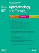
The clarity of a corneal graft can also provide long-term benefits over an opaque material such as sclera or pericardium. A previous study reports that e-beam treated corneas retained about 90% optical clarity relative to fresh corneas even after extended storage at room temperature [15]. In situations when the device is assessible, the clear cornea offers the surgeon the ability to visualize and inspect the device placement and sutures under the grafts (Fig. 1i, ii). Improved cosmesis can also be achieved with a clear corneal graft depending on the position of the implanted drainage device (Fig. 1ii, iii).
Fig. 1
Representative photographs of e-beam irradiated cornea and gamma irradiated sclera for glaucoma drainage device (GDD) patch grafts. i Intraoperative photograph of an e-beam irradiated corneal graft used to cover the GDD tube. Note the visible suture viewable under the clear corneal patch graft. ii Postoperative slit lamp photograph of an irradiated corneal graft covered by conjunctiva, 5 weeks after GDD surgery. The outlined area indicates the approximate position of the graft and the arrowheads point out sub-graft sutures which can be seen and inspected through the clear graft. iii A slit lamp photograph of a sclera patch graft used to cover a GDD
×
There are several limitations in our study. First, this is a retrospective analysis with a relatively short follow-up period. Loss of follow-up and incomplete electronic health record documentation may have underestimated erosion rates in our analysis. Additionally, our follow-up period may have been too short to detect erosions as this complication can occur well past our follow-up period. However, prior studies have found erosions as early as the first postoperative month [17] and 6.4 months [18] following GDD surgery. Lastly, if an erosion event occurred but was not surgically corrected, then the erosion would not have even been captured in our study. However, this would be highly unlikely, as the standard of care for an eroded graft and exposed GDD implant is surgical correction to avoid infection and endophthalmitis. Further studies with longer follow-up periods are underway to better characterize graft erosion rates in GDD surgery. Future prospective studies investigating surgical indications, complications other than erosion, and visual acuity, among other items, will further contribute to this area of research.
Conclusions
The comparable performance of e-beam irradiated corneas relative to scleral patch grafts for GDD surgery provides surgeons with another viable alternative to scleral tissue, while the sterile grafts provide an additional level of safety over fresh tissue or those preserved in glycerin.
Anzeige
Acknowledgements
We thank the participants of the study.
Funding
This study was funded by the National Eye Institute (Bethesda, MD; P30EY010572) and an unrestricted grant from Research to Prevent Blindness (New York, NY). All authors had full access to all of the data in this study and take complete responsibility for the integrity of the data and accuracy of the data analysis. The article processing charges were funded by the authors.
Authorship
All named authors meet the International Committee of Medical Journal Editors (ICMJE) criteria for authorship for this article, take responsibility for the integrity of the work as a whole, and have given their approval for this version to be published.
Disclosures
Khoa D. Tran is an employee of Lions VisionGift (Portland, OR). Corrina Patzer is an employee of Lions VisionGift. Shandiz Tehrani is a volunteer/unpaid member of the Lions VisionGift Medical Advising Committee. Ross M. Passo, Zachary B. Hoskins, Beth Edmunds, John C. Morrison, Mansi Parikh, and Hana L. Takusagawa have nothing to disclose.
Anzeige
Compliance with Ethics Guidelines
All procedures were in accordance with the Institutional Review Board of Oregon Health and Science University (FWA00000161; IRB00000471) and with the Helsinki Declaration of 1964, as revised in 2013, concerning human and animal rights. Springer’s policy concerning informed consent has been followed. Informed consent was obtained from all human subjects.
Data Availability
The datasets generated during and/or analyzed during the current study are available from the corresponding author on reasonable request.
Open Access
This article is distributed under the terms of the Creative Commons Attribution-NonCommercial 4.0 International License (
http://creativecommons.org/licenses/by-nc/4.0/
), which permits any noncommercial use, distribution, and reproduction in any medium, provided you give appropriate credit to the original author(s) and the source, provide a link to the Creative Commons license, and indicate if changes were made.
Bei Reizdarmsyndrom scheinen Diäten, wie etwa die FODMAP-arme oder die kohlenhydratreduzierte Ernährung, effektiver als eine medikamentöse Therapie zu sein. Das hat eine Studie aus Schweden ergeben, die die drei Therapieoptionen im direkten Vergleich analysierte.
Ob bei einer Notfalloperation nach Schenkelhalsfraktur eine Hemiarthroplastik oder eine totale Endoprothese (TEP) eingebaut wird, sollte nicht allein vom Alter der Patientinnen und Patienten abhängen. Auch über 90-Jährige können von der TEP profitieren.
Wenn unter einer medikamentösen Hochdrucktherapie der diastolische Blutdruck in den Keller geht, steigt das Risiko für schwere kardiovaskuläre Ereignisse: Darauf deutet eine Sekundäranalyse der SPRINT-Studie hin.
Insektenstiche sind bei Erwachsenen die häufigsten Auslöser einer Anaphylaxie. Einen wirksamen Schutz vor schweren anaphylaktischen Reaktionen bietet die allergenspezifische Immuntherapie. Jedoch kommt sie noch viel zu selten zum Einsatz.
Update Innere Medizin
Bestellen Sie unseren Fach-Newsletter und bleiben Sie gut informiert.