Leucine-rich glioma-inactivated protein 1 (LGI1) antibody-mediated encephalitis is a rare subtype of autoimmune encephalopathy, which is associated with autoimmunity against the neuronal plasma membrane proteins. The characteristic symptoms of this disease are memory dysfunction, seizures, faciobrachial dystonic seizures, cognitive deficits, neuropsychiatric disturbances, and intractable hyponatremia. The diagnosis of this disease mainly depends on the presence of anti-LGI1 antibody in serum or cerebrospinal fluid of patients. LGI1 antibody encephalitis has been reported mostly in adults, with rare occurrences in children.
Case presentation
In this report, we described a 4-year-old girl with typical seizures. Seizure types included focal seizures and generalized tonic-clonic seizures. The electroencephalogram findings showed focal discharges. Brain magnetic resonance imaging (MRI) showed normal. The cerebrospinal fluid (CSF) levels of cells, glucose, and chloride were within the normal range, and the culture did not reveal growth of any pathogen. Test of serum LGI1-Ab was positive, while the tests for autoimmune encephalitis antibody series in CSF were negative. The seizures of the patient were completely controlled after the therapy of immunoglobulin, methylprednisolone and antiepileptic drugs (AEDs), and the mental state almost returned to normal.
Conclusion
To our knowledge, the patient described here may be the youngest case of LGI1 antibody encephalitis reported to date. Children with the LGI1 antibody-associated encephalitis may present only with single symptoms such as epileptic seizures and have good response to the therapy of immunoglobulin, methylprednisolone and antiepileptic drugs. Our case report will provide hints for pediatricians in the diagnosis and treatment of LGI1-antibody encephalitis.
Hinweise
Junxia Luo and Jianguo Shi contributed equally to this work.
The original online version of this article was revised: ‘Ethics approval and consent to participate’ and 'Consent for publication' sections have been modified. Ethics approval has also been added to Background section.
The leucine-rich glioma inactivated 1 (LGI1) antibody-mediated encephalitis is a subtype of autoimmune encephalopathy, which is associated with the generation of antibodies against the neuronal plasma membrane proteins [1]. The characteristic symptoms of this disease include memory dysfunction, seizures, faciobrachial dystonic seizures (FBDS), cognitive deficits, neuropsychiatric disturbances, and intractable hyponatremia [2]. The diagnosis of LGI1 antibody-mediated encephalitis is based on the presence of LGI1 antibody in the serum or cerebrospinal fluid (CSF). LGI1-antibody encephalitis has been reported predominantly in adults, yet rarely in children.
In this report, we describe a 4-year-old Chinese girl with LGI1-antibody encephalitis who experienced fever over the course of 10 days and suffered multiple episodes of seizure, with abnormal electroencephalogram (EEG) findings during the natural course of the disease. To our best knowledge, this is the youngest case of LGI1-antibody encephalitis reported to date.
Anzeige
This study was approved by the Institutional Ethics Committee of Qilu Children’s Hospital of Shandong University and informed consent has been obtained from the guardian of patient prior to analysis.
Case presentation
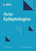
The case was a 4-year-old Chinese girl who was admitted to our hospital with generalized tonic-clonic seizures. She had an approximately 10-day history of discontinuous fever before the seizure onset. At the beginning of the course, she only experienced generalized tonic-clonic seizures, which lasted about 10 min and were then relieved. After hospitalization, she experienced frequent panic attacks with hypermotor seizure, while the heart rate was normal, and no auro or disturbance of consciousness occurred. This condition self-resolved within a few minutes but occurred with a frequency of dozens of times a day. No apparent positive signs were identified on a nervous system examination; however, she showed mild dysphoria, irritability, insomnia and decreased level of consciousness. Video electroencephalogram (VEEG; Fig. 1) revealed continuous epileptiform discharges in the left anterior region. In addition, two focal seizure onsets from the left anterior region were detected. Tumor marker and paraneoplastic neuronal antibody (anti-Hu, anti-Ri, anti-Yo, anti-Ma2/Ta, anti-CV2, and anti-Amphiphysin) tests were negative. The CSF levels of cells, glucose, and chloride were within the normal range, and CSF culture did not reveal growth of any pathogen. Test of serum LGI1-Ab was positive (titer, 1:30; Fig. 2), whereas the test for CSF LGI1-Ab was negative. Moreover, the tests for autoimmune encephalitis antibody series in CSF were negative. Brain magnetic resonance imaging (MRI) did not reveal abnormalities (Fig. 3). Serum sodium levels were within the normal range throughout the disease course. Abdomen ultrasound and chest radiography showed no signs of tumor.
Fig. 1
Video electroencephalography recordings. a EEG background is normal. Interictal EEG showed left forehead spike waves, sharp waves, and spike (or sharp) slow wave complexes. b Ictal EEG shows rhythmic sharp slow wave complexes in the left anterior head region, followed by low-voltage fast activity and rhythmic spike waves; these were contaminated by movements or muscle artifacts, and evolved to the left hemisphere, with occasional insertion of slow waves. The whole procedure lasted for 1–2 min. Moreover, scared facial expressions, hypermotor seizure were observed clinically (lasting for about 1–2 min). EEG, electroencephalogram
Fig. 2
The result of anti-leucine-rich, glioma inactivation-1 antibody. A 4-year-old girl with positive serum anti-leucine-rich, glioma inactivation-1 antibody on indirect immunofluorescence test (green fluorescence)
Fig. 3
MRI showed unremarkable findings before (a) and after (b) treatment
×
×
×
The patient was treated with intravenous immunoglobulin (1 g/kg per day for 4 days) (divided into two doses) and methylprednisolone pulse therapy (50 mg/kg per day for 3 consecutive days; 3 times total with intervals of 4 days; total course lasting 21 days), in combination with antiepileptic drugs (AEDs) (levetiracetam 0.2 g bid, clonazepam 0.25 mg bid, and oxcarbazepine 0.15 g bid). The patient had good response to immunotherapy, with no recurrence of seizure after the first stage of immunotherapy. Simultaneously, her panic attacks subsided. The prednisolone (15 mg; daily oral) therapy was administered subsequently. The dosage was gradually reduced throughout the treatment for 3 months. At 15 days after discharge, the VEEG examination was performed again and had normal results, except the presence of mild sharp waves in the left forehead (Fig. 4). During the 60-day follow-up, seizures did not recur, and the mental state of the patient returned to the level before disease onset.
Fig. 4
EEG recordings obtained after half a month. a EEG background appears normal. b Interictal EEG shows few spike waves and sharp waves in the sleep stage in the left forehead. EEG, electroencephalogram
×
Discussion
According to the Chinese expert consensus on the diagnosis and management of autoimmune encephalitis, the criteria for definite diagnosis of LGI1-antibody encephalitis are as follows: (1) having acute or subacute onset with progressive aggravation; (2) presenting with clinical features of limbic encephalitis or FBDS; (3) having normal leukocyte count or mild lymphocyte reaction on CSF examination; (4) showing abnormal brain MRI signals in the bilateral or unilateral medial temporal lobe; (5) showing abnormal EEG activity; and (6) tested positive for serum and/or CSF anti-LGIl antibody. The case presented here had an acute onset of the disease and experienced frequent panic attacks, which is a clinical feature of limbic encephalitis. These symptoms, combined with the abnormal EEG findings and positive anti-LGIl antibody in the serum and CSF, supported the diagnosis of LGI1-antibody encephalitis.
Anzeige
The LGI1-antibody encephalitis occurs predominantly in males, with a mean onset age of ~ 60 years (typical range, 30–80 years) [3]. Pediatric cases have been rarely reported [4]. Schimmel reported in 2017 a 14-year-old boy who was diagnosed with the LGI1-antibody encephalitis [5], and Zhang et al. reported an 8-year-old Chinese boy with the symptom of reduced night-time sleep in 2018 [6]. Therefore, to our best knowledge, the patient reported here is the youngest case reported to date.
Autoimmune encephalitis is a type of inflammation in the central nervous system. Studies have reported that the LGI1-antibody encephalitis is the second most common form of autoimmune encephalitis [7]. There is a high incidence of epileptic seizures during the acute phase of autoimmune encephalitis. In this context, the seizures may be an acute or induced symptom, and can be considered as autoimmune seizures, which has a prevalence of 60–100% [8]. According to the 2014 edition of Epilepsy Usability Definition by the International League Against Epilepsy (ILAE) [9], the acutely provoked or acute symptomatic seizures at this stage cannot yet be defined as epilepsy. In 2017, the ILAE further proposed a new classification of epilepsy, the “epilepsy of immune etiology”, for patients whose epilepsy “results directly from an immune disorder in which seizures are a core symptom of the disorder” [10]. Multiple frequent seizure semiology and subclinical seizures associated with temporal and frontal discharges have been reported in the LGI1-antibody encephalitis patients [11], and in our case the discharges were also concentrated in the frontal lobe. Some cases also showed frequent early seizures of encephalitis as clinical manifestations [12]. Consistently, the case in our report also displayed seizures as the first symptom.
In addition, the symptoms, the presence of antibody, and the treatment response of this case were all similar to those of adult patients. However, our patient did not show FBDS or hyponatremia, which are hallmark symptoms in adult patients with LGI1-antibody encephalitis [13]. Therefore, the diagnosis of LGI1-antibody encephalitis should be considered if pediatric patients display acute and progressing unexplained frequent episodes of seizures, in order to prevent misdiagnosis and missed diagnosis. In fact, seizures are extraordinarily frequent in the acute, inflammatory-provoked phase of many types of antibody-mediated encephalitis, especially in the LGI1-antibody encephalitis, but in most patients the seizures are not sustained and will resolve after the encephalitis remission [8]. However, a recent study showed that after 2 years’ follow-up, 14% of patients with LGI1-antibody encephalitis still had seizures and an additional 14% were still on AEDs after the encephalitis was cured [1]. Some scholars have suggested that the epileptiform seizures in the acute phase of autoimmune encephalitis cannot be diagnosed as epilepsy, instead, it should be followed up for at least 1 year to detect the presence of persistent seizures to determine the continued use of AEDs [8]. It is currently believed that the abnormalities in EEG and brain imaging are the most predictive factors for epilepsy after autoimmune encephalitis [14]. In this report, the patient showed normal results of brain imaging, and her seizures and EEG abnormality were improved as her encephalitis was controlled. However, as the follow-up time was not long enough in this report, we would continue to follow up to observe the prognosis of this patient.
Conclusion
The LGI1-antibody encephalitis is a newly discovered autoimmune disease in recent years, which is characterized by specific FBDS and hyponatremia and responds well to immunosuppressive therapy. Here we reported a 4-year-old Chinese girl with LGI1-antibody encephalitis, who may be the youngest patient reported in literature. Children with the LGI1 antibody-associated encephalitis may present only with single symptoms such as epileptic seizures while other symptoms of encephalitis may not appear, particularly in younger age groups. This study demonstrates that early diagnosis and treatment have more benefits for seizure control and improvement of mental recovery. The diagnosis of this disease should be confirmed by anti-LGI1-antibody detection, cranial MRI examination, VEEG and other assistant examinations to avoid missed diagnosis. Our case report will provide hints for pediatricians in the diagnosis and treatment of LGI1-antibody encephalitis.
Acknowledgements
We thank the affected individuals and their families for participating in this report.
Ethics approval and consent to participate
This study was approved by the Institutional Ethics Committee of Qilu Children’s Hospital of Shandong University and informed consent has been obtained from the guardian of patient prior to analysis.
Consent for publication
All authors and the guardian of patient agreed for the publication of this study.
Competing interests
The authors have declared that they have no competing interests.
Anzeige
Open AccessThis article is licensed under a Creative Commons Attribution 4.0 International License, which permits use, sharing, adaptation, distribution and reproduction in any medium or format, as long as you give appropriate credit to the original author(s) and the source, provide a link to the Creative Commons licence, and indicate if changes were made. The images or other third party material in this article are included in the article's Creative Commons licence, unless indicated otherwise in a credit line to the material. If material is not included in the article's Creative Commons licence and your intended use is not permitted by statutory regulation or exceeds the permitted use, you will need to obtain permission directly from the copyright holder. To view a copy of this licence, visit http://creativecommons.org/licenses/by/4.0/.
Der Einsatz von Antipsychotika gegen psychische und Verhaltenssymptome in Zusammenhang mit Demenzerkrankungen erfordert eine sorgfältige Nutzen-Risiken-Abwägung. Neuen Erkenntnissen zufolge sind auf der Risikoseite weitere schwerwiegende Ereignisse zu berücksichtigen.
Eine ältere Frau trinkt regelmäßig Sennesblättertee gegen ihre Verstopfung. Der scheint plötzlich gut zu wirken. Auf Durchfall und Erbrechen folgt allerdings eine Hyponatriämie. Nach deren Korrektur kommt es plötzlich zu progredienten Kognitions- und Verhaltensstörungen.
Mit einem Neurotrophin-Rezeptor-Modulator lässt sich möglicherweise eine bestehende Alzheimerdemenz etwas abschwächen: Erste Phase-2-Daten deuten auf einen verbesserten Synapsenschutz.
Ein hohes soziales Niveau ist mit die beste Versicherung gegen eine Demenz. Noch geringer ist das Demenzrisiko für Menschen, die sozial aufsteigen: Sie gewinnen fast zwei demenzfreie Lebensjahre. Umgekehrt steigt die Demenzgefahr beim sozialen Abstieg.
Update Neurologie
Bestellen Sie unseren Fach-Newsletterund bleiben Sie gut informiert.